PHERAstar FSX
Powerful and most sensitive HTS plate reader


Barry Whyte is Application Scientist and Science Writer at BMG LABTECH in the United States. He has PhD and Bachelor of Science (BSc) degrees in biochemistry from the University of Bristol in the United Kingdom and more than 20 years of experience in the life sciences and science communications. Over the years, Barry has worked on three continents and traveled widely. He enjoys building on his international work experience and learning new ways to help scientists advance their research.
A receptor-ligand interaction involves the binding of a molecule (ligand) to a site on a receptor. Receptors are detection and cell signaling molecules inextricably linked to the activities of cells1. Ligands are diverse molecules with the ability to interact with receptors. They may be endogenous (naturally occurring) molecules or synthetic ligands such as drugs.
Receptor-ligand interactions are crucial on two fronts. First, they play important roles in the biology of the cell by triggering a cellular response or altering receptor activity. Secondly, receptor-ligand interactions are often important targets for drug discovery and development due to their pivotal role in cell biology.
In this blog, we look at receptor-ligand interactions: what they are, where receptor-ligand interactions occur, and how they are studied. We also look at the benefits offered by a microplate reader to investigate receptor-ligand interactions and provide some examples of specific applications. The different detection options for receptor-ligand interactions are highlighted.
.
Receptor-ligand interactions are fundamental molecular events involved in the molecular biology of the cell. Receptors come in many different forms and are found at diverse locations within a cell, but they typically operate through a very specific interaction with ligands. Receptors may show a strong preference for one ligand. Others are less selective.
Some receptors involved in receptor-ligand interactions, such as G-protein coupled receptors (GPCRs), are found exclusively on the external membrane of the cell. Other receptors that bind ligands are located within the cell as we will see later. Ligand-gated ion channels, which are formed by specific arrangements of protein subunits that create functional channels for ion passage upon ligand binding, are an example of receptor types that may occur on the external membrane or on a membrane within the cell.
As mentioned earlier, a receptor-ligand interaction occurs when a molecule or ligand binds to a specific site on a receptor, an event that may trigger a cellular response or alter receptor activity. The binding event in a receptor-ligand interaction is typically non-covalent, relying on hydrogen bonds, electrostatic interactions or Van der Waals forces and these forces are reversible which means that they are dynamic interactions.
Ligands themselves come in many shapes or sizes. They include small molecule ligands (neurotransmitters, metabolites and most drugs), peptide ligands (hormones, neuropeptides, chemokines), protein ligands (growth factors, cytokines), lipid-derived ligands (steroids, cannabinoids), nucleic acid ligands (RNA, DNA fragments), and ions or inorganic ligands (calcium, sodium). Water-soluble ligands bind to the extracellular domains of cell-surface receptors, initiating signaling pathways related to hormones, neurotransmitters, and growth factors. This diversity of ligands is reflected in the different types of receptors. Proteins, including many receptors and ligands, are composed of amino acids, and the specific sequence and arrangement of these amino acids determine their structure and function in receptor-ligand interactions. 1-3
Operationally, the binding of ligands to a receptor in a receptor-ligand interaction alters receptor activity or triggers a response within a cell. This receptor-ligand interaction leads to changes that alter other molecules, subsequent molecular processes and biological pathways downstream of the initial point of interaction. In cell signaling, signaling cells produce and release ligands, which then travel to and bind specific receptors on target cells, initiating a response. These interactions are essential for many biological processes and can be measured using cell-based or in vitro assays, many of which can be performed on a microplate reader. 1
Receptor-ligand interactions range from hormones regulating metabolism to neurotransmitters involved in neuronal function. These molecular encounters govern how cells respond to their environment. Vision, smell and taste, to name just a few outputs, arise from cascades of signaling pathways within cells set in motion by receptor-ligand interactions. Understanding how tight, quickly and with what consequences a ligand binds to a receptor is a fundamental part of not only understanding biology but also ensuring the success of the drug discovery process. Quantifying the binding affinity of a ligand is essential, as it determines the potency of the ligand. The extent of a physiological response is often proportional to the amount of ligand bound to its receptor in a receptor-ligand interaction and to receptor–ligand kinetics.
The physiological response to receptor-ligand interactions is a finely tuned process that underpins many biological processes essential for life. When a ligand binds to its specific receptor—whether on the cell surface or within the cell—it initiates a cascade of molecular events that ultimately shape the function and fate of the target cell (Fig. 1).
For most water-soluble ligands, such as peptide hormones or neurotransmitters, the journey begins at cell surface receptors embedded in the plasma membrane. Here, ligand binding induces a conformational change in the receptor’s structure, activating its intracellular domain. This activation sets off a signal transduction pathway, often involving a series of protein-protein interactions and the recruitment of signaling molecules like G-proteins or kinases. These signaling molecules propagate the signal through the cell, amplifying the response and engaging downstream effectors such as ion channels or transcription factors.
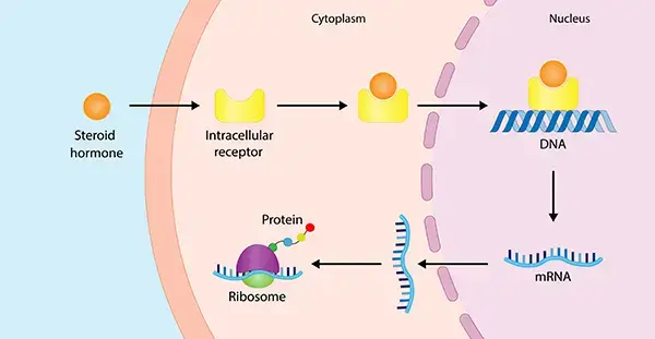
In contrast, hydrophobic ligands—such as steroid hormones—can diffuse directly across the plasma membrane and interact with intracellular receptors. These intracellular receptors often function as ligand-activated transcription factors, binding to specific regions of DNA and regulating the expression of certain genes. This direct modulation of gene expression can lead to long-term changes in cellular metabolism, growth, or differentiation, illustrating how small molecules that act as ligands can have profound effects on cellular processes.
The magnitude and nature of the cellular response due to receptor-ligand interactions depend on several factors, including the free ligand concentration, the total concentration of receptors, and the binding affinity between ligand and receptor. High-affinity interactions, characterized by a low dissociation constant (Kd), typically result in more robust and sustained signaling, while lower affinity interactions may produce more transient or subtle effects. The presence of co-receptors, inhibitory ligands, or other molecules can further modulate the response, either enhancing or dampening the signaling output.
These complex biomolecular and receptor-ligand interactions—spanning proteins, nucleic acids, and small molecules—are central to the regulation of gene expression, cellular metabolism, and other vital cellular processes. For example, the activation of transcription factors following receptor-ligand binding can lead to the upregulation or repression of genes involved in cell proliferation, migration, or survival. In this way, receptor-ligand interactions serve as the molecular switches that control many biological processes and physiological responses.
Ultimately, the study of physiological responses to receptor-ligand interactions not only deepens our understanding of fundamental biology but also drives the development of new therapies for diseases ranging from heart disease to cancer. By leveraging high-performance microplate readers and advanced detection technologies, scientists can dissect these molecular events with precision, paving the way for innovative drug discovery and improved health outcomes.
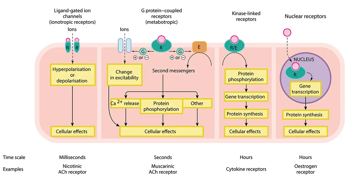
Cell receptors come in many forms and operate in different locations of the cell (on one level either extracellular or intracellular) as we hinted earlier. Several broad categories are recognized, each mediating different signaling pathways. These include GPCRs, ion channel receptors, kinases (e.g. receptor tyrosine kinases), nuclear receptors, and intracellular receptors. Here we describe the main categories of receptors and give a few examples of applications on microplate readers suitable for each group (see also summary in Table 1).
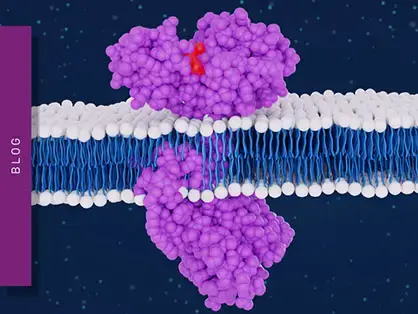
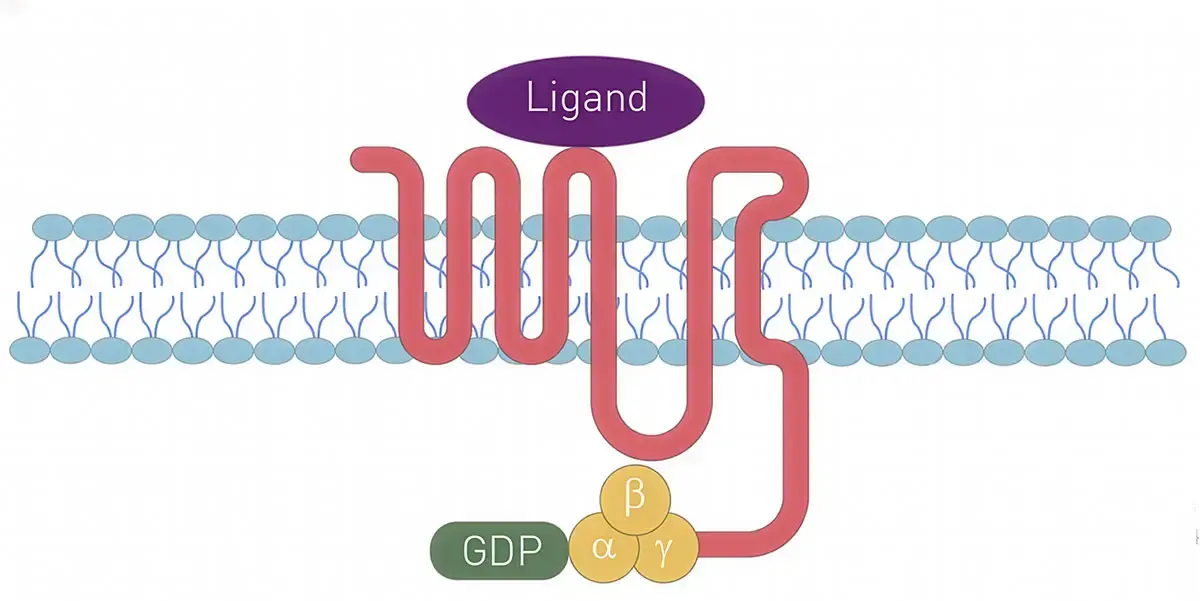
GPCRs are signaling proteins found in the membrane that surrounds a cell (Fig. 2). They are seven-pass transmembrane domain receptors engaged in receptor-ligand interactions that allow the cell to detect an extracellular signal and convert that signal into an intracellular signal. The N-terminus of the receptor resides on the outside of the cell and is followed in turn by seven α-helices that pass through the cell membrane. The extracellular part of the receptor binds the ligand or stimulus. The end of the receptor or C-terminus sits within the cell. The intracellular C-terminus is crucial for interacting with GTP-binding proteins or G-proteins and sets in motion the signaling cascade within the cell. 4 More than 30% of FDA-approved drugs target GPCRs.5
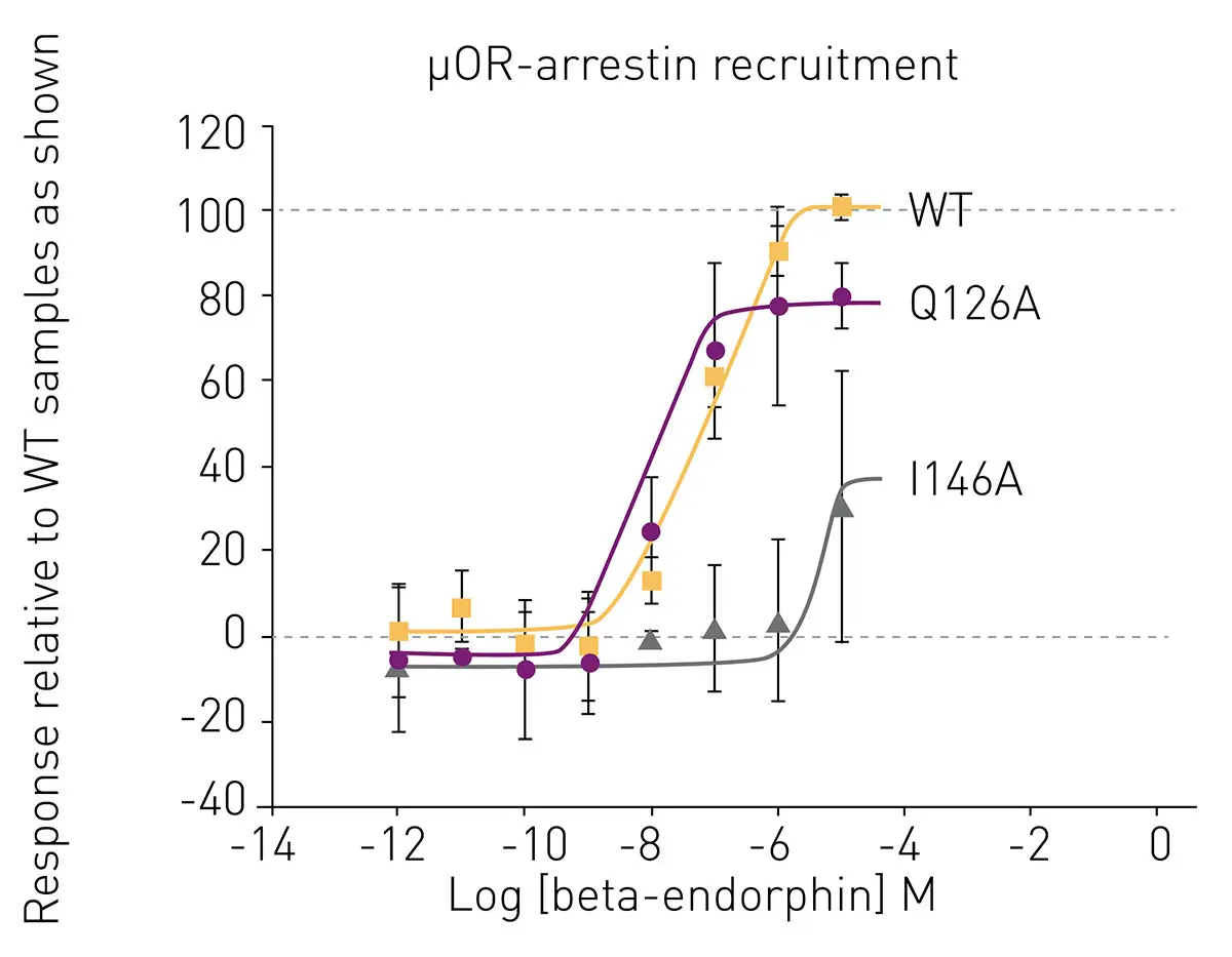
In the application note AN383: Structure-guided monitoring of the human opioid receptor family using BRET assays G protein dissociation and β-arrestin recruitment assays were used to facilitate a framework for the rational design of safer opioid drugs that target GPCRs. Bioluminescence Resonance Energy Transfer (BRET) assays, a type of luminescence assay, were employed to investigate wild-type and mutant proteins of the human opioid receptor family using the TRUPATH platform. TRUPATH is an open-source biosensor platform that can be used to interrogate GPCR transduction. BRET assays were performed on the PHERAstar® FSX (Fig. 3). The Simultaneous Dual Emission detection halves read time and improves data quality for all dual emission assays including BRET as the reader can detect two emission wavelengths (e.g., donor and acceptor) in a single measurement.
 The application note AN386 Competition assay using Fluorescence Polarisation to determine the Residence Times for Calcitonin and AMYR agonist, AM833 describes competition assays on the PHERAstar® FSX that do not require receptor or compound labelling and which are useful to study GPCRs. Competition assays are used to determine if two or more molecules bind to the same site and allow the binding affinity to be determined. A labeled ligand that is known to bind a target is incubated with that target in the presence of unlabeled ligand. If the competing ligand binds to the same site, it will displace the labeled ligand in a concentration-dependent manner. Amylin and calcitonin are peptide hormones that act through GPCRs. The AMYR agonist AM833 used in the study represents a promising novel alternative for the treatment of obesity. The high sensitivity in fluorescence polarization and simultaneous dual emission of the PHERAstar® FSX allowed for very fast kinetic measurements. The use of fluorescence polarization for Kon and Koff rate determination in competition assays represents a useful alternative strategy to existing methods for evaluating agonists and antagonists of GPCRs.
Cannabinoid receptors are GPCRs that lead to diverse cellular events mediated through different cell signaling pathways. In the application note AN388 Differential binding of ∆9-tetrahydrocannabinol derivatives to type 1 cannabinoid receptors (CB1), CB1 receptor-ligand interactions were detected with the PHERAstar® FSX at high sensitivity. The study made use of CELT-335, a dual (CB1/CB2) fluorescent ligand that serves as a Time-Resolved Fluorescence Resonance Energy Transmission (TR-FRET) acceptor compatible with terbium. Natural cannabinoids differentially bound to CB1 and CB2 receptors resulting in qualitatively different effects. The binding affinities determined using the TR-FRET method compared favorably to those determined by radioligand binding assays.
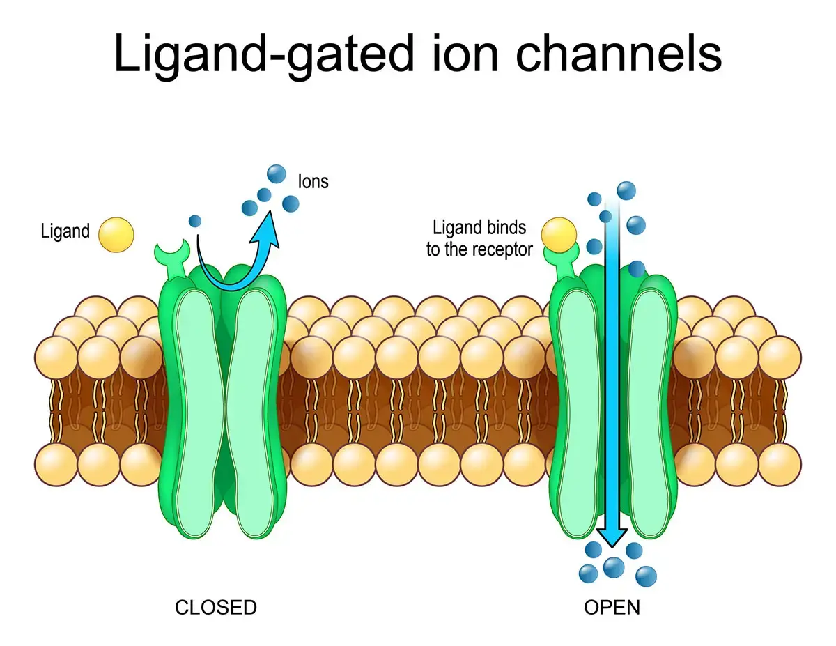
Ion channel receptors directly control the flow of ions like calcium or sodium across cell membranes (Fig. 4). They represent another broad category of receptors found on the cell surface or within the cell. The binding of a ligand (e.g. a neurotransmitter or small molecule) to an ion channel receptor can open or close the channel. Ion channels open and close quickly (in microseconds to milliseconds) and are important because they convert chemical or physical signals into rapid electrical and ionic changes ensuring fast communication, responses, and control of homeostasis within the cell. Without ion channels, thought, movement or reflexes would not be possible.
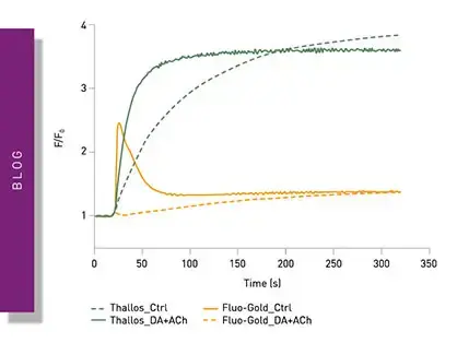
Examples of ion channel receptors include the nicotinic acetylcholine receptor, calcium channels controlling the release of neurotransmitters and muscle contraction, as well as taste receptors. In the application note AN402 Adaptation of a potassium channel assay to enable easier high throughput screening use of the Brilliant Thallium Snapshot assay enabled an endpoint HTS read on the PHERAstar FSX in 1536-well plates (Fig. 5). Ion channels are involved in many diseases, but treatments that target ion channels are lacking. The fluorescence-based assay described in this application note was shown to be a cost-effective, accurate alternative to potassium channel assays requiring kinetic imaging readers.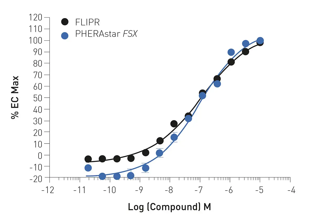
Receptor tyrosine kinases are another group of receptors that play significant roles in growth, development and disease (Fig. 6). These receptors are located in the cell membrane and involve ligands such as growth factors. Ligand binding promotes receptor dimerization. Examples include the epidermal growth factor receptor, platelet-derived growth factor receptor, the insulin receptor family, and the vascular endothelial growth factor receptor. Receptor tyrosine kinases are important because they translate extracellular growth and environmental cues into long-lasting cell signaling events that may control cell fate including cell proliferation, survival, differentiation, metabolism and migration.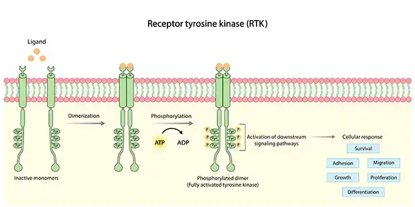
The application note AN317 NanoBRET™ assay quantitatively evaluates VEGF binding to the VEGFR2 in real-time in living cells describes a novel labelling technique that fluorescently labels the tyrosine kinase receptor VEGFR for use in NanoBRET interaction studies. The assay acquires quantitative information on ligand-receptor interaction which is suitable for molecular pharmacology studies of VEGFR (Fig. 7). The PHERAstar simultaneously detects luciferase and fluorophore signal for accelerated kinetic reads.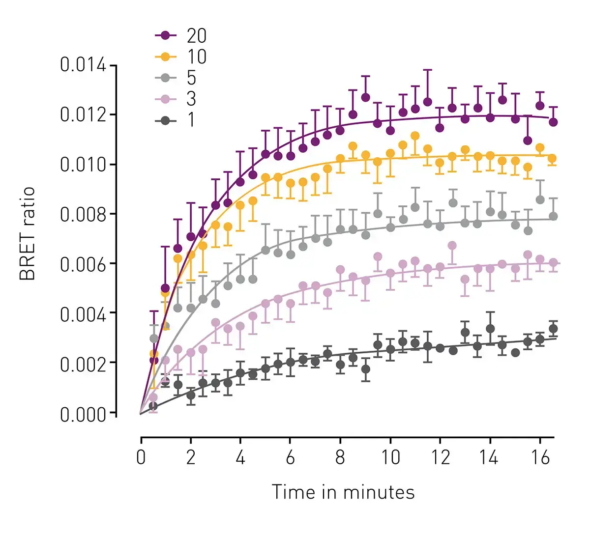
Nuclear receptors are ligand-activated transcription factors that may occur in the cytoplasm (in the unbound state) or nucleus of the cell. They do not signal via second messengers like GPCRs but act at the level of gene regulation. These intracellular receptors bind small, hydrophobic ligands that can cross the plasma membrane including steroid hormones (Fig.8), thyroid hormones or retinoids. Examples of this type of receptor include the estrogen receptor, glucocorticoid receptor or the androgen receptor. When they bind a ligand, these receptors typically translocate to the nucleus where they bind DNA and regulate gene expression. Nuclear receptors are important because they allow direct coupling of small cellular signals to gene regulation thus allowing cells to make coordinated, long-term physiological responses related to development, metabolism and homeostasis.
Fluorescence and luminescence-based (Fig. 9) nuclear receptor assays are highlighted in AN392 Identification of androgen-disruptors using a cell-based androgen receptor dimerization assay. Here an androgen receptor dimerization assay (AR2) enables screening of agonists, antagonists and xenobiotic metabolites. The AR2 receptor dimerization assay is a viable alternative to existing toxicology assays for androgen disruptors. Detection using NanoLuc® Binary Technology (NanoBiT®) was performed on the CLARIOstar® Plus microplate reader.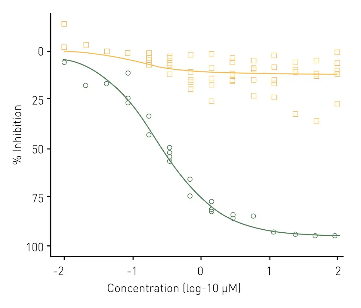
Table 1. Selected overview of some BMG LABTECH application notes for receptor-ligand interactions.
The applications highlighted earlier for the different groups of receptors are testament to the diversity of ways available to study receptor-ligand interactions, which are a subset of broader macromolecular interactions critical to understanding biological processes. Functional receptor activation is usually assessed by fluorescence and luminescence measurements, with careful attention to the composition of the solution in which receptor-ligand interactions are measured, as it can significantly impact assay results. If kinetic information is required or live cell interactions are involved then fluorescence, Förster´s Resonance Energy Transfer (FRET), or BRET luminescence assays are all options.
The choice of method, such as ultracentrifugation, gel filtration, or equilibrium dialysis, for separating or analyzing receptor-ligand complexes before studying receptor-ligand interactions can influence the accuracy and suitability of binding measurements depending on the experimental context. Direct binding affinity of receptors to ligands is most often measured by fluorescence polarization, FRET, Time-Resolved Fluorescence Resonance Energy Transfer (TR-FRET) or endpoint assays like AlphaScreen and AlphaLISA®. Multi-mode microplate readers that are specifically designed to support these technologies in parallel are versatile platforms to study receptor-ligand interactions. Other options for investigation of receptor-ligand interactions also exist. Isothermal Titration Calorimetry (ITC) allows for quantitative, label-free analysis of macromolecular interactions, providing valuable thermodynamic data. Additionally, nanoanalytical tools like atomic force microscopy and optical tweezers have increasingly been used to analyze receptor function.
While we have already highlighted some of the technological features of a microplate reader that facilitate receptor-ligand interaction studies for specific applications, here we summarize some of the key technology assets for performance.
Dual‑emission detection is often used to measure indirect assays that serve as valuable proxy measurements for receptor-ligand interactions. The Simultaneous Dual Emission detection, which is available on the PHERAstar FSX, halves read time and improves data quality for all dual emission assays such as FRET, TR-FRET, fluorescence polarization, and BRET, as the reader can detect two emission wavelengths (e.g., donor and acceptor) in a single measurement. This helps improve accuracy of measurements for binding curves and competition assays, reducing well-to-well variability and improving precision.
The PHERAstar FSX, VANTAstar and the CLARIOstar Plus include Enhanced Dynamic Range (EDR) technology for superior performance in a single luminescence or fluorescence run. This ensures accurate signal quantification across low to high concentrations of ligands and targets without running into the risk of signal saturation.
The PHERAstar FSX and the CLARIOstar Plus offer a sampling rate of 100 data points per second. This is particularly advantageous for resolving fast kinetic processes and for generating higher‑density datasets of interaction events.
The ability to inject reagents and simultaneously detect a signal is likewise useful for fast reactions and kinetic measurements. Injectors are therefore great assets for reading in kinetic well mode since they offer the best performance in terms of speed and throughput (sampling rate). An injector should offer low dead volumes and the ability to back flush for precious reagents. All BMG LABTECH microplate readers come with up to two injectors per reader and are compatible with plate formats with up to 384 wells.
High throughput is beneficial when profiling large ligand libraries against different receptors. Support for 96-, 384-, 1536- and 3456-well microplate formats helps accelerate screening campaigns.
BMG LABTECH readers offer excellent robotic integration capabilities, multi-user control, digital signature and FDA 21 CFR part 11 compliance. Their robotic software interface makes them easy to integrate into all leading robotic platforms.
The PHERAstar FSX is the reader of choice for high-throughput screening. For assay development, the CLARIOstar® Plus is BMG LABTECH`s high-end monochromator-based reader. The VANTAstar offers similar flexibility to the CLARIOstar Plus, as well as EDR technology and Atmospheric Control Unit compatibility (see below) for controlled live cell conditions in a more streamlined format.
In the application note AN403 Identification of peptide ligands for orphan GPCRs by measuring Ca2+-induced luminescence in transfected cells the VANTAstar® with injectors was used for cell-based assays looking at GPCR deorphanization. This type of study provides new insights into the evolution, physiological roles and therapeutic potential of orphan GPCRs. Cell-based assays with promiscuous/chimeric G-proteins are useful to identify ligands for a huge variety of GPCRs. The VANTAstar multimode-microplate reader offers an option for a reagent injector compartment that includes a built-in magnetic stirrer heating plate ideally suited for performing cell-based assays that enable GPCR deorphanization and which enable the necessary manual steps to be reduced and the process to be standardized.
The Atmospheric Control Unit from BMG LABTECH provides researchers with a system that uniquely enables control of both the oxygen and carbon dioxide concentrations in an independent manner. Receptor-ligand interaction studies may be performed in live cells and atmospheric control is therefore another useful feature for these studies.
Most microplate readers can control CO2 and O2 levels in the range of 1-20%. In some cases, they can decrease O2 to 0.1%. The CLARIOstar® Plus with ACU can reach 0.1% O2 and can read at 100 datapoints per second which makes it an ideal selection for plate and well mode kinetic measurements that require specific atmospheric conditions and rapid sampling.
Temperature can affect kinetic binding parameters. All BMG readers allows precise and stable incubation up to 45°C. The Advanced Assay Stability (AAS) system on the PHERAstar FSX maintains constant temperatures between 18 and 45°C by automatically heating or cooling the measurement chamber. This prevents environmental fluctuations from affecting assay performance, enabling consistent conditions throughout entire batches or screening campaigns.
The number of studies using receptor-ligand interaction assays on BMG LABTECH microplate readers continues to grow at pace. These studies are benefitting from more options for advanced kinetic measurements, the adoption of live cell and native-context assays, improvements to in vitro assays, as well as better integration of measurements to predict toxicology and in vivo pharmacology for early drug discovery. High throughput discovery workflows are improving all the time and with these improvements come efficiencies and better use of resources.
Whatever your requirements, BMG LABTECH has the microplate reader for your endpoint and kinetic mode assay applications.
The PHERAstar FSX was specifically conceived for screening campaigns and is your go-to reader for the fastest high-performance high-throughput screenings.
Both the VANTAstar® and CLARIOstar Plus allow for wavelength flexibility and include Enhanced Dynamic Range technology for superior performance in a single run of endpoint and kinetic mode assays. They also offer increased light transmission and sensitivity courtesy of Linear Variable Filter Monochromators™ and different filter options for endpoint and kinetic mode assays.
All BMG LABTECH microplate readers have exceptionally fast reading capabilities for endpoint and kinetic mode assays, which, together with the well mode function, ensure that sample reactions are recorded from start to finish without losing any important data points. In addition, the Omega series, VANTAstar, CLARIOstar Plus, and PHERAstar FSX microplate readers can be equipped with up to two reagent injectors that can offer the very best options for detection at the time of injection for endpoint and kinetic mode assays.
Collectively, BMG LABTECH multi-mode readers combine high-quality measurements with miniaturised assays, short measurement times, and offer considerable savings on materials and other resources for endpoint and kinetic mode assays.
Powerful and most sensitive HTS plate reader
Most flexible Plate Reader for Assay Development
Flexible microplate reader with simplified workflows
Binding constants quantify the strength of a binding reaction between a biomolecule and its target (ligand). But how do you measure them and what can you do with them?
Receptor-ligand kinetics is the study of the rates at which receptors and ligands interact, bind and dissociate. Learn why these types of measurements are important and how to measure them.
Redox processes play an important role in cell homeostasis. Read here, how to monitor cellular redox changes with roGFP using the most sensitive, high-performance plate readers from BMG LABTECH.