Introduction
Ion channels are the second largest family of membrane proteins with at least 400 members. They are responsible for numerous normal physiological functions throughout the body serving as the means to transport ions across the cell membrane1.
Channelopathies are the collection of genetic diseases that are caused by mutation of ion channel proteins. Therapies that target ion channels could be beneficial to a wide variety of diseases including cancer and autoimmune diseases. Despite extensive drug discovery efforts only a small number of ion channel family members have been successfully targeted for treatment. Herein we describe an approach to expand the utilization of ion channel assays that will be beneficial in the search for additional therapies1.
Thallium-sensitive dye-based methods have made it possible to measure monovalent ion channels such as potassium channels. However, they typically employ kinetic imaging readers that are not common laboratory equipment. Here, a modified potassium channel assay is demonstrated, that can be detected with the PHERAstar FSX 2 with comparable results to kinetic modality readers.
Assay principle
Cells expressing G protein-gated inwardly rectifying potassium channels (GIRK) were loaded with thallium as surrogate for potassium and Thallos, a thallium sensitive dye (Figure 1). 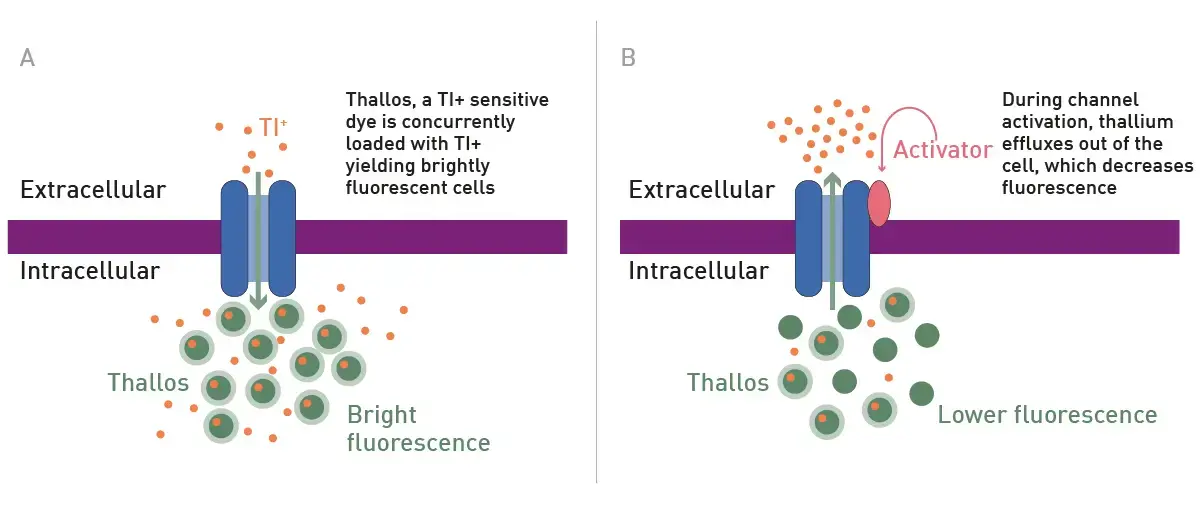
A) The loading step B) Compound activation of a GIRK leads to efflux of thallium (Tl-) and decreased fluorescence signal.
Materials & methods
- CHO-G12 cells and Brilliant Thallium Snapshot Flex kit (Ion Biosciences)
- 1536, black, TC-treated, microplates (#782078, Greiner)
- LOPAC 1280 Library (Sigma-Aldrich)
- PHERAstar FSX (BMG LABTECH)
- for the origin of other chemicals and reagents please see2
Experimental Procedure
To seed CHO-G12 cells, the appropriate concentration was prepared to result in indicated cell number and 2.5 µL was dispensed to each well. Plates were centrifuged at 500 g for 5 min, followed by 24 h in an incubator (37°C, 5% CO2). 2.5 µL of Brilliant Thalium Snapshot loading solution was dispensed to all wells. Plates were centrifuged at 500 g for 5 min, followed by 30 min incubation at 37°C and 5% CO2.
A T0 read was performed using the settings indicated in the table below, followed by addition of 30 nL. Following the indicated incubation es were read again (Tf). For detailed information on the method please see 2.
Instrument settings
|
Fluorescence Intensity, end point
|
||
|
Optic settings
|
Optic System
|
Top Optic
|
|
Optic Module
|
FI 485 520
|
|
| Gain |
400 |
|
|
Focal height
|
7.7 |
|
|
General settings
|
Flashes
|
1 |
Results & Discussion
The optimization of the endpoint readout version of this assay began with understanding the effect of cell numbers. The initial assessments shown in Figure 2 were performed at the 5-minute time point.
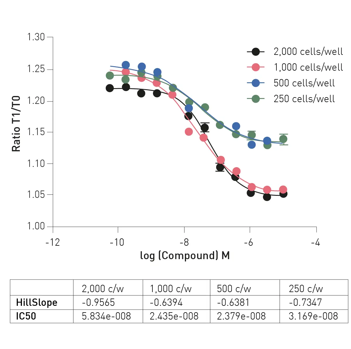
The example data in Figure 2 indicates that 1000 cells/well exhibit good assay quality metrics. Extending the compound incubation time to 10 min showed additional improvement in assay quality (data not shown). Figure 3 shows the comparison between the PHERAstar FSX and an imaging reader under the optimized conditions.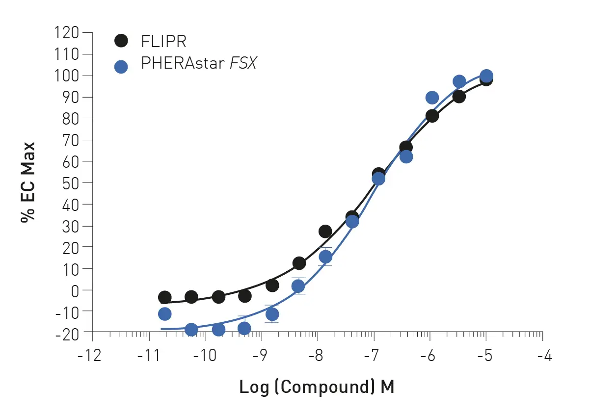
As further proof of concept the adapted assay was employed in a pilot screen using the LOPAC 1280 compound library. Each plate contained a quality control to determine plate Z’ and only plates with a score of 0.5 or higher were deemed acceptable. Comparison of triplicate screening plates showed reproducibility (data not shown).
Following the pilot HTS, one compound exhibited confirmed dose responsiveness (Fig. 4). Zimelidine dihydrochloride is a known serotonin reuptake inhibitor.
Conclusion
We show the adaptation of Snapshot technology for an endpoint potassium channel assay. The LOPAC pilot screening revealed no new hits, which is in line with what was anticipated. This simplified assay approach should prove useful for larger diversity library HTS campaigns which is now enabled to a much greater number of screening labs that may not have access to kinetic imaging readers.
References
- Hutchings, C.J., et al. Ion Channels as therapeutic antibody targets. MABS. (2019) 11(2): 265-296
- Smith, E., et al. Protocol for kinetic mode potassium channel assays on common plate readers and microscopes. SLAS Discovery (2024) 29: 100148