PHERAstar FSX
Powerful and most sensitive HTS plate reader


This author profile refers to work created by Dr Andrea Krumm during her tenure at BMG LABTECH - the Microplate Reader Company, where she served as an Applications Specialist contributing scientific content, application notes, and technical expertise. Dr Krumm is no longer employed at BMG LABTECH, but her published materials remain available here for reference and archival purposes. Dr Andrea Krumm is a biotechnology specialist and product manager known for her work in analytical instrumentation and biopharmaceutical analysis. She studied biotechnology and later earned a PhD in cancer biology, focusing on topics such as DNA repair, epigenetics, and tumor biology. After completing her doctorate, she spent several years as an Applications Specialist at BMG LABTECH, where she authored application notes, conducted workshops, and supported scientific customers. In 2020, Dr Krumm joined Tosoh Bioscience GmbH as Product Manager for Analytical Columns.
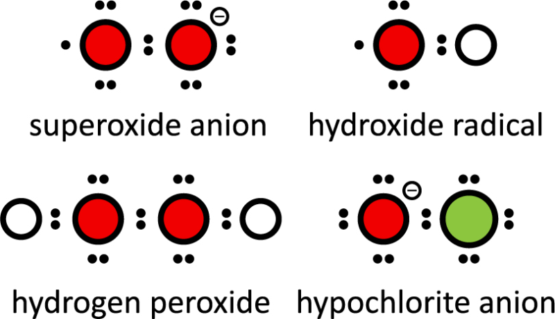
Reactive oxygen species (ROS) are oxygen‑derived oxidants that include both free radicals (e.g., superoxide O2•⁻, hydroxyl radical •OH, peroxyl radicals ROO•) and non‑radical species (e.g., hydrogen peroxide H2O2, hypochlorous acid HOCl). These unstable molecules are typically generated as a byproduct of the natural metabolism of oxygen. Closely related reactive nitrogen species (RNS), such as nitric oxide (NO•) and peroxynitrite (ONOO⁻), often participate in the same redox processes and signaling pathways. There are many different ROS, each with distinct chemical properties and biological roles. It is important to distinguish certain ROS, such as superoxide, peroxyl radicals, hydrogen peroxide, hydroxyl radicals, and peroxynitrite, as they have unique reactivities and effects within biological systems. Reactive species also include nitrogen species, which play significant roles in cellular signaling and oxidative stress.
When reactive oxygen species build up at high levels inside cells, also referred to as oxidative stress, they can damage DNA, RNA and proteins, and even cause cell death. ROS biomolecules have been implicated in a range of pathologies, including cancer, neurodegenerative diseases, atherosclerosis and the process of ageing2. Arachidonic acid, a polyunsaturated fatty acid present in cell membranes, is a major target of lipid peroxidation by ROS, leading to the formation of signaling molecules and protein adducts that contribute to disease processes. Cells counterbalance this with antioxidant defenses, including superoxide dismutases (SODs), catalase, glutathione (GSH)–dependent systems such as glutathione peroxidases (GPx), and peroxiredoxins. These convert dangerous free radicals to harmless molecules, such as water3.
Low levels of reactive oxygen species have been found to have useful and beneficial effects. For example, they play an important role in regulating cellular signaling and gene expression. Evidence suggests that reactive oxygen species may modulate cell differentiation, including hematopoietic differentiation involving stem cell production, in both physiological and pathological (disease) conditions4.
There are many intracellular sources of reactive oxygen species, including cell mitochondria (e.g. products of mitochondrial metabolism) and NADPH oxidases (NOX enzymes) linked to neutrophils2, white blood cells, which produce high levels of ROS as part of their defense role. A wide range of enzymes also produce ROS, including xanthine oxidase, nitric oxide synthase, cyclooxygenases, cytochrome P450 enzymes and lipoxygenases. Within biological systems, mechanisms such as Fenton chemistry—where ferrous or cuprous ions catalyze the formation of highly reactive hydroxyl radicals from hydrogen peroxide—and redox cycling, which generates specific ROS like superoxide through compounds such as paraquat and quinones, further contribute to ROS production.
Oxidative stress is further induced by exogenous stimuli. For instance, alcohol leads to formation of reactive oxygen species during its degradation and induce ROS production by activation of cytochrome P450 enzymes5. A second example of a drug inducing ROS is tobacco smoke. It contains radicals reacting with oxygen to form ROS6. An exogenous ROS-inducer that is less avoidable is ultraviolet light. The induction of reactive oxygen species increases oxidative damage to cellular components and thus contributes to the development of skin cancer7.
Oxidative damage measurement is fundamental for understanding how reactive oxygen species (ROS) impact biological molecules and cellular health. When ROS such as superoxide, hydrogen peroxide, and hydroxyl radical accumulate, they can trigger oxidative stress, leading to damage of nucleic acids, proteins, and lipids. This oxidative damage is closely linked to cell death, altered gene expression, and the progression of various diseases.
To detect and quantify oxidative stress in live cells, researchers rely on a range of specific fluorescent probes and reagents, and fluorescence microscopes and microplate readers. The Oxidative Stress Detection Reagent is widely used for visualizing ROS in live cells via fluorescence microscopy, providing high sensitivity and spatial resolution. For more targeted analysis, the Superoxide Detection Reagent enables the selective detection of mitochondrial superoxide—a major component of ROS production—helping to unravel the role of mitochondria in oxidative stress and cell death.
Nitric oxide (NO), another reactive species, plays a critical role in cellular signaling and can be measured using dedicated fluorescent probes. In cell culture experiments, ROS production can be induced by activating enzymes such as nitric oxide synthase or NOX enzymes, and the resulting oxidative damage can be monitored using these specific probes. The use of specific inhibitors, like NOX enzyme inhibitors, allows researchers to pinpoint the sources of ROS and better understand the mechanisms driving oxidative stress.
Lipid peroxidation, a hallmark of oxidative damage, can be detected using boronate probes and other specific fluorescent indicators. These probes help measure the extent of lipid oxidation, providing insights into membrane integrity and cellular health. To ensure assay specificity, negative controls such as ROS inhibitors (e.g., n-acetyl-l-cysteine) are essential for distinguishing true ROS signals from background or interference caused by other components in tissue culture media or assay buffers.
Advanced detection methods, including microplate reader-based assays and flow cytometry, enable high-throughput and quantitative measurement of ROS levels and oxidative damage in cultured cells. Microplate readers, such as those offered by BMG LABTECH, are particularly valuable for their ability to process large sample numbers with high sensitivity and reproducibility, making them ideal for drug discovery and redox biology research.
Despite the power of these assays, technical challenges such as background fluorescence and interference from other biological molecules can complicate ROS detection. Careful assay design, the use of specific fluorescent probes, and the inclusion of appropriate controls and inhibitors help overcome these obstacles, ensuring reliable measurement of oxidative damage and redox status.
As reactive oxygen species impact on many biological events, they are studied extensively. Assays to measure ROS belong to the category of cell-based assays and are used to report on immune responses, to monitor the balance of ROS and antioxidants or to study their role in cancer development. However, detecting ROS levels can be difficult as they are often in the nanomolar range and have very short half-lives. General indicators are often used as broad, non-specific tools for the initial assessment of oxidative stress, providing an overall view of ROS activity when more specific detection methods are unavailable.
The most common reagent used for measuring ROS is 2',7'-dichlorodihydrofluorescein diacetate (H2DCFDA). It is intracellularly trapped by esterase activity that removes lipophilic blocking groups. Upon oxidation of the probe by ROS the resulting dichlorofluorescein (DCF) exhibits high green fluorescence. Different derivatives of H2DCFDA are employed for reactive oxygen species measurements as they have specific characteristics: carboxy-H2DCFDA displays an improved cellular retention whereas the fluorinated molecule (H2DFFDA) proves more stable when exposed to light. HeLa cells are commonly used as a model system for monitoring ROS production in these assays.
A probe that selectively detects superoxide generated in mitochondria during oxidative phosphorylation is MitoSOX™ Red (ThermoFisher). The live cell reagent is targeted to mitochondria and its oxidation by superoxide increases its red fluorescence. Ex vivo measurements can also be performed, where ROS are assessed in tissue samples or slices outside the organism to reflect physiological relevance.
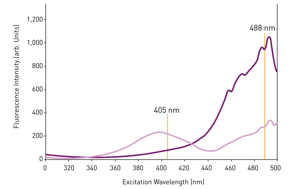
Apart from fluorescent probes added to the cell culture, genetically encoded probes may report on changes in reactive oxygen species directly in the cell at the location of interest. The fluorescent probe roGFP (redox sensitive green fluorescent protein) has different excitation wavelengths dependent on its redox state: reduced roGFP can be excited at 488 nm whereas oxidized roGFP is better excited at 405 nm (Fig. 2). A peroxidase that scavenges selectively H2O2 facilitates oxidation of roGFP and the ratio of measurements performed with both excitation wavelengths reports on the presence of H2O28. A similar approach is employed by the genetically encoded H2O2 sensor HyPer. The Biosensor thiazole orange in the antioxidant assay kit AOP-1 also functions as a biosensor to detect ROS. At the same time it also fills the role of a photosensitizer, generating ROS in living cells, which is a prerequisite to study the presence and effects of potential antioxidants. Horseradish peroxidase is commonly used as a catalyst in hydrogen peroxide assays, such as those utilizing Amplex Red, to enable sensitive detection of H2O2. Mitochondria targeted probes, such as MitoB and MitoP, allow for localized measurement of ROS within mitochondria, providing precise assessment of mitochondrial redox states. Organic dyes and small molecule probes are widely used for multiplexed and specific ROS detection in live-cell assays.
In the scientific talk "Real-time monitoring of redox changes in cells with a microplate reader", Bruce Morgan, Professor for Biochemistry at the University of Saarbrücken, Germany, discusses how redox-sensitive probes can be used to monitor redox enzyme activity. To address this question, roGFP2 was combined with glutaredoxin or glutathione. His team was then able to induce the oxidation of the signal molecule and to monitor in semi-high-throughput the change of roGFP fluorescence on a CLARIOstar Plus microplate reader. Quantifying the amount of ROS generated and using a ROS inhibitor as a control are important steps to confirm assay specificity and to understand the role of ROS in cellular processes.

Next to fluorescent methods, luminescent methods are available to detect reactive oxygen species. On the one hand, there are methods based on chemiluminescence. They employ a molecule such as luminol, lucigenin, Pholasin® or the recently described and more sensitive L-012 that emit light when transitioning to their oxidized state. As this process is induced by ROS, light emission can be directly translated to the presence of ROS9. On the other hand, enzyme-dependent luminescence (bioluminescence) detects ROS. The enzyme is a luciferase that converts a substrate under the emission of light. A precursor of the substrate is added to the sample and is ROS-dependently converted into the luciferase substrate. Thus the luminescent signal correlates with ROS levels10. Another example is highlighted in this application note, that employed Promega’s luminescent ROS-Glo assay for the detection of ROS in cell-based assays.
These popular methods to study and measure levels of reactive oxygen species are based on fluorescent or luminescent read-outs. This makes it possible to use a microplate format allowing running 96 up to 1536 samples at once and performing the detection in microplate readers. These devices record light signals in the tiny reactions taking place in the wells of a microplate.
Detection modes: depending on the assay chosen to detect reactive oxygen species the ideal microplate reader needs to cover either luminescence, fluorescence or both if the flexibility running different assays should be given. However, if you would like to combine the reactive oxygen species measurement with other assays, further detection modes may need to be covered as well. An example would be the combination of ROS-measurements with absorbance-based viability assays such as WST. BMG LABTECH offers a range of multi-mode plate readers capable of measuring luminescence, fluorescence, absorbance and advanced detection modes.
Sensitivity and throughput: The sensitivity the ideal microplate reader needs to offer is dictated by the plate format that is used and by the ROS-assay itself. For instance high density plate formats (1536 well) have a lower reaction volume and lower signal intensities. This requires highest sensitivity of the microplate reader and a fast reading of the plate. The PHERAstar FSX microplate reader is designed to meet exactly these requirements.
The most commonly used reactive oxygen species assay, the H2DCFDA assay, gives high signal intensities when used in 96-well or 384-well format and with sufficient material, for instance when using immortal cell lines. For instance, the signals can be easily recorded by all BMG LABTECH multi-mode readers. Increased sensitivity is required by genetically encoded probes such as roGFP since expression levels are limited. Researcher Prince Saforo Amponsah from the Technical University Kaiserslautern explains in detail the challenge to read the fluorescent probe and how the CLARIOstar® assisted the measurements.

Accessories: Reactive oxygen species detection based on H2DCFDA, bioluminescence or roGFP is suitable to be kinetically measured in live cells. Depending on the time frame of interest, live cell measurements require a controlled gas atmosphere to allow long-term cell incubation. In particular this is a CO2 atmosphere of 5-10 % to guarantee a stable pH in carbonate buffered media. For the measurement of reactive oxygen species the surrounding O2 concentration plays a critical role. The atmospheric O2 level of 21 % alone is capable of inducing oxidative stress without further stimulation. Physiologic O2 concentrations that cells are exposed to in the human body are much lower ranging from 4 % in the brain and 10 % in the kidney11. Accordingly, reliable ROS measurements can only be achieved when measuring at lower O2 concentrations. The Atmospheric Control Unit (ACU) regulates both the CO2 and O2 concentration in the microplate reader and thus allows running ROS experiments at physiological O2. This module is available for CLARIOstar, VANTAstar and Omega microplate readers.
In this video Prof. Giovanni Mann gives a tour of his lab: Precision under physiological oxygen: How the CLARIOstar plate reader advances redox research at King’s College London.

If you are interested in real-life data measuring reactive oxygen species on BMG LABTECH readers, please feel free to have a look into our collection of peer-reviewed articles on this topic.
Overall, the measurement of ROS and oxidative damage is of considerable interest across biology, medicine, and pharmacology. By leveraging advanced detection reagents, specific inhibitors, and robust instrumentation, researchers can gain critical insights into ROS production, oxidative stress, and their effects on cellular function and disease progression.
An example of how these approaches are applied in practice is shown in a video interview with researchers at the Max Planck Institute for Polymer Research, who combine computational modeling, liposome based systems, and fluorescence based kinetic assays to study ROS driven lipid oxidation in biological membranes.

Powerful and most sensitive HTS plate reader
Most flexible Plate Reader for Assay Development
Upgradeable single and multi-mode microplate reader series
Flexible microplate reader with simplified workflows
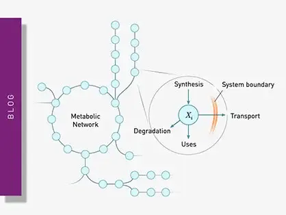
Explore metabolic flux analysis, its significance in research, and how microplate readers enhance measurements for insights.
Explore the vital role of mitochondrial metabolism in cellular function, its measurement techniques, and how microplate readers enhance research in this field.
Binding constants quantify the strength of a binding reaction between a biomolecule and its target (ligand). But how do you measure them and what can you do with them?
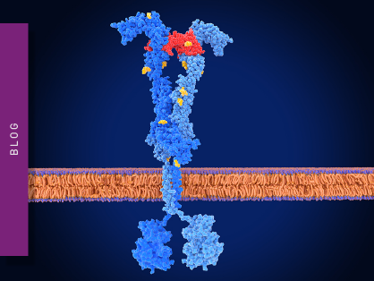
Receptor-ligand kinetics is the study of the rates at which receptors and ligands interact, bind and dissociate. Learn why these types of measurements are important and how to measure them.
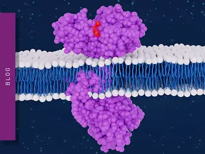
Receptor-ligand interactions are crucial for cell signalling. They are also important for drug discovery. How do microplate readers deliver benefits to both?
Gene reporter assays are sensitive and specific tools to study the regulation of gene expression. Learn about the different options available, their uses, and the benefits of running these types of assays on microplate readers.