NEPHELOstar Plus
Microplate nephelometer for light-scattering and turbidity measurements


Barry Whyte is Application Scientist and Science Writer at BMG LABTECH in the United States. He has PhD and Bachelor of Science (BSc) degrees in biochemistry from the University of Bristol in the United Kingdom and more than 20 years of experience in the life sciences and science communications. Over the years, Barry has worked on three continents and traveled widely. He enjoys building on his international work experience and learning new ways to help scientists advance their research.
Immunology is an important focal point for many research laboratories.
Scientists have many options available to measure immune-related molecules, but light scattering is often a first port of call when rapid quantitative measurements are needed.
Nephelometry is routinely performed in research laboratories and academic institutions as a first step to help investigate immunological diseases. In this blog, we look at some of the types of tests performed in research settings and discuss how microplate readers can help pave the way to diagnostic testing and related applications in immunology.
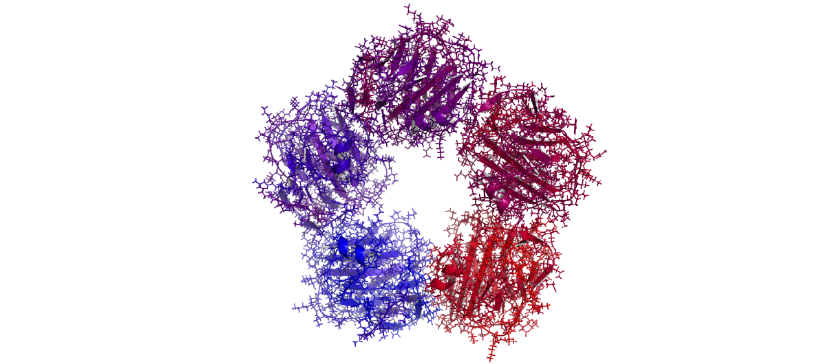
Nephelometry is a versatile technique that measures the intensity of light scattering by particulate matter in a liquid sample (Fig.1).
 In the research laboratory, nephelometry is often used for protein quantification related to immune responses or immune events. Experimental approaches might involve the use of animal model systems or specific in vitro cell-based assays where the levels of immunological molecules need to be determined to assess the impact of disease or infection.
This type of work is a precursor to assist or advance clinical decisions on different immunological diseases including autoimmune deficiencies, disorders like complement deficiencies, or the impact of various infections on immunity.
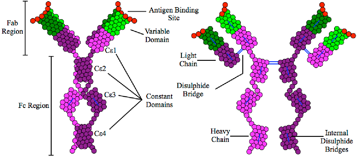
Allergy research is another area of interest since antigen-antibody immune complexes play a role in many allergic and autoimmune diseases. Some allergies raise the levels of IgE antibodies, and this includes responses to pollen, dust mites and animal dander (Fig. 2). Scientists in research laboratories can use nephelometry to look at changes in the levels of IgE which are a useful marker for the presence of allergies. Nephelometry can also support basic research into autoimmune diseases including rheumatoid arthritis, systemic lupus erythematosus and celiac disease.
 Definitive diagnoses require regulatory compliant clinical tests but nephelometry provides an invaluable tool for developmental research.
Nephelometry is a robust and reliable technique known for its high precision and accuracy. Quantitative measurements of light scattering allow for precise and reproducible quantification of molecules of interest. Such measurements can also be easily automated which allows for high throughput analysis of large numbers of samples. This makes it suitable in research settings where a high volume of samples needs to be processed efficiently and accurately.
Methods of measuring immune parameters using nephelometry are divided into two types: endpoint and kinetic assays. These types of assays differ in how the measurements are obtained and the type of information they provide.
Endpoint nephelometry, which is also referred to as static nephelometry, is a method where the intensity of scattered light is measured at a fixed time point after the reaction between antibody and antigen has started. Kinetic nephelometry, in contrast, is a method where the change in intensity of scattered light is measured continuously over time after the reaction between antigen and antibody has begun. In this method, the sample is continuously measured for the intensity of scattered light at regular time intervals using the nephelometer.
In many cases, endpoint nephelometric measurements provide sufficient information about the immunological reaction under study. In some situations, however, kinetic nephelometry is preferred due to its ability to provide more information about the dynamics of immune reactions. These situations may include scenarios where a researcher may wish to look at the time dependency of a reaction, monitor complex reactions, detect rapid changes, look at the dynamics of sample stability or determine the rates of reactions.
The choice of an endpoint or kinetic assay is made on a case-by-case basis depending on the requirements of the experiment or type of clinical information needed.
The precipitin or immunoprecipitation test is commonly used to detect the presence of specific antibodies or antigens in a biological sample. Specifically, it can be used to detect the presence of antigens in biological samples, to confirm the presence of specific antibodies, or determine the specificity of antibodies or antigens. The formation of immune complexes between antigens and antibodies gives rise to a precipitate which scatters light and gives a quantitative signal.
A precipitin test involves several steps. First the biological sample, for example blood, plasma or tissue extract, is collected and prepared for testing. Preparation may involve diluting the sample to a concentration that gives an optimal light scattering signal or removing any contaminating substances. Next the sample is mixed with specific antibodies known to bind to the target antigen. The mixture is incubated for enough time to allow the formation of the antibody-antigen complex. Nephelometry is subsequently used to detect the immune-complex precipitates according to their ability to scatter light. The amount of light reaching the nephelometer detector is directly proportional to the quantity of protein-antigen in the sample. Quantification of antibody-antigen is achieved by comparing the reading from the sample with the values on a standard curve.
Nephelometry can be used to quantify the levels of a wide range of proteins from biological samples in a research setting. 1,2 These proteins may include immunoglobulins, complement factors, rheumatoid factors, lipoproteins, immune complexes, and antigens linked to disease and infections. A quantitative measurement allows for precise and reproducible assessment of the levels of these different types of proteins and an opportunity to look at how these levels may change over time.
Quantitative nephelometry on a microplate is routinely used to measure the levels of immunoglobulins under different conditions including disease or infection. In a research laboratory, this type of measurement can support the development of new diagnostic tests.
Nephelometry can be used to specifically measure the levels of IgG, IgM and IgA which can be compared with the expected concentration ranges for these molecules in the body. Increased or decreased levels of these immunoglobulin molecules is useful information to help develop new diagnostics. For example, decreased levels of IgG are known to occur in different types of cancer (e.g. leukemia, myeloma), conditions like preeclampsia (high blood pressure during pregnancy), or chemotherapy treatment with different drugs.
Light scattering can be used to measure the levels of C-reactive protein in different samples (Fig. 3). C-reactive protein is an acute phase protein produced by the liver. Infection with bacteria, systemic inflammation and internal organ failures can all lead to elevated levels of C-reactive protein. For example, the levels of C-reactive protein can increase up to 1000-fold after the onset of inflammation and can decrease rapidly when inflammation is resolved. The measurement of the levels of C-reactive protein in biological samples is therefore a good marker for inflammation and one that can be performed with a microplate reader.
Nephelometry can also be used to determine the concentrations of complement components (e.g. C3, C4), part of the immune system involved in inflammation and defense against pathogens. There are two ways to measure the levels of complement using nephelometry. Complement can be measured directly by measuring the levels of complement components. Alternatively, it can be measured indirectly by detecting the levels of complement activation products.
Nephelometry is a useful way to detect and measure albumin in biological samples for research purposes. Albumin is a protein typically found in the blood. If it is present in urine, it may indicate that the kidney is not working properly and could be a sign of early-stage disease. The presence of albumin in urine is known as albuminuria or proteinuria and may or may not be directly related to disease. Some levels of proteins in urine are not uncommon but persistently high levels of protein may be a sign of kidney disease. If albumin is detected in the urine by nephelometry further tests are required to confirm the presence of disease.
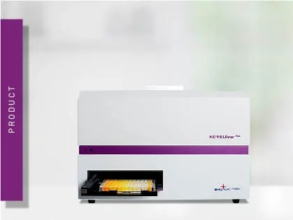
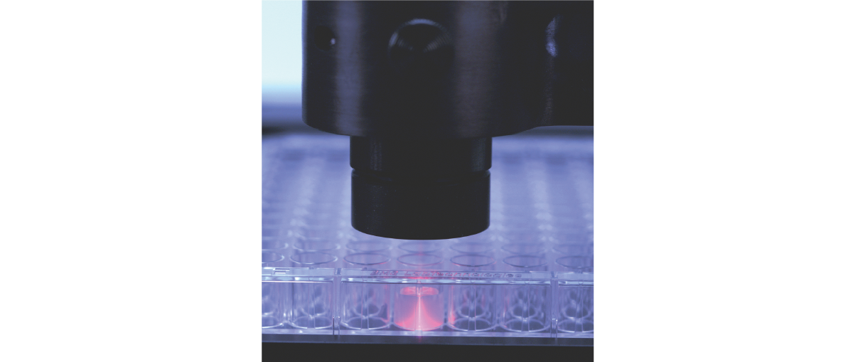
Nephelometry is a highly sensitive technique that can detect and measure minute changes in the concentration or size of particles in a sample. This makes it well suited for studying low levels of antigens, antibodies, or immune complexes in biological samples, even when the analyte of interest is present at low abundance. Microplate-based nephelometric detection is a fast, scalable, sensitive and accurate way to measure the concentration of particulate matter in a sample. The detection principles of the NEPHELOstar® Plus microplate nephelometer are shown in Fig. 4.
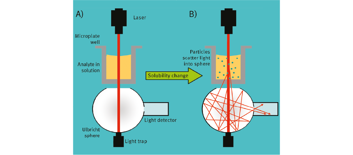 The NEPHELOstar Plus from BMG LABTECH is the microplate reader of choice for measuring light scattering and is the ideal partner for sensitive and accurate immunology tests performed at scale on a low volume of samples. As the world’s only laser-based microplate nephelometer available today it was developed to meet the high-throughput requirements of a wide range of immunological assays. The high-intensity light source of the NEPHELOstar Plus is a laser at 635 nm. The principles of nephelometry and instrument design of the NEPHELOstar Plus are described in more detail in the Microplate Reader ABC article on the BMG LABTECH web site.
Nephelometry can be readily used to make light scattering measurements on a microplate with low volumes of sample at high sensitivity. The application note Improving throughput for assessing nephelometric turbidity units (NTUs) using the NEPHELOstar® Plus describes how light scattering measurements are amenable to miniaturized assays on a high-throughput laser-based microplate nephelometer.
Results were obtained with a read time of less than 2 seconds per sample with significant reductions in sample volumes compared to similar detection devices. The NEPHELOstar Plus from BMG LABTECH was able to detect signals with a Limit of Detection comparable to industry standard instrumentation but provides higher throughput using smaller sample volumes.
In the application note Measurement of rheumatoid factors by NEPHELOstar® Plus microplate reader laser nephelometry was shown to be a reliable method for the measurement of rheumatoid factors. Rheumatoid factors are immunoglobulins that react to antigenic sites on the Fc region of human or animal immunoglobulin G. They are more commonly found in patients with rheumatoid arthritis compared to individuals in the general population and are useful disease markers to assist basic and clinical research.
Nephelometric testing of rheumatoid factor levels is based upon antigen-antibody reactions. Rheumatoid factors bind to IgG to form immune complexes. As the number of antigen-antibody immune complexes increases, the solution becomes visibly cloudier. The NEPHELOstar Plus microplate reader is used to pass a light beam through the well to measure the turbidity of the sample. The more rheumatoid factors present, the more immune complexes are formed and the more light scattering will occur. The amount of light scattered is directly proportional to the number of immune complexes and therefore the amount of rheumatoid factor particles suspended in the sample.
The NEPHELOstar Plus plate reader was demonstrated to accurately determine both rheumatoid factor antibody status and titer (Fig.5). Thus, this rheumatoid factor assay is a viable and inexpensive assay that can be used in medium to high throughput laboratories.
The NEPHELOstar Plus from BMG LABTECH is the microplate reader of choice for measuring light scattering and is the ideal partner for sensitive and accurate immunology tests performed in a research setting at scale. All BMG LABTECH microplate readers have exceptionally fast reading capabilities and the NEPHELOstar Plus is no exception. By combining high performance with miniaturized assays and short measurement times, the NEPHELOstar Plus offers considerable savings on time, materials and other resources.
If you want to learn more about different applications for the NEPHELOstar Plus you can read further details at Microplate Reader ABC article and Drug solubility: why testing early matters in drug discovery.
The NEPHELOstar Plus is available for research purposes only.
Configure your microplate reader and get an initial recommendation!
Microplate nephelometer for light-scattering and turbidity measurements
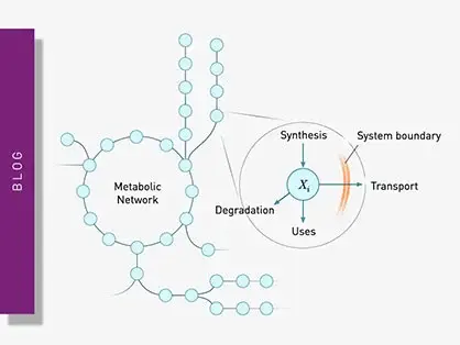
Explore metabolic flux analysis, its significance in research, and how microplate readers enhance measurements for insights.
Explore the vital role of mitochondrial metabolism in cellular function, its measurement techniques, and how microplate readers enhance research in this field.
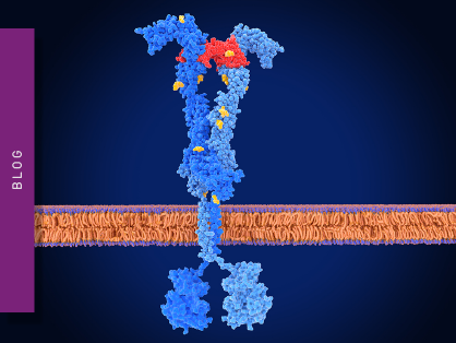
Binding constants quantify the strength of a binding reaction between a biomolecule and its target (ligand). But how do you measure them and what can you do with them?
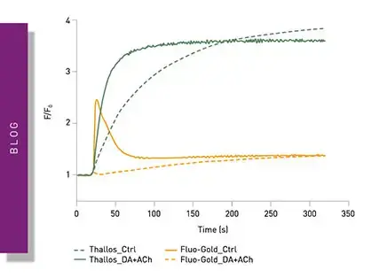
Receptor-ligand kinetics is the study of the rates at which receptors and ligands interact, bind and dissociate. Learn why these types of measurements are important and how to measure them.
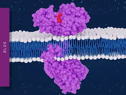
Receptor-ligand interactions are crucial for cell signalling. They are also important for drug discovery. How do microplate readers deliver benefits to both?
Gene reporter assays are sensitive and specific tools to study the regulation of gene expression. Learn about the different options available, their uses, and the benefits of running these types of assays on microplate readers.