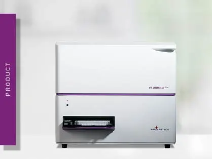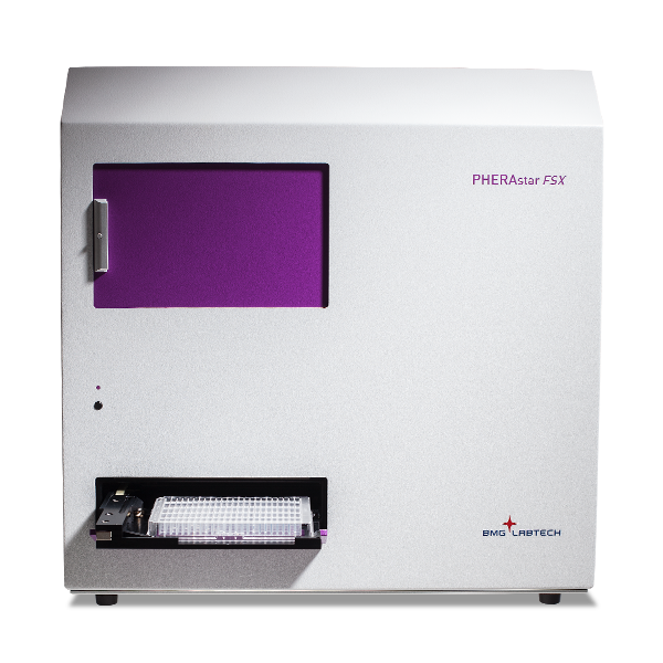
PHERAstar FSX
Powerful and most sensitive HTS plate reader





Barry Whyte is Application Scientist and Science Writer at BMG LABTECH in the United States. He has PhD and Bachelor of Science (BSc) degrees in biochemistry from the University of Bristol in the United Kingdom and more than 20 years of experience in the life sciences and science communications. Over the years, Barry has worked on three continents and traveled widely. He enjoys building on his international work experience and learning new ways to help scientists advance their research.

Cell proliferation assays provide crucial insight for scientists in the life sciences. They find many uses in basic research, including different scale applications in biochemistry, cell biology, metabolism, and molecular biology, as well as in large-scale screening assays for drug development.
Available in formats that support a range of throughputs, cell proliferation assays are indispensable to examine the characteristics of cells and to assess the utility of these cells for specific experiments and applications. In this context, they serve as a robust platform for scientific discovery and drug testing.
Cell proliferation and their widely used counterpart cell viability assays have some significant differentiating features that can impact research outcomes (for an in-depth look see the BMG LABTECH blogs Cell viability assays – Measure how happy your cells are and Cell-based assays on the rise). In this blog, we focus on the distinctive features of cell proliferation assays and examine how to select the most appropriate assays for different experiments and applications.
Cell proliferation in its strictest sense is what scientists refer to when they talk about cell division (Fig. 1). Assays that measure cell proliferation answer the specific question of whether cells are increasing in number (i.e. dividing) and give important information on cell counts. Since dividing cells are viable cells, these assays can also be classified as viability assays but the unique trait being measured is cell division.
Students of the life sciences are often taught that living organisms have several core characteristics.1 Some of the frequently cited fundamental characteristics of cells include organization and structure, the ability to use or generate energy (metabolism), the capacity to regulate their environment (homeostasis), cell growth, cell development, heritable characteristics (genetics), reproduction (cell division), as well as adaptation and evolution. The importance of cell division and cell growth is reflected by their inclusion in this list as two separate entries.![Figure 1: Cell proliferation assays. [BW2.1]Cell proliferation is one of several cellular processes that researchers study using an array of cell-based assays.](https://www.bmglabtech.com/hubfs/1_Webseite/5_Resources/Blogs/cell-prolifiration-assays-fig1.webp)
The traditional tried-and-tested ways to study cell proliferation include direct cell counting either manually with a hemocytometer or using electrical impedance-based cell counters. They also include DNA synthesis measurements that track new DNA synthesis during S-phase with radioactive or non-radioactive analogs such as BrdU and EdU as well as the use of protein markers such as Ki-67 and proliferating cell nuclear antigen (PCNA) expressed in dividing cells.
Table 1 summarizes each of these methods and highlights some of their advantages and disadvantages. Due to the constraints of using some of these approaches, improvements have been sought to workflows including ways to save time and resources and options to avoid reliance on radioactive labeling. Flow cytometry can also quantify proliferation across multiple generations through carboxyfluorescein succinimidyl ester (CFSE) dye dilution. We will come back to these more recent approaches later in this blog.
Table 1. Traditional cell proliferation assays: advantages and disadvantages
| Method |
What is measured | Main advantages |
Main disadvantages |
| Manual or automated cell counting | Direct counting of cell numbers versus time | Direct measurement; straightforward concept and workflow | Labor intensive; low throughput; operator variability |
| 3H-Thymidine incorporation | Incorporation of radioactive thymidine into newly synthesized DNA during cell division | High sensitivity; traditional gold standard | Radioactive method; resource intensive; personnel training and safety requirements |
| Bromodeoxyuridine (BrdU) incorporation | Incorporation of bromodeoxyuridine into newly synthesized DNA during cell division | Non-radioactive method; compatible with microscopy and flow cytometry | Requires DNA denaturation for antibody access |
| 5-Ethynyl-2’-deoxyuridine (EdU) incorporation | Incorporation of 5-ethynyl-2’-deoxyuridine into newly synthesized DNA during cell division | Fast detection; no DNA denaturation needed; sensitive and reproducible | Cost and resources |
| DNA content assay | Fluorescent quantification of total cellular DNA | Sensitivity; microplate compatibility | Specificity of signal with live and dead cells needs to be considered in some cases |
| Clonogenic (colony forming) assay | Single cell development into a colony | Measures longer term proliferative capacity | Labor intensive, slow, low throughput |
| Ki-67 staining | Detection of proliferation-specific nuclear protein | Useful in tissue and cancer studies due to indicator of active cells in cell division cycle | Staining and analysis requirements |
| Flow cytometry | DNA content measurements in cell-cycle phases or tracking division generations by fluorescent dye dilution | High information content | Complex workflow; specialized equipment |
It is important to recognize that the terms cell proliferation and cell viability sometimes get used interchangeably which can lead to confusion. This confusion has been exacerbated since scientists have developed a formidable array of cell-based assays that fall into these categories. These assays have provided many insights into biology which underline their utility and the need to be clear on the merits of both.2
A recent paper published in the journal NPJ Breast Cancer highlighted the risks of being unaware of the distinction, using a metabolic assay as an endpoint in drug screening when it was more appropriate to use a cell proliferation assay that measures DNA content.3 The authors documented that earlier large-scale screens to identify which cancer cells are most sensitive to widely used CDK4/6 inhibitors have been somewhat misleading due to an overreliance on metabolic-type proliferation assays. CDK4/6-inhibited cells arrest in the G1 phase of cell cycle but continue to grow and produce more mitochondria. These cells show no inhibition in terms of signal in ATP-based proliferation assays. In fact, these signals continue to increase over time as the cells grow. If a DNA-based assay is used, however, it becomes clear that cell division has been affected. Thus, cells appear to have “proliferated” using ATP-based endpoints, even though they have not. ATP assays and other endpoint assays can be confounded by changes in metabolic activity or cell death, so proper controls are needed for reliable assay results. Most viability assays, including metabolic assays, should not be used exclusively as endpoints for cell proliferation.
Many cell proliferation assays can be performed on a microplate reader with significant savings in resources and time. In the next section, we look at the options for cell proliferation assays in the context of which assays are amenable to measurement on a microplate reader. This is an area in progress, but where innovation is making a difference.
Table 2 shows some of the more recently developed cell proliferation assays that measure cell count and which are also suitable for measurement on a microplate reader. Significantly, like the traditional methods performed without microplates these assays measure the rate of cell division, DNA synthesis or changes in cell count over time, properties that do not just scale with cell growth but reflect cell division.
As can be seen, the use of 5-ethynyl-2’-deoxyuridine (EdU)-type methods, a thymidine analog used to monitor proliferation by labeling newly synthesized DNA, either as antibody-based detection that requires separation of the DNA duplex or more friendly chemical labeling methods that do not require DNA duplex separation, are gaining traction above the conventional use of bromodeoxyuridine (BrdU)-based methods that are not microplate friendly. EdU detection uses click chemistry to attach fluorescent probes, making it well suited to quantify cellular proliferation in microplate-based formats and a suitable assay method for measurements on plate readers.
Table 2. Some of the more recent options for cell proliferation assays.
| Cell Proliferation Assay |
Cell Proliferation (cell count) |
Cell growth | Note | Detection mode | Example |
| CyQUANT Cell Proliferation Assay | ✓ | ✓ | DNA content assay (fluorescent dye specifically binds to DNA) | Fluorescence | CLARIOstar |
| CyQUANT NF (No Freeze) Cell Proliferation Assay | ✓ | ✓ | Eliminates need for cell freezing and simplifies workflow | Fluorescence | CLARIOstar |
| CyQUANT Direct Red Cell Proliferation | ✓ | ✓ | Red format can be used in live cells for continuous measurements | Fluorescence | |
| Click-iT EdU Microplate Cell Proliferation Assay (modified base 5-ethynyl-2’-deoxyuridine) | ✓ | ✓ | DNA synthesis assay (modified base incorporation assay with fluorescent azide dye detection) | Fluorescence | |
| Lumit® hKi-67 Immunoassay | ✓ | ✓ | Proximity-based luciferase assay for hKi-67 protein biomarker found in dividing cells | Luminescence |
Collectively, cell proliferation assays can be measured by absorbance, fluorescence or luminescence outputs. In contrast, live cell imaging and live cell analysis approaches can monitor cell proliferation in real time and reveal cellular dynamics over the course of an experiment. Some cell analysis platforms are also label-free, including impedance-based systems, and are useful when continuous live cell assays are preferred over endpoint assays. Here we provide a few examples of cell proliferation assays from recent publications that involve fluorescence and luminescence detection.
In the blog Metabolic flux: the dynamic flow of biochemical reactions, we describe how researchers developed a computational tool to simulate intracellular metabolic fluxes in human cells. In this study, cell counts (Fig. 2) were crucial for calculating the metabolic flux data of the amino acids in the different phases of cell growth, where flux estimates can vary with growth rate and changing expression across phases of culture.4 These data were used to improve the performance of the predictive power of the genome-scale metabolic models described in the paper.
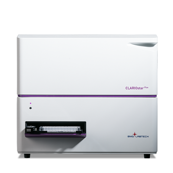
Absolute cell counts were measured at 22, 26, 30, 46, 50, and 54 h after seeding using the CyQUANT™ Cell Proliferation Assay kit from ThermoFisher Scientific with a CLARIOstar® Plus microplate reader. CyQUANT uses a DNA-binding fluorescent dye whose signal increases when bound to cellular nucleic acids. It estimates proliferation via the cell number or amount of DNA and is not based on the cell’s metabolic status. As a DNA content assay, it gives a more accurate handle on cell proliferation compared to other traditional colorimetric methods. The CLARIOstar is one of three microplate readers from BMG LABTECH that offers Enhanced Dynamic Range (EDR) measurements. The signal intensity is often hard to predict in cell-based assays in advance especially in time course measurements. In these types of assays, researchers benefit from EDR which automatically determines the optimum dynamic range without the need for manual intervention.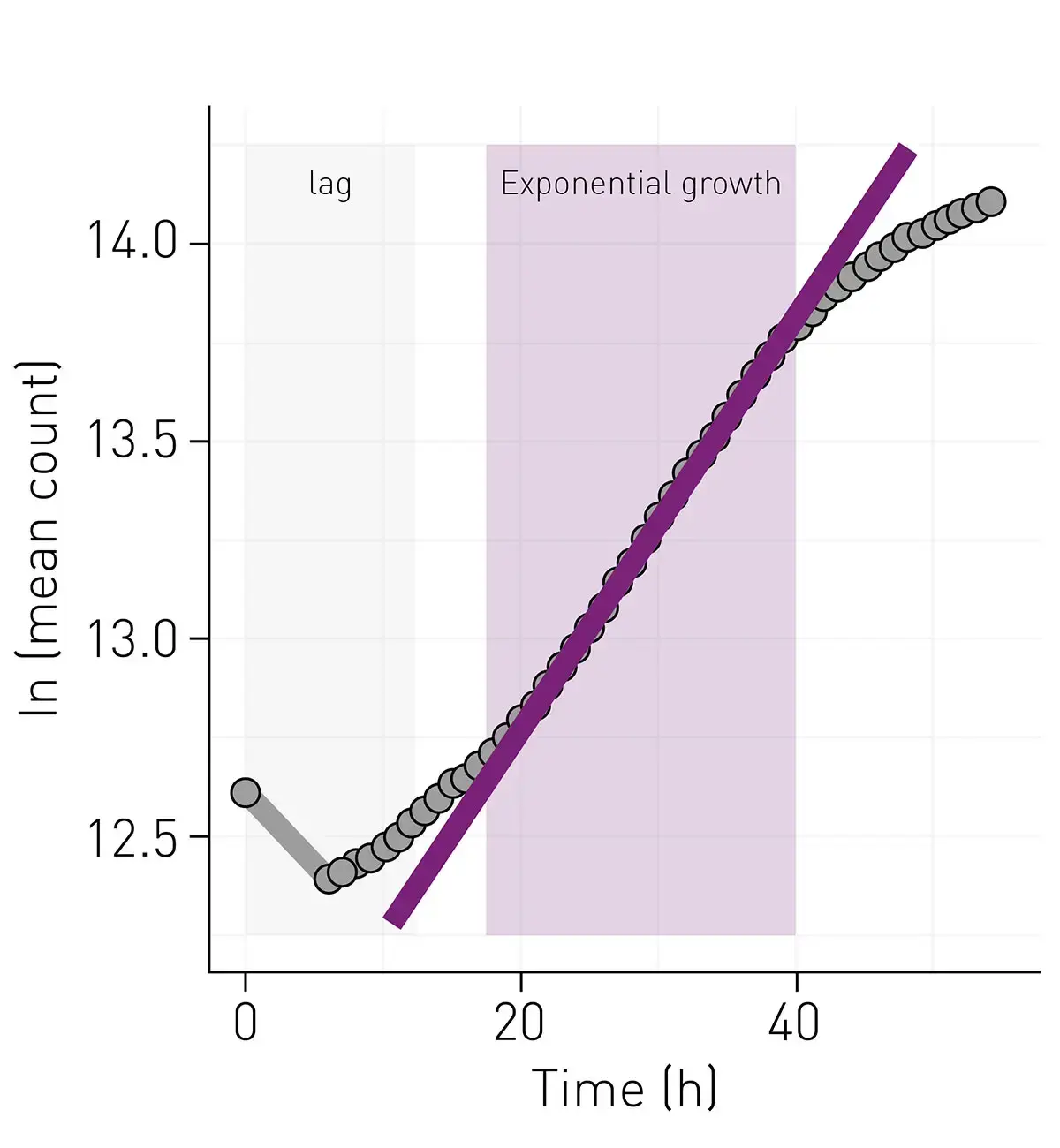
In the paper, “Copper drives remodeling of metabolic state and progression of clear cell renal cell carcinoma” researchers investigated the metabolic responses to copper accumulation during the growth of clear cell renal cell carcinoma.5 Clear cell renal cell carcinoma is the most common type of renal cancer found in humans. High levels of copper have been reported in blood samples and tissues in various cancers. The study in question found elevated levels of copper and increased allocation of copper to cytochrome c oxidase, an enzyme indispensable for aerobic mitochondrial respiration. A crucial part of the investigation was to look at the impact of copper metabolism upon cancer cell proliferation. The number of cancer cells increases as the cells divide and the cancer progresses. CyQUANT NF (No Freeze) Cell Proliferation Assays were therefore performed on a CLARIOstar using 96-well plates by measuring the fluorescence emission at 530 nm after excitation at 485 nm.
In addition to the EDR feature mentioned earlier that facilitates the automatic selection of the dynamic range for different experiments, the CLARIOstar includes an LVF Monochromator that provides wavelength flexibility with filter-like sensitivity for any type of fluorescence and luminescence measurements. Both features are assets for cell-based assays. The CyQUANT NF Cell Proliferation Assay is a non-toxic, high-throughput fluorescence-based screening method for live cell analysis of proliferation without cell lysis or fixation. It combines the cell-permeant dye used in CyQUANT Cell Proliferation Assays with a membrane permeabilization reagent in a way that eliminates the need for cell freezing or prolonged incubations. As part of this study, cell proliferation was measured to determine the impact of different copper levels on cellular metabolism (Fig. 3).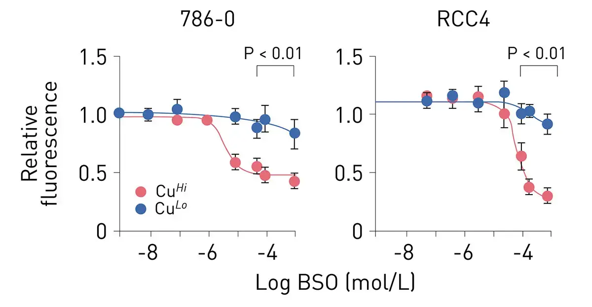
Overall, the study demonstrated that copper mediates metabolic reprogramming by enhancing mitochondrial cytochrome c oxidase function, detoxifying copper-associated oxidative stress, and fueling cancer cell proliferation.
In the paper “Neuron-specific protein network mapping of autism risk genes identifies shared biological mechanisms and disease-relevant pathologies”, researchers were interested in looking at the impact of cell signaling networks on disease mechanisms linked to autism spectrum disorder.6 They were able to show that neuron-specific protein-protein interaction networks provide a scalable method to reveal disease mechanisms for autism spectrum disorder. The protein-protein interaction network resource and screening system is capable of being applied more broadly to additional autism risk genes to identify disease mechanisms that are not captured with current approaches. As part of the study, the researchers needed to measure oxygen consumption rates to analyze cellular respiration. They measured cellular respiration using live-cell metabolic assays in mouse cortical neurons. However, to work out the impact of these interactions on metabolism it was necessary to normalize the results to the number of cells per well. For this purpose, CyQUANT™ Cell Proliferation Assays were carried out on a CLARIOstar microplate reader.
As these applications in cancer, metabolism and neuroscience show, cell proliferation assays can be readily used on microplates to support scientific discovery.
Microplate readers offer distinct benefits for researchers looking to perform cell proliferation assays that directly quantify DNA content, DNA synthesis, mitotic markers or proxy methods for cell counting. Perhaps the biggest benefit is scale. A microplate reader offers simultaneous measurement of tens to hundreds of experimental conditions, allows researchers to test multiple drug concentrations, look in detail at time courses (kinetic measurements), and facilitates measurements of replicates and controls. Readers can be used for example to study growth factor responses, oncogene-driven cell proliferation, anti-proliferative drug effects, gene perturbations (CRISPR, siRNA), and how external signaling proteins, hormones, or nutrient formulations stimulate or inhibit tissue regeneration. The use of a microplate reader helps reduce human variability. Fluorescence, luminescence and absorbance outputs can be normalized and statistically analyzed which helps to improve reproducibility.
Many of the cell proliferation assays described in this blog offer high sensitivity, which is useful when looking at stem cells, primary cells, rare cell populations or working with miniaturized formats. Measurement of growth curves rather than just endpoint measurements help distinguish cytostatic effects, delayed proliferation, transient responses as well as rebound growth.
Unlike in some other assays, microplate formats require relatively small volumes which translate into low reagent and cell consumption. This is especially useful for patient-derived samples and use of specialized cell types (e.g. immune cells). Proliferation measurements can be carried out in parallel to other measurements (multiplexing with for example metabolic assays, toxicity measurements or gene reporter assays). For large-scale assays microplate readers offer easy integration with automation systems including liquid handlers and robotic tools as well as incubators such as atmospheric control units. These workflows also support high throughput screening of large compound sets, including metabolic or ATP-based formats, and can identify candidates that halt cancerous growth under defined culture conditions.
Cell proliferation assays require a microplate reader that delivers flexibility and performance over a wide range of experimental conditions. We have highlighted a few of these technological features in the applications selected in this blog. Additional technological features that deliver benefits to cell-based experiments that may be encountered are summarized in Table 3.
Table 3. Summary of benefits offered by microplate readers for cell proliferation measurements.
|
Feature |
Benefit |
Available on |
Note |
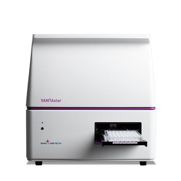
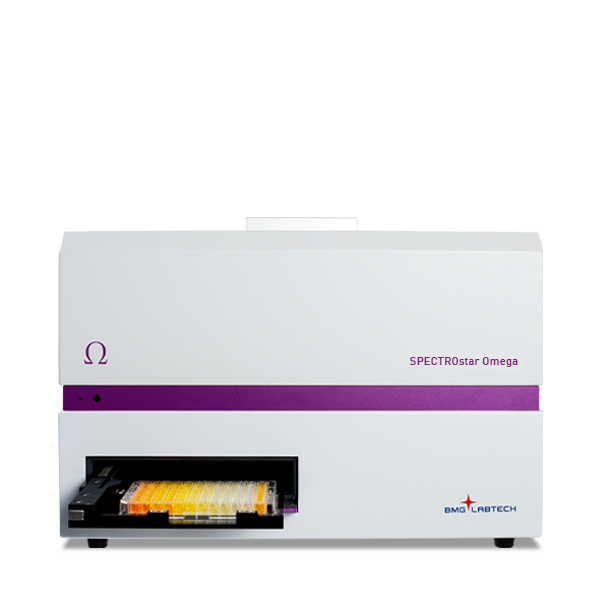
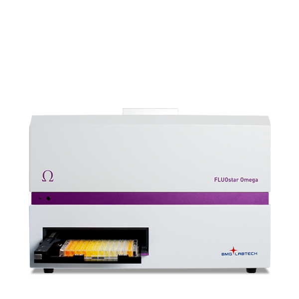
| Atmospheric Control Unit | Independently control oxygen and carbon dioxide concentrations for cell-based assays (as low as 0.1% oxygen on the CLARIOstar Plus) | VANTAstar, CLARIOstar Plus, Omega series | Beneficial for live cell assays including cell proliferation assays and other cell-based assays with specific requirements for the surrounding atmosphere. |
| LVF Monochromator | Provides wavelength flexibility with filter-like sensitivity for fluorescent and luminescent assays | VANTAstar, CLARIOstar Plus |
|
| Enhanced Dynamic Range | Ensures accurate signal quantification across low to high concentrations of ligands and targets without running into the risk of signal saturation | PHERAstar FSX, VANTAstar, CLARIOstar Plus | Particularly beneficial for kinetic assays where signal intensity builds up over time. |
| Incubation and shaking | Optimize assay conditions with precise temperature incubation and shaking control, ensuring best results for cell-based and enzymatic applications | All BMG LABTECH readers offer accurate temperature regulation up to 45°C (some devices even offer the option of temperature regulation up to 65°C). The VANTAstar, CLARIOstar Plus, the Omega series and the SPECTROstar Nano can be equipped with an incredibly robust transport system for shaking 24/7 where required | Three shaking modes with adjustable speed up to 700 rpm (optionally to 1100 rpm) provide optimum aeration settings for cells that require agitation. |
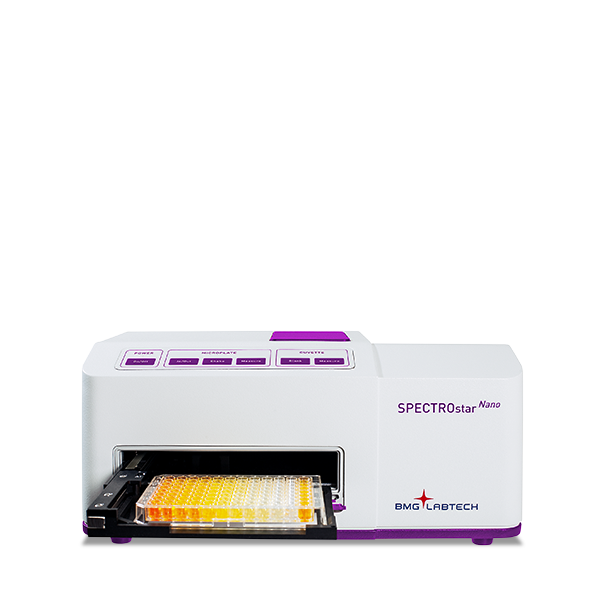
| Well scanning | Well scanning ensures uniform signal detection for cell-based and heterogeneous assays | PHERAstar FSX, VANTAstar, CLARIOstar Plus, Omega series, SPECTROstar Nano | |
| Reagent injectors | Reagent delivery to any plate format from 6-384 wells: control of injection timing, injection speed, delivery volume. Ability to inject different volumes in each well of a plate | The PHERAstar FSX, CLARIOstar Plus, and the Omega series allow simultaneous injection and measurement. The VANTAstar can be equipped with a modular injection unit. |
In conclusion, cell proliferation assays, where cell numbers are crucial, are best served by DNA-content assays or other biomarkers that reflect whether the cell is actively dividing. In rigorous experiments, researchers may often pair a cell viability assay with a proliferation/cell count assay. In clinical pathology, immunohistochemistry on tissue biopsies using Ki-67 or PCNA can help estimate tumor grade and growth speed. Proliferation assays can also support chemosensitivity testing by evaluating patient tumor cells treated ex vivo with different regimens, including repeat readouts from the same sample.
Microplate readers offer scalable, quantitative and reproducible measurements of changes in cell number or cell cycle progression at high throughput with much greater efficiency than microscopy or manual cell counting.
Collectively, BMG LABTECH multi-mode readers combine high-quality measurements with miniaturised assays, short measurement times, and offer considerable savings on materials and other resources.
Configure your microplate reader and get an initial recommendation!
Powerful and most sensitive HTS plate reader
Most flexible Plate Reader for Assay Development
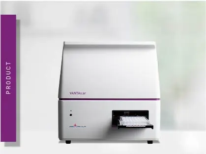
Flexible microplate reader with simplified workflows
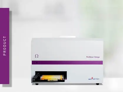
Upgradeable single and multi-mode microplate reader series
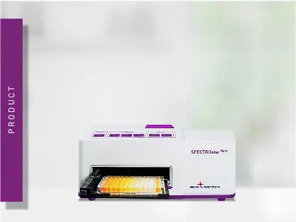
Absorbance plate reader with cuvette port
Explore metabolic flux analysis, its significance in research, and how microplate readers enhance measurements for insights.
Explore the vital role of mitochondrial metabolism in cellular function, its measurement techniques, and how microplate readers enhance research in this field.
Learn about applications for bacterial metabolism on a microplate reader.
Life in the depths of the ocean operates under extreme conditions. Find out how proteins from deep-sea luminescent organisms are useful for measurements on microplate readers.
Second messengers play a pivotal role in signal transduction events in cells. But how do you measure these small, transiently lived molecules and how can microplate readers help?