SPECTROstar Nano
Absorbance plate reader with cuvette port


Tobias Pusterla’s scientific background spans veterinary biotechnology, cancer cell biology, and the molecular mechanisms underlying inflammation‑driven tumorigenesis. After graduating in Veterinary Biotechnology at the University of Milan, Italy, he worked in mouse mutagenesis before completing a Ph.D. in Cellular and Molecular Biology through a joint program between the Open University of London, UK and the San Raffaele Scientific Institute, Milan, Italy. He later conducted postdoctoral research at the German Cancer Research Center (DKFZ) in Heidelberg, Germany, focusing on tumor biology, the tumor microenvironment, and the role of chronic inflammation in cancer development. His scientific work has contributed to understanding how damage‑associated molecular signals drive immune activation, cell migration, inflammation, and tumorigenesis, helping to clarify fundamental pathways linking cellular stress responses to physiological and pathological outcomes. After more than 13 years of research experience, he joined BMG LABTECH in 2013. Here, he oversees global marketing activities, including the creation of scientific content and the coordination of application support.
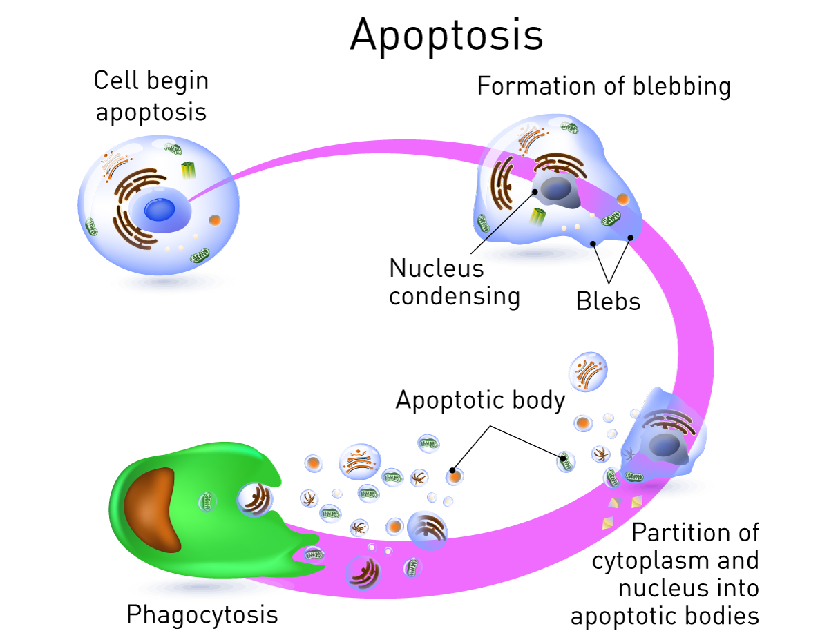
Cell death is a normal process in multicellular organisms, playing an important role in homeostasis. There are two types of cell death: necrosis (or accidental cell death), and apoptosis, a form of programmed cell death (PCD) characterized by specific morphological changes. Apoptosis is responsible for the removal of damaged or unnecessary cells throughout the lifecycle, including normal cell turnover, cell loss during embryogenesis – for instance, removing cells between fingers – negative selection by the immune system and nervous system development1. Cell death is a highly regulated process involving complex signaling pathways, essential for maintaining cellular homeostasis and normal physiological functions.
Apoptosis is one of the most investigated processes in cell biology which means many assays are available for researchers to choose from. These assays cover a wide range of markers for different stages of complex pathways. As a result, there are many apoptosis assays described in the literature.
There are two key pathways in which apoptosis can occur – the caspase (extrinsic) cascade and the mitochondrial (intrinsic) pathway. The extrinsic pathway involves the activation of death receptors on the cell surface, which recruit adaptor proteins to transduce death signals and initiate the apoptotic process.
Dysregulation in one or both pathways is linked with several human diseases, including cancers, stroke, and neurodegenerative conditions such as Alzheimer’s. Dysregulation is normally due to a disruption of the balance between pro-and anti-apoptotic proteins, changes to caspase activity or altered death receptor signaling. It is therefore vital to determine the mechanism of action of apoptotic dysregulation to understand the pathogenesis of a disease.
Caspase enzymes are proteolytic enzymes that play a central role in the apoptotic process by cleaving specific cellular substrates during cell disassembly. The apoptotic process is tightly controlled by multiple signaling pathways. It is therefore vital to determine the mechanism of action of apoptotic dysregulation to understand the pathogenesis of a disease.
Understanding the complex regulation of cell death is also essential for the development of effective therapeutics and apoptosis is therefore one of the most widely investigated processes in drug development today2.
The complexity of the pathways involved in apoptosis means that different markers can be used to study the process. Which pathway and stage of apoptosis to follow will vary depending on your specific application, so it is important to pick the best markers for your apoptosis assay. Some assays are specifically designed to detect apoptosis induction and monitor the onset of programmed cell death.
The mitochondrial membrane potential assures ATP production during oxidative phosphorylation. As an early event during apoptosis, the mitochondrial permeability transition pore (MPTP) opens and depolarizes the membrane potential. Measuring depolarization is achieved using the JC-1 indicator. JC-1 fluoresces red if present as aggregates in healthy mitochondria. With a decrease in mitochondrial membrane potential, JC-1 shifts to monomers that have a green fluorescence (Figure 2). The ratio of red to green fluorescence is monitored on a microplate reader and decreases along with the mitochondrial membrane potential. The application note “The CLARIOstar with ACU exposes cells to ischemia-reperfusion conditions and monitors their oxygenation” and monitors their oxygenation” describes the use of JC-1 to measure mitochondrial membrane potential.
Similarly, the fluorescent dyes Rhodamine 123 or TMRE (tetramethylrhodamine ethyl ester perchlorate) can be used to measure mitochondrial membrane potential. Both substances accumulate in the healthy mitochondrial membrane and cannot bind if the membrane potential is disrupted. During this apoptosis assay, cells are stained with Rhodamine 123 or TMRE and excess dye is removed by washing. The fluorescence intensity is measured on a microplate reader and loss of membrane potential which occurs during apoptosis is reported by a decrease in fluorescence.
Cytochrome c – a mitochondrial protein – plays roles in both cellular metabolism and apoptosis4. The protein interacts with apoptotic protease activating factor-1 (Apaf-1) in the cytosol to initiate the proteolytic maturation of caspase-9 and the death protease caspase-3. During early apoptosis in the course of the opening of the mitochondrial permeability transition pore (MPTP), cytochrome c is released into the cytosol and is used as a marker for PCD. Cytochrome c in cytosolic and mitochondrial cell extracts is quantified by Western blot or microplate readers to measure apoptosis.
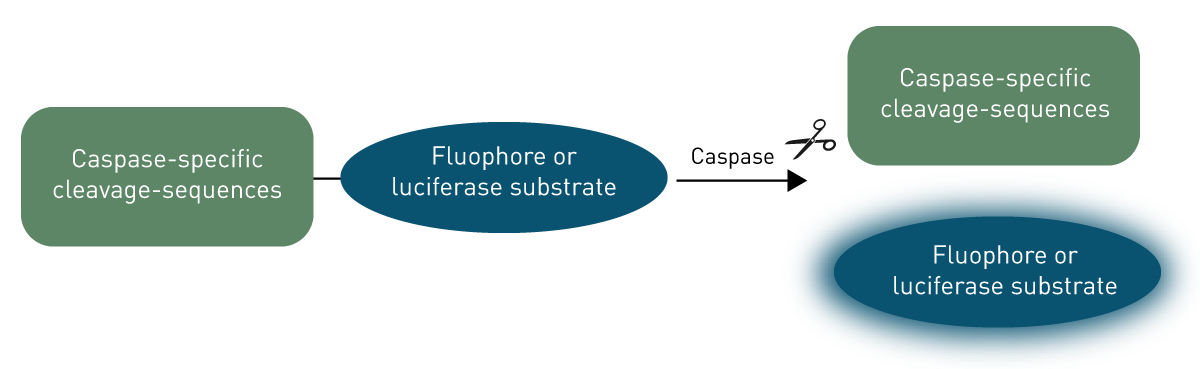
Caspases belong to a large family of proteins that are involved with inflammation or apoptosis. They are constitutively expressed in most cell types as inactive zymogens that require proteolytic processing before they can be fully active. In humans, initiator caspases (caspase-2, -8, -9, and -10) are signaling proteins that initiate the process of apoptosis by activating effector caspases through proteolytic processing. Effector caspases – such as caspase-3, -6, and -7 – are vital for apoptosis. They direct cell death itself by cleaving downstream targets including many vital cellular proteins and breaking up the nuclear scaffold and cytoskeleton. They also activate deoxyribonuclease (DNase), which further degrades nuclear DNA, causing cell death2. Artificial caspase substrates can be designed that contain cleavage sequences specific to different caspases (Figure 3).
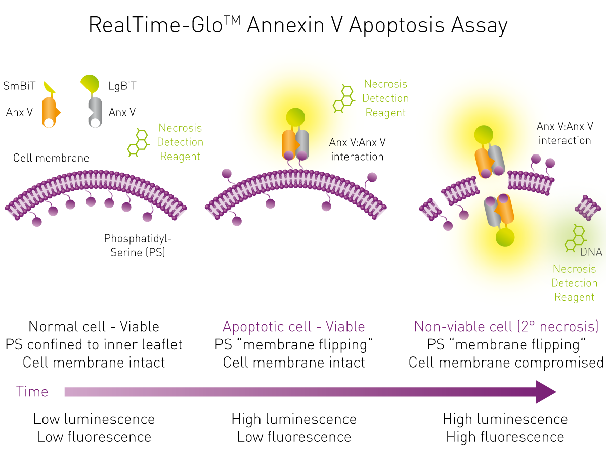
The appearance of phosphatidylserine (PS) residues on the surface of a cell can be used to measure apoptosis. In healthy cells, PS is located on the cytoplasmic surface of the plasma membrane. During early PCD, structural changes to the plasma membrane occur, including translocation of PS from the inner to the outer leaflet of the plasma membrane. Maintaining plasma membrane integrity is crucial for cell health, and loss of this integrity is a key indicator of apoptosis.
PS can therefore act as a marker for phagocytosis by macrophages without the release of pro-inflammatory cellular components2. Annexin V – a cellular protein – has a strong Ca2+-dependent affinity for PS. It can therefore be used as a probe for PS on the outer leaflet of the plasma membrane and serve as a way to detect cells undergoing apoptosis3. A luminescence-based apoptosis assay can be used to study annexin V binding to PS on the outer membrane. Annexin V is linked to two different, non-functional luciferase parts. If apoptosis occurs, the two linked annexin V moieties bind to each other as they interact with PS exposed on the cell and both luciferase parts come together to form a functional luciferase (figure 4). Hence, the induction of apoptosis produces luminescence that can be quantified by a luminescence plate reader. Details are found in the application note “Real-time assessment of apoptosis and necrosis”.
Another approach utilises biocytometry to detect apoptotic cells specifically in a subpopulation of cells. A luminescent reporter signal is generated upon binding of bioparticles to specific antigens on the cell surface.
DNA fragmentation occurs when genomic DNA is cleaved. This type of cleavage of genomic DNA is a process that occurs towards the end of apoptosis. DNA fragmentation is a key marker of late-stage apoptosis.
Assays labelling the ends of genomic fragments, such as a dT-mediated dUTP Nick End Labeling (TUNEL) assay, can use colorimetric or fluorescent detection to study apoptosis in cells1. The enzyme terminal deoxynucleotidyl transferase (TdT) adds nucleotides to 3'-hydroxyl termini. It adds fluorescently labelled nucleotides or horseradish peroxidase-linked nucleotides, producing a fluorescent or colorimetric signal. The further apoptotic DNA fragmentation progresses, the more nucleotides are included and the higher is the signal. Analysis is either done using light microscopy to visualize DNA fragmentation and structural changes during late-stage apoptosis, or in a microplate reader.
TUNEL assays compatible with BMG LABTECH microplate readers are available from various suppliers (Table 1).
Morphological and biochemical features such as nuclear chromatin compaction and DNA fragmentation distinguish apoptotic cells from necrotic cells. DNA fragmentation assays like TUNEL help differentiate between these forms of cell death.
More specific approaches also use extracellular ATP to detect immunogenic cell death- driven apoptosis. This was successfully demonstrated in the following application note: Extracellular ATP measurement in real time using living cells exploiting the incubation features of the CLARIOstar.
Real-time apoptosis assays and multiplexing
Traditional experimental approaches to study apoptosis are often time-consuming, labour intensive, expensive, and may not generate sufficient data. Conventional endpoint methods to measure cell health are not efficient when multiple assays are required to study different time points. The use of resources is very high in these cases. As no single parameter fully defines cell death in all systems, using several different approaches has advantages. Running different assays sequentially – studying one parameter followed by another in different plates – is one solution, but the increased time and costs of consumables are major constraints, especially as cell culture is already relatively expensive. Cell-based assays are also especially vulnerable to variations in cell growth and metabolism between cells in different plates.
Measuring in real-time lowers resource costs and reduces the need for multiple assays with different endpoints. This ensures that no time points are missed and data are continuously collected. Multiplexing provides the opportunity to measure several parameters in the same well, minimizing the consumption of valuable test compounds while saving time and reducing consumable costs. These assays are especially useful when studying cell viability such as apoptosis and necrosis, allowing specific stages to be recognized in the process and improving assay results. In real-time apoptosis assays, cell shrinkage and membrane integrity are important indicators that can be monitored alongside other parameters.
The real-time nature of the results allows the easy capture of information that would require extensive effort using previous apoptosis assay techniques. Additionally, fewer cells are needed to acquire the same amount of data, and measurements are more precise. In this way, apoptosis assays can be combined with other related assays – for instance, cell viability and cytotoxicity assays – to provide more information about mechanisms of cell death on a single sample5,6.
Given the number of different markers to study different stages of apoptosis, it is no surprise that a huge variety of assays exist from different companies. Below is a small selection of available microplate-based kits and their markers.
|
Apoptosis marker
|
Commercial kits
|
Detection mode
|
|
Mitochondrial membrane potential
|
JC-1 Dye (Mitochondrial Membrane Potential Probe; ThermoFisher Scientific)
JC-10 Mitochondrial Membrane Potential Assay Kit (Microplate; abcam)
Rhodamine 123 and TMRE are available from common life science chemicals providers
|
Fluorescence
|
|
Cytochrome c release
|
Human Cytochrome C ELISA Kit (SimpleStep ELISA by abcam)
Cytochrome C Human ELISA Kit (ThermoFisher Scientific)
|
Absorbance
|
|
Caspase activity
|
Caspase 3 Assay Kit, Colorimetric (SigmaAldrich)
Caspase-Glo 3/7 (Promega)
Molecular Probes™ EnzChek™ Caspase-3 Assay-Kit (ThermoFisher Scientific)
|
|
|
Phosphatidylserine on membrane surface
|
RealTime-Glo™ Annexin V Apoptosis and Necrosis Assay (Promega)
|
Luminescence (apoptosis), fluorescence (necrosis
|
|
DNA fragmentation
|
Click-iT® TUNEL Alexa Fluor® (ThermoFisher Scientific)
Cell Meter(TM) TUNEL Apoptosis Assay Kit (AAT Bioquest)
|
Fluorescence
|
Apoptosis assays are especially important for evaluating how tumor cells respond to therapies and for understanding cancer biology, as detecting changes in the apoptotic pathway within tumor cells can provide critical insights into treatment effectiveness and disease progression.
Cell viability assays require a microplate reader that is sensitive and can generate a lot of data. BMG LABTECH’s CLARIOstar Plus and VANTAstar with Atmospheric Control Unit (ACU) offer the control needed for cell viability assays, regulating temperature, CO2, and O2 levels to ensure cells are maintained in an appropriate environment. The system offers the best sensitivity of its class and features the Enhanced Dynamic Range technology, as well as multiple shaking, well scan, and reagent injectors, making it the ideal platform for any live cell-based assay. This enables novel methodologies to be carried out and real-time data to be captured, offering a true walk-away solution5,7.
Not sure what marker or apoptosis assay to use or what is best to measure for your application? Contact BMG LABTECH for all your apoptosis queries, and we can point you in the right direction.
Absorbance plate reader with cuvette port
Powerful and most sensitive HTS plate reader
Most flexible Plate Reader for Assay Development
Upgradeable single and multi-mode microplate reader series
Flexible microplate reader with simplified workflows
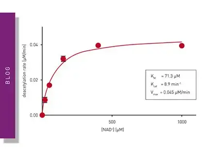
Learn about applications for bacterial metabolism on a microplate reader.
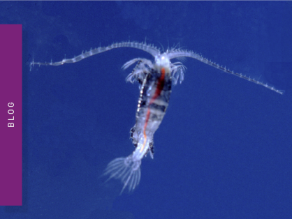
Life in the depths of the ocean operates under extreme conditions. Find out how proteins from deep-sea luminescent organisms are useful for measurements on microplate readers.
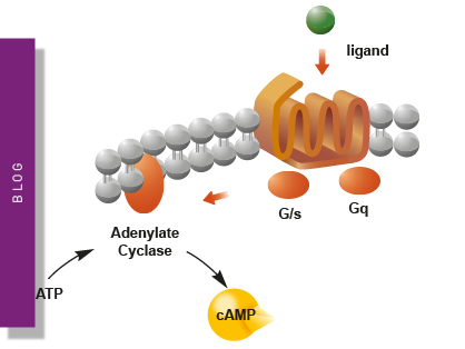
Second messengers play a pivotal role in signal transduction events in cells. But how do you measure these small, transiently lived molecules and how can microplate readers help?
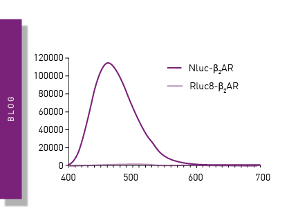
NanoBRET is used to analyse binding events, signaling pathways and receptor trafficking in live cells and has significantly expanded the range and applications of BRET assays.
Find out about the different types of cell-based assays and why they have become an indispensable tool in a such a broad variety of disciplines in this blog article.
Read here about the various threats associated with mycoplasma contamination and find out about methods to prevent, detect, and eliminate mycoplasma in your cell culture.