Introduction
Apoptosis, programmed cellular death, is often dysregulated in a variety of conditions including cancer, autoimmune disorders and neurodegenerative diseases. Current methodologies for measuring apoptosis do not facilitate high-throughput, cell-specific apoptosis profiling within complex samples, leading to averaged readouts that may not accurately represent the diverse responses within different cell subpopulations. With the shift from animal models, the analysis of complex sample matrices becomes increasingly critical 1.
The biocytometry kits provided by Sampling Human, Inc.2, were designed for the targeted monitoring of apoptosis across various cell types. The comprehensive and userfriendly system can be evaluated with any multi-well luminometer and provides a highly sensitive detection of apoptotic events in mixed cell cultures.
Assay principle
The biocytometry assay for the readout of cell-specific apoptosis operates by targeting cells displaying both phosphatidylserine (PS), a well-established marker of early apoptosis3, and additional cell-specific antigen marker(s), in this case the epithelial marker EpCAM. Individual bioparticles expressing antibody mimetics bind to cells in a sample displaying corresponding antigen markers (Fig. 1). Only when bioparticles of both types (PS and EpCAM) are present on the same cell is production of the luminescent reporter triggered, ignoring partial matches (EpCAM only or PS only) and cell debris. The resulting luminescence signal is proportional to the number of targeted cells. The biocytometry kits provided by Sampling Human, Inc.2, were designed for the targeted monitoring of apoptosis across various cell types. The comprehensive and userfriendly system can be evaluated with any multi-well luminometer and provides a highly sensitive detection of apoptotic events in mixed cell cultures.

Materials & methods
- Jurkat (#ab275468, Abcam) and HaCaT cell line (#300493, CLS)
- Culture medium RPMI with 5 % serum and 5g/L glucose (Thermo Fisher scientific)
- 100 mM DTT (#D0632, Sigma-Aldrich),
- 96-well U-shaped suspension plate, white (#267350, Thermo Fisher Scientific)
- 96-well flat bottom plate, transparent, TC-treated (#92096, TPP)
- Biocytometry kits for EpCAM+ and EpCAM+/PS+ (#100020 and #100022, Sampling Human)
- VANTAstar® with injector (BMG LABTECH)
Experimental procedure
Cell culture and apoptosis induction
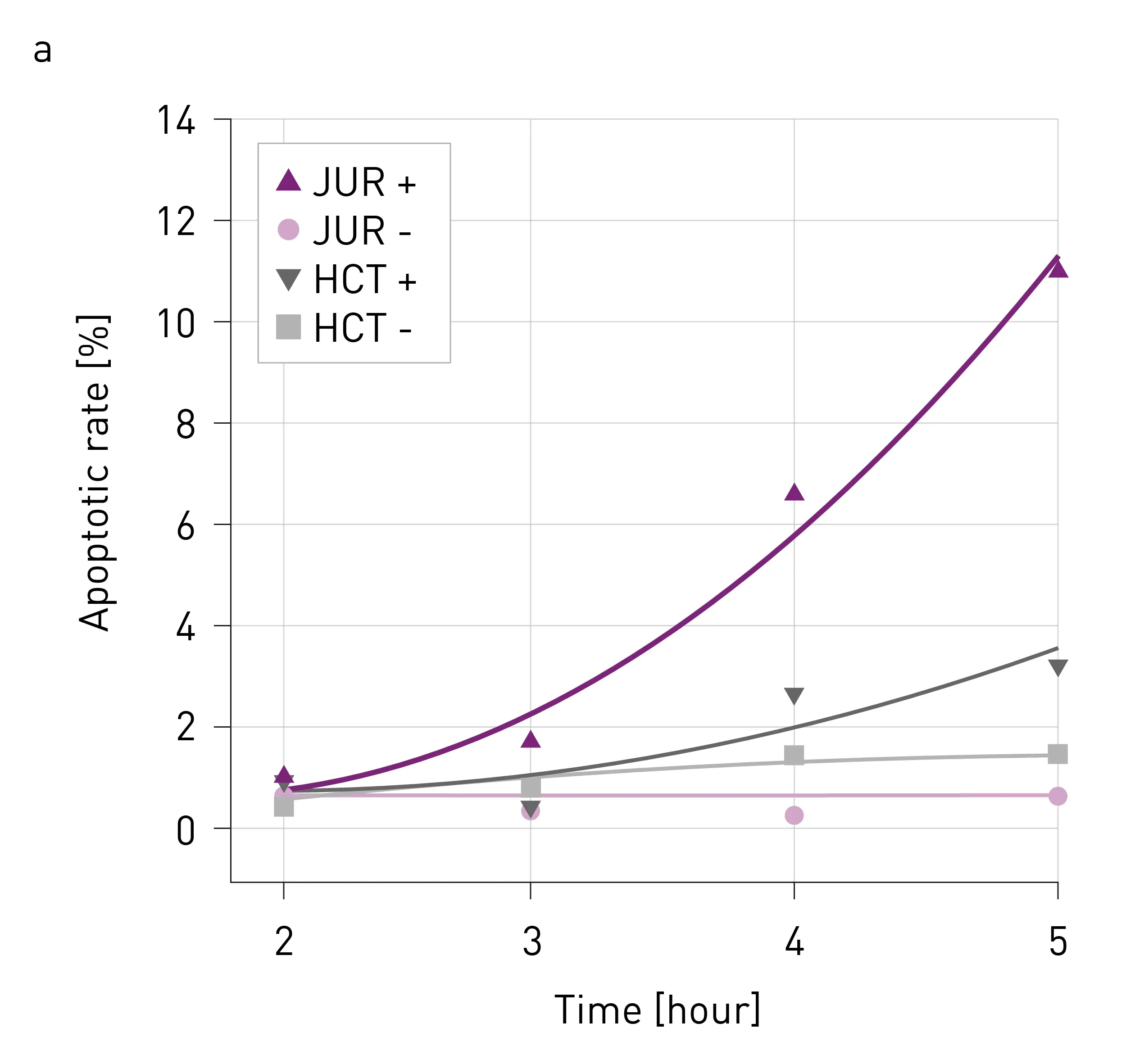
To confirm the suitability of the biocytometry method to determine the time course of apoptosis HaCaT and Jurkat cells were first treated with 0.5mM DTT in monocultures. Following the apoptotic rate was monitored via PS over a period of 5 hours.
For the co-culture experiments, the HaCaT and Jurkat cells were seeded in white suspension plates at 20,000 per well in both mono-cultures and co-cultures. All cultures were treated with 2.5 mM DTT to induce apoptosis, followed by incubation for 2 hours at 37 °C and 5% CO2.
Biocytometry
The biocytometry assay was conducted according to the manufacturer’s instructions (Fig. 2), utilizing three technical replicates for both EpCAM and EpCAM/PS kits to analyze samples from induced and non-induced co-cultures. The ratio of quantified events was used to determine the specific apoptosis rate in the HaCaT cell line. Additionally, induced Jurkat mono-culture was analysed to evaluate the assay’s specificity.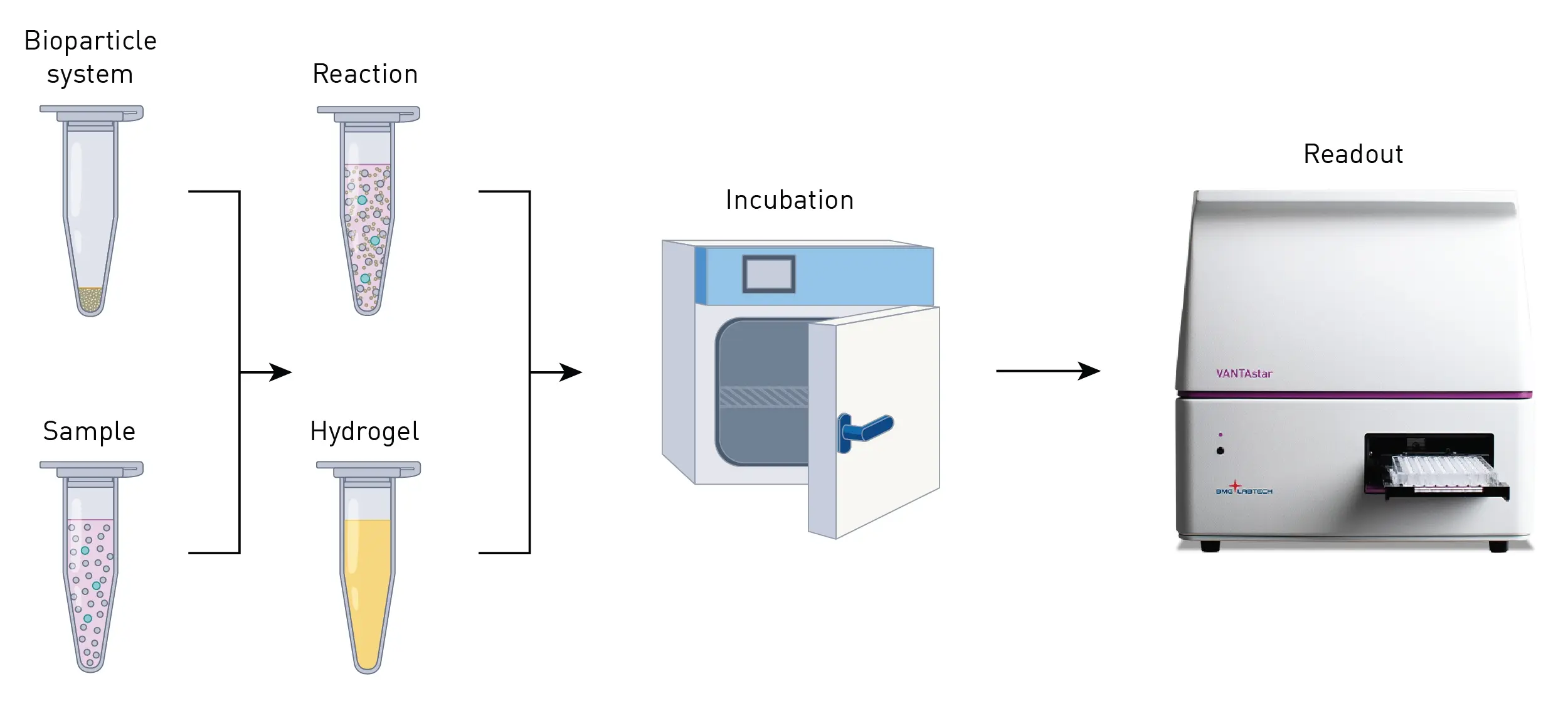 Readout
Each plate was transferred to the VANTAstar that was pre warmed to 37 °C. Luminescence substrate was mixed with Luminescence buffer in the ratio 1:40. The mix was injected, and the plate was subsequently read with the following parameters.
Instrument settings
|
Optic settings
|
Luminescence, plate mode kinetic
|
|
|
Emission
|
No filter |
|
|
Optic
|
Top Optics |
|
|
General settings
|
Gain | EDR |
| Measurement interval time | 5 s | |
| Focal height | height | |
| Number of cycles | 2 | |
|
Kinetic settings
|
Cycle time | 441 |
| Injection | 200 μL in cycle 1 | |
| Shaking | 700rpm for 180s after cycle 1 | |
|
Incubation
|
37 °C | |
Results & Discussion
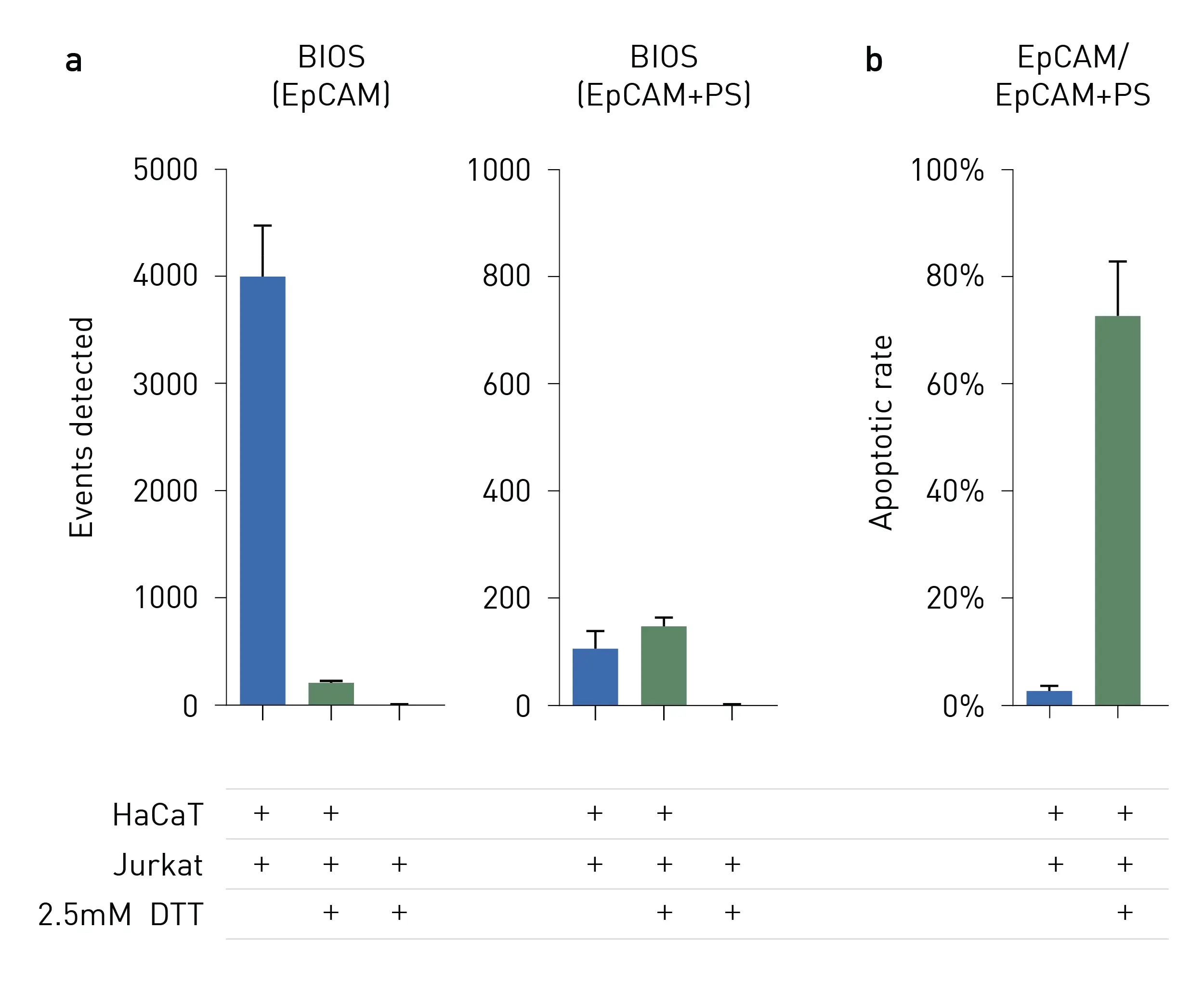
Biocytometry facilitated the quantification of global apoptotic events over time (Fig. 3) and enabled the identification of apoptosis in specific cell subpopulations in mixed cultures (Fig. 4). The response of HaCaT cells to DTT, evidenced by a signifi cant reduction in EpCAM+ events, was captured using the EpCAM biocytometry kit (Fig. 4a, left panel). The apoptosis ratio for HaCaT cells was then calculated by normalizing the events detected with the EpCAM/PS kit against those obtained with the EpCAM kit alone (Fig. 4b). Following DTT exposure, HaCaT cells exhibited a marked increase in their apoptosis rate, from 2.6% to 72.4%. Notably, Jurkat mono-cultures yielded no detectable signal.
Conclusion
Sampling Human’s biocytometry kits offer a very sensitive and user-friendly option for the analysis of cellular immunophenotypes on the single-cell level. The method represents a promising alternative to cell cytometry with lower equipment requirements, as the readout is done on a luminometer. The biocytometry kits feature a homogenous workflow, enabling streamlined assessment of cell-specific apoptosis rate without the need for wash steps. The readout of the experiment with the VANTAstar provides users with a true walk away solution of the last workflow steps including the optimal signal detection. With the VANTAstar’s injector module, the luminescence substrate and buffer mix can be injected into the microplate with high reproducibility and low time expenditure. The on board shaking function and temperature control allow optimal mixing of the substrate mix into the temperature sensitive hydrogel. Further, the EDR function ensures reliable detection at a large range of signal intensities in the same run.
References
-
Han, J.J. FDA Modernization Act 2.0 allows for alternatives to animal testing. Artif. Organs 47, 449-450 (2023).
-
Cienciala, M. et al. Massively parallel identification of single-cell immunophenotypes. bioRxiv (2024).
-
Lee, S-H et al. Phosphatidylserine exposure during apoptosis refl ects bidirectional traffi cking between plasma membrane and cytoplasm. Cell death and differentiation vol. 20,1 (2013).