SPECTROstar Nano
Absorbance plate reader with cuvette port


Dr. Ann Cathrin Volz is a trained nutritionist with deep expertise in cell biology, biomaterials and food science. She studied Molecular Nutritional Sciences at the University of Hohenheim, where she completed her PhD developing a serum-free in vitro model of adipose tissue. During her scientific training, she combined academic biochemistry with practical laboratory work in bioengineering. After completing her doctorate, she spent two years as a postdoctoral researcher at Reutlingen University, where she focused on transferring her knowledge of tissue engineering from biomedicine to the field of in vitro agriculture. In 2020 Ann-Cathrin joined BMG LABTECH as an Applications Specialist, where she authors application notes, conducts workshops, and supports scientific customers.
With a size of 0.2 - 0.8 µm, mycoplasmas represent the smallest free-living organisms 1. They belong to the mollicutes class within Gram-positive bacteria. As they lack a cell wall (see figure 1), they cannot be eliminated by the use of standard antibiotics like many other bacteria2.
Their genome only comprises 0.58 - 2.2 Mb DNA, which explains their limited biosynthetic capabilities 1,3. Consequently, mycoplasma maintenance and growth depend on the availability of nutrients and metabolites from host cells. Although around 195 mycoplasma species have been identified, just 6 species are responsible for 95% of all mycoplasma contaminations in cell culture: M. orale, M. arginini, M. fermentans, M. hyorhinis, M. hominis, and A. laidlawii 4.

The nasty thing about mycoplasma contamination is, without specific testing, it will stay unnoticed. Mycoplasma contaminations are not visible to the naked eye. Moreover, due to the limited size and comparably low doubling rate of these bacteria, contaminated cell cultures do not show a reduced pH or obvious turbidity. However, mycoplasma contaminations may exist and persist despite the regular use of antibiotics and may significantly impact cellular events. In case you noticed reduced proliferation or transfection efficiency, morphological changes, or cell aggregation, you should consider a mycoplasma contamination test.
In contaminated cell cultures, these bacteria compete with your target cells for nutrients, and further on exposing them to undesired metabolites. They influence the levels of DNA, RNA, and protein synthesis, alter gene expression, and impact cell membrane, organelles, cell morphology, and antigenicity. Thereby they can inhibit cell growth, cellular metabolism, and may cause cell death (figure 2) 1,5,6,7,8. Therefore, undiscovered mycoplasma contamination fundamentally calls into question the relevance and validity of your results.

Humans, pigs, and cattle represent potential hosts for mycoplasma contaminations 9. The main infection paths are through direct or indirect contact with infected lab staff, reagents like a fetal bovine serum, or through the culture of infected cell lines. To prevent mycoplasma contamination, proper aseptic cell culture technique should be applied including routine cleaning of all used devices like incubators, sterile workbenches, or water baths 11,12. It is also recommended to dedicate separate media and reagents for each individual cell line and only handle one cell line at a time 2. However, the most effective method is to closely follow a testing routine in defined intervals (monthly/quarterly) and strictly quarantine cell lines introduced to the lab until tested negatively.
Mycoplasmas stop proliferating at -196°C - they do however survive and may be carried from one vial to the other 10. It has to be kept in mind, that cells in cryovials can be contaminated via the liquid phase of nitrogen during the storage in a joint vessel.
There are basically two categories of mycoplasma contamination tests. You can either directly look for mycoplasmas e.g. by culturing and detecting the whole organism, or you can make use of some subcellular units of mycoplasma by testing for specific gene expression or enzyme activity 13. Table 1 summarizes the most popular testing methods used for the detection of mycoplasma contamination. Many testing methods lack the ability to identify specific species. However, this feature is irrelevant in most cases since it is first and foremost a matter of general detection of potentially contaminated cell lines.
Table 1: mycoplasma detection methods
| Testing method | Testing principle | Advantages | Limitations | Ref |
| Microbiological culture (reference method for new methods). | Supernatant of suspected cultures is transferred to a specific medium and subsequently to agar. After about 2 weeks, mycoplasma contaminations result in visible colonies in a “fried egg” appearance. | Cheap, sensitive, specific | Time-consuming, complex, highly dependent on user experience, specific identification only possible for some species, slow. | 1,2,4 |
| DNA staining. | Suspected cell cultures are stained with a non-specific fluorescent nucleic acid stain (e.g. DAPI), mycoplasmas can be identified as smaller spots close to eukaryotic cells, but with some distance to their nucleus. | Cheap, rapid, simple | Unspecific, low sensitivity, subjective, no species identification. | 2,15,16 |
| Biochemical detection of enzymes. | Mycoplasmas produce enzymes, which are not present in eukaryotic cells. These enzymes catalyze the turnover of a substrate, resulting in luciferase activity/luminescence, readable on a luminescence plate reader. The use of a biochemical assay for the detection of mycoplasmas is described in the application notes 130 and 281 with the example of MycoAlertTM. | Rapid, simple, cheap, specific | No species identification, results may vary between cell lines, luminescence microplate reader required | 2,15 |
| Gene detection via PCR (current gold standard). | Mycoplasma contaminations are analyzed by PCR amplification of specific gene sequences coding for mycoplasma 16S rRNA. Different mycoplasma species can be identified with specific primers addressing 16S - 23S intergenic regions. Amplified DNA is usually quantified with fluorescent dyes, either after their separation by gel electrophoresis, during amplification in a real-time PCR, or by measuring end-point fluorescence intensity on a fluorescence plate reader. | Very sensitive, specific, fast, objective results, commercial kits available. |
Optimization depends on experience, risk to detect false positives/false negatives requires RNAse-free handling, thermocycler required. |
2,8 |
| Detection of surface antigens with ELISA | Antibodies against mycoplasma surface antigens are used to capture the subunits and quantify them mostly by reading absorbance on an absorbance microplate reader. | Simple, cheap, suitable for HTS | Absorbance microplate reader required | 2 |
| RNA/DNA hybridization | Probes are hybridized with mycoplasma RNA or DNA. They can be compatible with a specific species, group, or universally for all mycoplasmas. Signal is detected as dots of hybridized material on blots or as detectable emission from liquid hybridization. | Specific, rapid, cheap |
Dependent on equipment and experience requires RNAse-free handling | 17 |
| Gene detection via isothermal amplification | Like in PCR, mycoplasma DNA is amplified and subsequently detected – however, in isothermal conditions and using 4 - 6 primers. Since a lower and constant temperature is sufficient, the method can also be carried out without a thermocycler. Thanks to their incubation feature, BMG LABTECH’s microplate readers CLARIOstar Plus and the Omega series allow performing isothermal amplification including the quantification step in the same device. | See PCR, no thermocycler required, faster runtime | Requires RNAse-free handling | 18 |
It is a pity, but the best way to get rid of mycoplasma contamination is to dispose of the contaminated cell cultures with all associated consumables and thoroughly clean all used equipment 2,11.
Nevertheless, some options for mycoplasma eradication are available. These are mostly based on the application of combined antibiotics including tetracyclines and macrolides for several weeks to inhibit protein synthesis8,19. The application of antibiotics to contaminated cell cultures is often associated with side effects like cytotoxicity, growth inhibition, loss of cellular characteristics, and clonal selection20. However, the risks associated with treatment with antibiotics may be considered acceptable if the affected cell line is either very expensive, time-consuming to establish, or even irreplaceable. Novel approaches combine antibiotics with non-antibiotic treatments and promise to directly kill the common mycoplasma species.
MycoAlertTM is a registered Trademark of Lonza.
Absorbance plate reader with cuvette port
Powerful and most sensitive HTS plate reader
Most flexible Plate Reader for Assay Development
Flexible microplate reader with simplified workflows
Explore metabolic flux analysis, its significance in research, and how microplate readers enhance measurements for insights.
Explore the vital role of mitochondrial metabolism in cellular function, its measurement techniques, and how microplate readers enhance research in this field.
Learn about applications for bacterial metabolism on a microplate reader.
Life in the depths of the ocean operates under extreme conditions. Find out how proteins from deep-sea luminescent organisms are useful for measurements on microplate readers.
Second messengers play a pivotal role in signal transduction events in cells. But how do you measure these small, transiently lived molecules and how can microplate readers help?
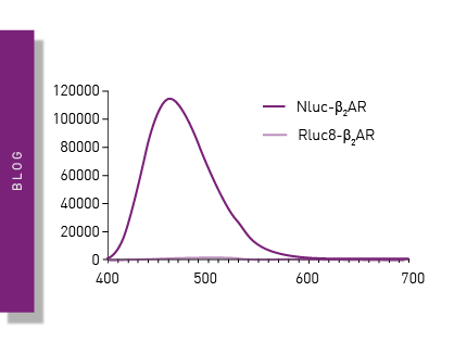
NanoBRET is used to analyse binding events, signaling pathways and receptor trafficking in live cells and has significantly expanded the range and applications of BRET assays.