Introduction
Most commercially available antibody pairs have been designed to be used in sandwich ELISA assays. They offer the flexibility of miniaturizing experiments to 384- and even 1536- well formats for higher throughput demands. Abcam’s matched antibody pairs (MAPs) are well-characterized, recombinant antibodies with highly sensitive reagents that cover a wide variety of targets. MAPs consist of an unlabelled capture antibody, a biotinylated detection antibody and a protein standard. In order to enhance assay performance, alternative measurement techniques and different secondary anti-body labels can be used. As an example, Time-Resolved Fluorescence (TRF) detection uses long-lifetime fluorophores, known as lanthanides, that emit light over a longer period after excitation (microseconds as opposed to nanoseconds). This helps reduce background noise by delaying the start of the measurement until after the background signal has decayed. The measurement of assay signal using non-visible light spectra can increase dynamic range and is not limited to the same extent by saturation.
Here we describe how Abcam’s MAP kits can be used in a 384-well TRF immuno-assay (DELFIA® TRF – PerkinElmer) and read on the PHERAstar® FSX for the detection of IRAK4, a protein kinase involved in cell signaling as part of the immune response..
Assay Principle
In the DELFIA® TRF Assay, the MAP binds to the analyte in the sample and is then incubated with streptavidin-Europium in place of a traditional label such as HRP. Bound streptavidin-Europium is detected using the DELFIA® enhancement solution which liberates bound Europium and promotes the formation of a micellar chelate solution. The Europium chelate is excited at 337 nm and emits at 615 nm (Fig. 1).
Using the PHERAstar FSX this can be read using either a Xenon flash lamp or laser for excitation. In this application note the laser was used to maximise assay signal and sensitivity.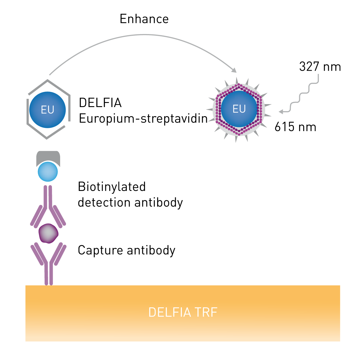
Materials & Methods
- Black 384 NUNC Maxisorp™ plates
- Human IRAK4 Matched Antibody Pair kit (ab218182)
- DELFIA® Europium-labelled streptavidin
- DELFIA® assay buffer
- DELFIA® enhancement solution
- PHERAstar® FSX (BMG LABTECH)
Experimental procedure
Assay performed in total volume of 50 µl according to MAP kit protocol (1) using black 384-well NUNC MaxisorbTM plates. The plate was incubated for 30 mins with streptavidin-Europium diluted according to manufacturers’ recommendations in DELFIA® assay buffer. After the addition of 50 µL of DELFIA® enhancement solution, time-resolved fluorescence was read on BMG LABTECH PHERAstar FSX equipped with a TRF laser with both non-optimized and optimized settings.
Instrument settings
| Optic settings
|
Time resolved fluorescence (TRF), top optic |
|
| Optic module | 337 615 (Ex: 337 nm; Em: 615 nm) |
|
| Excitation source | laser | |
| Number of flashes | 5 | |
| Integration start | 400 µs | |
| Integration time | 400 µs | |
|
General settings |
Settling time | 0 s |
Results & Discussion
The PHERAstar FSX is equipped with a laser excitation source, and photon counting detectors with an extended red detection range for TRF measurements. Combined with Z-height optimization for both top and bottom optics, this ensures the highest performance and sensitivity in any assay. The MARS data analysis package provided with the PHERAstar FSX is capable of automatically generating a calibration curve, from which the concentration of unknown samples can be determined. Calculation templates can also be saved to a protocol, meaning that future data analysis of test runs can be fully automated.
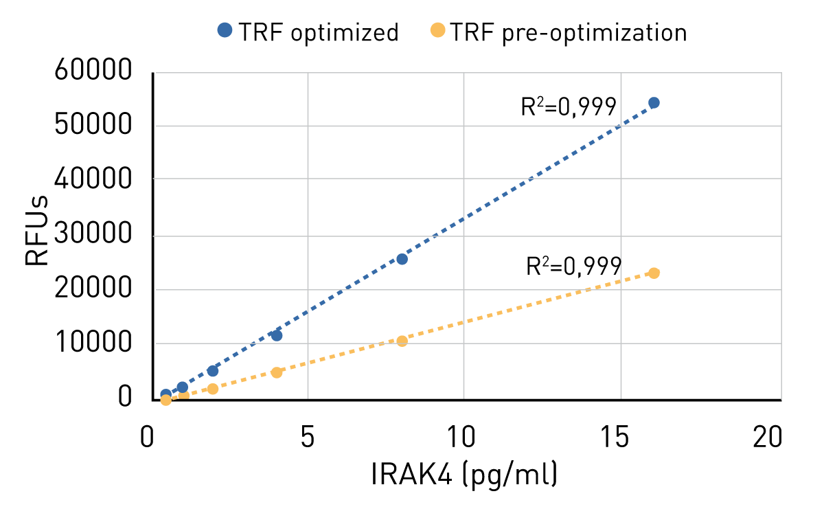
The optimal focal height for fluorescent measurements is dependent on the volume of sample used and dimensions of the microplate. The PHERAstar FSX is capable of automatically selecting the optimal Z-height for the optic when reading the plate from top or bottom. The curve in Fig. 2 shows the samples read in a 384-well plate before (yellow) and after (blue) optimization of the Z-height for the new microplate format. This adjustment improved both signal and signal to blank.
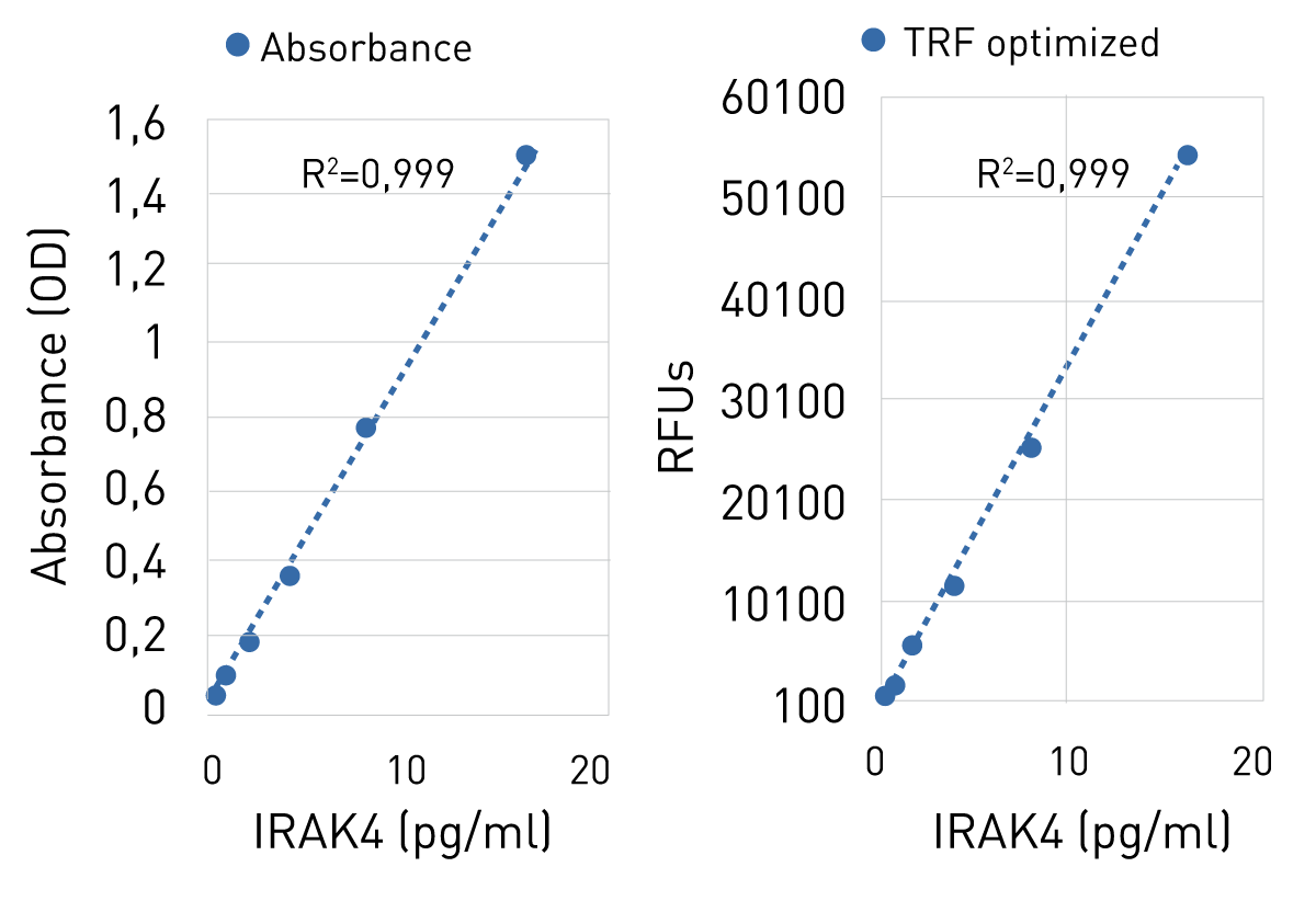
When compared to data previously obtained using the same MAP kit in a traditional absorbance based ELISA assay in a 96-well plate (Fig. 3), the TRF-based assay in a 384-well format with Human IRAK4 MAP kit shows a similar if not extended working range, as saturation was not reached. The linearity is given for both assays with an R² of 0.999.
Conclusion
Abcam’s recombinant MAPs offer enhanced flexibility in the type of detection method used for plate-based immunoassays. This application note demonstrates that MAPs are suitable to use in miniaturized TRF immunoassays, providing sensitivity, specificity and reproducibility of results. This combined with the 384-well format make MAP kits particularly well suited for high-throughput screening. Additional detection methods can be achieved with Abcam’s PBS-only formulations, which provide even greater flexibility. The PHERAstar FSX is capable of optimizing read conditions including focal height, and when coupled with a laser-based excitation source and photon-counting detection, maximizes sensitivity and signal window. These optimizations and features are particularly important when miniaturizing to 384-well microplates for high-throughput screening.
References
- How to use Matched Antibody Pair kit – Protocol (abcam)