Introduction
The NADPH oxidase family member, NOX2, has been extensively studied based on its association with phagosomes1. Among its functions, NOX2 has been shown to play a central role in regulation of antigen presentation on dendritic cells. More specifically, NOX2 is involved in alkalinization of dendritic cell phagosomes, thus limiting protease activity, resulting in only partial degradation of the antigen. The antigen then undergoes further processing before it is presented to CD8+ T cells. More recently it was shown that NOX2 undergoes activation mediated by PKCδ leading to pinocytosis of antigens2.
NOX2 activity is known to be regulated by numerous cytosolic factors (Figure 1). In order to more completely understand the regulation of, and to seek out possible inhibitors of, NOX2 activity, the appropriate tools must be employed. Here, we describe the use of a chemiluminescent probe suitable for the detection of NOX2 activity in a microplate-based assay.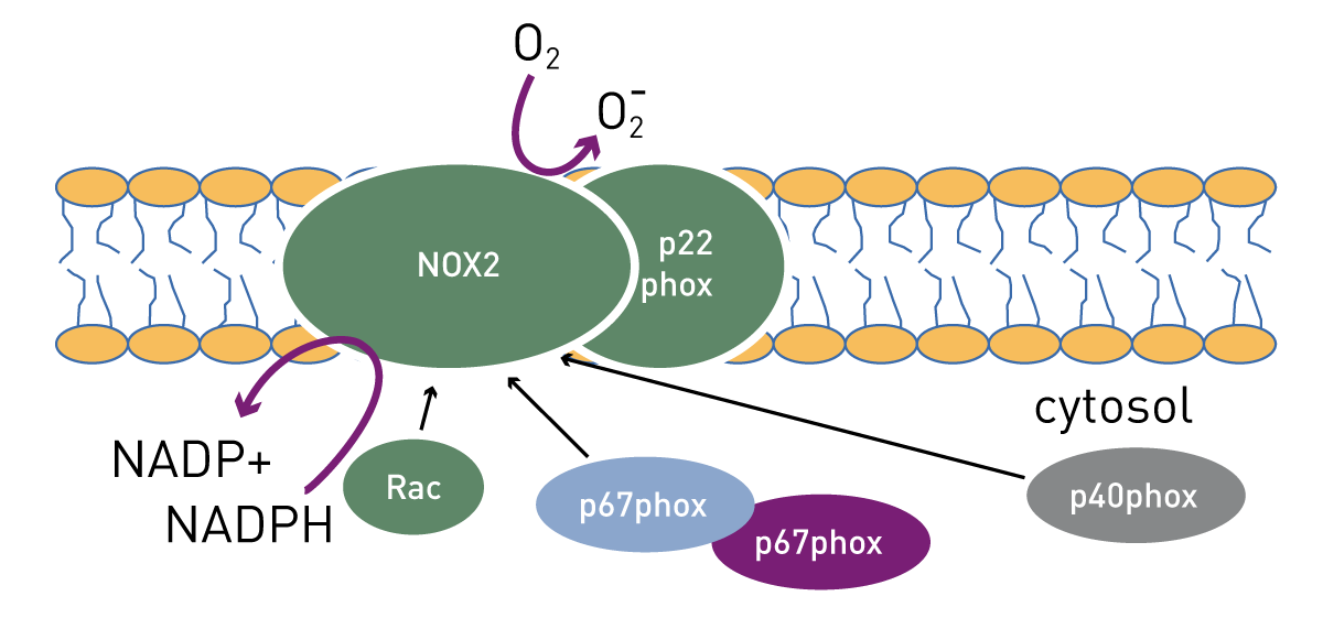
Assay Principle
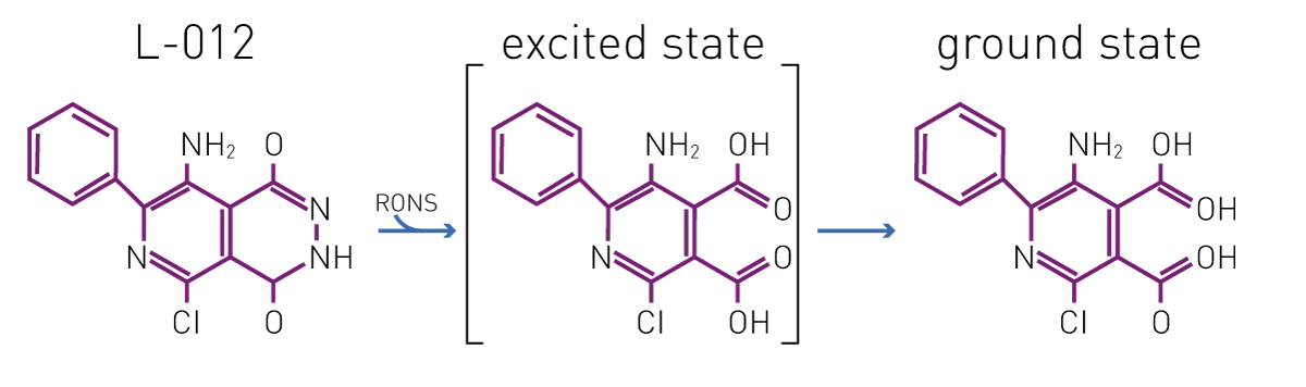
The luminol analog L-012 has been shown to be more sensitive at detecting ROS than its predecessor3. Furthermore, it has a higher luminescence yield compared to other superoxide probes as luminol and lucigenin3. Figure 2 shows the proposed reaction from interaction between L-012 and reactive oxygen (and nitrogen) species.
Materials & Methods
- CLARIOstar
- L-012 from Wako Chemical (120-04891)
- other chemicals obtained from commercially available sources
Experimental Procedure
Bone Marrow-Derived immature Dendritic Cells (BMiDCs) were prepared from wild type and NOX2 knockout mice and CD11c positivity determined as described in Singla et al2. Fifty thousand cells/well were plated in sterile phosphate buffered saline (PBS) containing 400 µM L-012. Cells were stimulated with 1 µM PMA (phorbol 12-myristate 13-acetate) and in some cases, further addition of superoxide dismutase (SOD) at a concentration of 150 U/mL provides confirmation of specificity of this test for O2-. Luminescence was read as indicated below. Evaluation was performed using BMG LABTECH’s MARS data analysis software.
CLARIOstar settings
|
Optic settings
|
Luminescence, plate mode kinetic
|
|
|
Gain
|
3500 | |
|
Kinetic settings
|
Number of cycles |
60
|
| Cycle time | 120 s | |
|
Measurement interval
|
0.2 s
|
|
| Incubation |
37 °C
|
|
Results & Discussion
Figure 3 shows the kinetic response of BMiDCs over a 2 hour period based on the light intensity produced by L-012 chemiluminescence. In vehicle treated cells a stable low level of light output is observed. Treatment with the protein kinase C activator PMA induces a strong increase in luminescent signal that peaks at ~1 hour. PMA stimulation in the presence of SOD fails to elicit a response indicating that the light production by L-012 is dependent on the presence of O2- in this system.
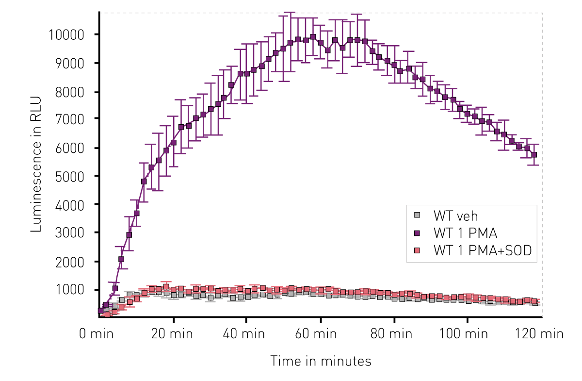 Figure 4 compares WT BMiDCs with BMiDCs from NOX2 KO background based on their ability to promote light output from L-012 chemiluminescence. After 1 hour of treatment, PMA stimulated a greater than 9-fold increase in RLU in WT cells compared to vehicle. By contrast cells from the Nox2 KO did not respond to PMA stimulation.

Additional results indicated that PKC δ is the predominant PKC isoform present in BMiDCs and that plays a functional role in PMA-dependent O2- production.
Conclusion
The results indicate that L-012 is a very useful tool for the detection of ROS using a microplate reader-based assay. Sensitive detection using the CLARIOstar aids in the utility of this assay format. The results shown here indicate that there is a PKC-dependent activation of NOX2 in dendritic cells leading to O2- production important for their role in immune response.
References
- Bedard, K et al. The NOX Family of ROS-Generating NAPH Oxidases: Physiology and Pathophysiology. Physiol. Rev. (2007) 87: 245-313.
- Singla, B et al. PKCδ-Mediated Nox2 Activation Promotes Fluid-Phase Pinocytosis of Antigens by Immature Dendritic Cells. Front. Immunol. (2018) 9: Article 537.
- Brandes, R.P. et al. Nox family NADPH oxidases: Molecular mechanisms of activation. Free Radic. Biol. Med. (2014) 76: 208-226.