SPECTROstar Nano
Absorbance plate reader with cuvette port

Besides vaccination and drug treatment, diagnostic viral detection methods, which detect acute infections with viruses, such as influenza, are a central tool to fight viral spread and help to contain viral infection 1. The use of a polymerase chain reaction (PCR) to detect specific viral genomic sequences during an ongoing infection with viruses is still considered to be the gold standard among available virus detection methods, despite its limitations.
Especially in acute outbreaks of an infection with viruses, features such as high-throughput capability, ease of use and low turn-around time play a major role besides the general demand for precision, accuracy, and specificity in virus assays.
Microplate readers meet these requirements and can cover a wide range of viral detection methods used for the analysis of molecular and cellular mechanisms taking place during an infection with viruses objectively and quantitatively.
Viral detection methods can identify a virus infection in a plethora of ways, by the analysis of different components and functions of the virus. These targets range from viral genome, over expressed antigens, and enzyme activity to virus infectivity (fig. 1).
Microplate readers allow for the coverage of a great variety of assays to detect viruses by using different detection modes, which are either available in dedicated single-mode readers or combined to a versatile platform in multi-mode readers. BMG LABTECH offers customised solutions for both categories. This blog provides an overview of the various viral detection methods which can be used for the analysis of viruses in different detection modes on microplate readers.
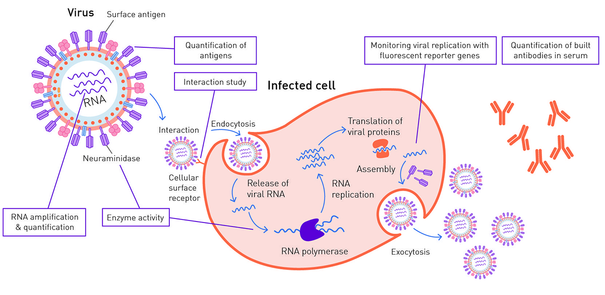
In absorbance measurements, the part of light which is taken up (absorbed) by a sample is analysed and quantified. Enzyme-Linked Immunosorbent Assays (ELISAs) use antibodies and can be applied as a virus detection method for the quantitative analysis of viral antigens present in serological or swab samples. This virus detection method is based on the use of specific antibodies. These antibodies recognise and bind viral antigens. Although, the final output can also be of luminescent or fluorescent nature, ELISAs are mainly used with a colorimetric readout coupled to the used antibodies.
Besides the gold standard PCR, other viral detection methods are also based on the amplification of nucleic acids of viruses. One of them is the isothermal loop-mediated amplification (LAMP) assay. Unlike PCR, it can be run isothermally at constant 65°C, and is a virus detection method that can also be measured in real time with a microplate reader equipped with absorbance or fluorescence detection and an incubation function up to 65°C. For a detailed description of a colorimetric LAMP assay applied as a virus detection method addressing a SARS-CoV-2 infection as an alternative to PCR, see our application note: LAMP assay for detecting SARS-CoV-2 RNA using an absorbance-based colorimetric readout.
The process in which a substance absorbs light and consequently emits light of a longer wavelength is called fluorescence. In combination with intercalating fluorescent dyes, microplate readers are well suited to quantify nucleic acids by fluorescence measurement, e.g., after their amplification by PCR (see also: DNA quantification using absorbance (A260) and fluorescent methods (Qubit™ and Quant-iT™/PicoGreen™)). When using fluorescent probes, fluorescence microplate readers can also be used to evaluate fast PCR approaches which use innovative heating technology to accelerate the PCR runtime: AN370: NextGenPCR™ evaluated with a fluorescence microplate reader accelerates testing for SARS-CoV-2 viral RNA.
Next to the amplification of viral DNA/RNA via PCR or respective alternatives, the activity of viral enzymes can be exploited as a virus detection method to detect an ongoing infection with viruses. Neuraminidase is a key glycoprotein on the surface of influenza viruses, leading to virion release from the infected cell in vivo and in culture. The enzymatic activity of neuraminidase can be measured with a virus detection method using a synthetic substrate, which releases a fluorophore or chemiluminescent cleavage product 2,3 .
Viral enzymes can also be tested as targets for anti-viral drugs. For instance, NSP14, an exonuclease of the coronavirus family, is currently investigated as a COVID-19 target as discussed by Joao Pisco in this interview:

In luminescence, a light signal is produced by the energy conversion of a molecule, independent of an excitation source. The most frequently used source in life science approaches is chemiluminescence, where the emitted light is derived from a chemical reaction.
Viral replication is an important step in the viral infection process. In vitro it is usually monitored by the expression of fluorescent or luminescent reporter genes by viruses, which are introduced for the infection of a cell culture. This virus detection method is typically used to assess the effectiveness of antiviral compounds to fight an infection. The higher the number of virions in cell culture, the higher the amount of expressed reporter protein and its according fluorescence signal will be during the infection.
This virus detection method was for instance used in the following app notes based on cell culture and infection in vitro with BMG LABTECH advantageous features like well-scanning and direct optic bottom reading: Studying the molecular mechanism of viral replication in real time using the CLARIOstar Plus with ACU; Antiviral assay based on expression of fluorescent proteins by the viruses.
Next to fluorescent or luminescent markers, cell viability can be used as an indirect measurement of virus replication. With increasing infection, cell viability decreases. By the use of cell viability or cell cytoxicity assays, viral cytopathic effect or its reduction by potential drugs can be quantified. In a similar way, bacterial cells can be employed to evaluate the antibacterial properties of bacteriophages using absorbance detection.
Time-resolved fluorescence (TRF) is based on a special group of fluorophores with prolonged emission. This allows a delayed detection of the specific signal after background signals and scattered excitation light have decayed. This sensitivity is further enhanced using the BMG LABTECH TRF-laser. In the immuno-assay format (e.g. DELFIA®), this viral detection method is often used to detect viral antigens with antibodies but also for the analysis of titres of antibodies in vivo.
The chemistry of TR-FRET also allows for a delayed detection of the emission signal. TR-FRET stands for Time-resolved Förster’s Resonance Energy Transfer. In FRET assays, energy is transferred from a donor, which is regularly excited by an external excitation source, to an acceptor, which in turn is indirectly excited and emits light of a higher wavelength 4. This transfer can only take place when the acceptor and the donor get into proximity and thereby offers the possibility of investigating the interaction of target molecules bound to fluorophores. Among virus detection methods, this is particularly useful for interaction studies, e.g. between virus and host cell components in culture, or for the evaluation of potential drugs at target sites, which can prevent infection with viruses. Since these assays can be measured in a homogenous format, they represent a well-suited virus detection method for automation supported culture attempts with viruses in high throughput.
Fluorescence polarization (FP) analyses fluorescence emission at different polarization planes. Since FP is known to increase with the molecular size of formed complexes, it is mainly used to study molecular interactions in solution.
Among other applications, FP represents a cost-effective virus detection method to quantify the activity of the RNA-dependent RNA Polymerase (RdRP), which plays an important role during infection with viruses. Here, a fluorescently labelled single strand RNA molecule is used for signal output of the virus detection method. Upon binding to RdRP, the resulting complex increases in size, provided that the RdRP of viruses synthesises a complementary strand, and the change in FP can be used for analysis to detect enzymatic activity. For more details on the application of this virus detection method see: Fluorescence polarization-based RNA synthesis assay.
Another detection mode which is mainly applied to study molecular interactions is AlphaScreen® (ALPHA for Amplified Luminescent Proximity Homogenous Assay). AlphaScreen is a bead-based technology which leads to a luminescent signal when potential interaction partners get into close proximity. The required energy transfer is achieved by the production of singlet oxygen, which provides very high signal amplification and allows to detect a signal with very high sensitivity.
Like BRET and (TR-)FRET, AlphaScreen can be used as a virus detection method to determine interaction of virus and cell components in culture, like e.g. in this app note, showing the development of an AlphaLISA protein-protein interaction assay to screen for re-purposed drugs as targeted disruptors5. As for TRF, BMG LABTECH also offers a special Alpha Technology Laser to optimize data output in these applications.
Absorbance plate reader with cuvette port
Powerful and most sensitive HTS plate reader
Most flexible Plate Reader for Assay Development
Upgradeable single and multi-mode microplate reader series
Flexible microplate reader with simplified workflows
Gene reporter assays are sensitive and specific tools to study the regulation of gene expression. Learn about the different options available, their uses, and the benefits of running these types of assays on microplate readers.
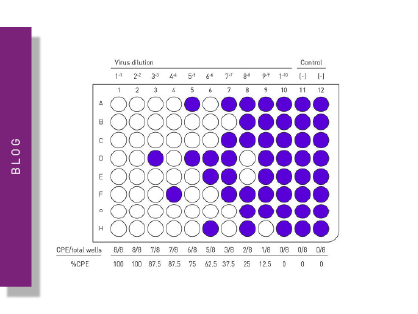
Do you want to study viral infectivity and screen for anti-virals? This blog post highlights the advantages of TCID50 assays for the determination of viral titers in microplate format.
This blog article highlights different approaches on how microplate readers can be used for the detection of viruses. Read more here.
The dangerous viral outbreaks of recent years, such as SARS-CoV-2, illustrate the intensive research that virology requires. Find out more about available virus assays here.
Vaccines are powerful tools to fight viral diseases. Read more about the science behind vaccines and what we can expect from vaccine development in the future.