PHERAstar FSX
Powerful and most sensitive HTS plate reader


Tobias Pusterla’s scientific background spans veterinary biotechnology, cancer cell biology, and the molecular mechanisms underlying inflammation‑driven tumorigenesis. After graduating in Veterinary Biotechnology at the University of Milan, Italy, he worked in mouse mutagenesis before completing a Ph.D. in Cellular and Molecular Biology through a joint program between the Open University of London, UK and the San Raffaele Scientific Institute, Milan, Italy. He later conducted postdoctoral research at the German Cancer Research Center (DKFZ) in Heidelberg, Germany, focusing on tumor biology, the tumor microenvironment, and the role of chronic inflammation in cancer development. His scientific work has contributed to understanding how damage‑associated molecular signals drive immune activation, cell migration, inflammation, and tumorigenesis, helping to clarify fundamental pathways linking cellular stress responses to physiological and pathological outcomes. After more than 13 years of research experience, he joined BMG LABTECH in 2013. Here, he oversees global marketing activities, including the creation of scientific content and the coordination of application support.
Homogeneous Time-Resolved Fluorescence (HTRF®) is a Time-Resolved FRET (TR-FRET) immunoassay developed by Cisbio (now part of Revvity, Inc.). This technology combines Time-Resolved Fluorescence (TRF) detection with Fluorescence Resonance Energy Transfer (FRET) and is mainly used for high-throughput drug screening.
FRET is a proximity assay that takes advantage of the energy transfer between two fluorophores (donor and acceptor) when they are close together. Upon excitation of the donor by a light source, the energy is transferred from the donor to the acceptor if the distance between the two is between 20 and 90 Å. The excited acceptor subsequently emits fluorescence at a given wavelength.
FRET is mainly used to analyze molecular binding events or interactions and can be employed to determine the binding affinity of two binding partners. These are assessed by coupling each binding partner with a fluorophore, and by detecting the level of energy transfer taking place.
Green Fluorescent Protein/Red Fluorescent Protein (GFP/RFP) and Cyan Fluorescent Protein/Yellow Fluorescent Protein (CFP/YFP) steady-state fluorescent FRET pairs have been widely used in fluorescence intensity assays. However, highly sensitive measurements are challenging with standard FRET. This method is negatively affected by background noise derived from scattered excitation light and autofluorescence.
Through the adoption of time-resolved fluorescence, TR-FRET allows the elimination of the short-lived steady-state background fluorescence by introducing a time delay between excitation and detection. This is made possible using long-lived fluorophores called lanthanides as donors.
Time-resolved FRET and HTRF assays rely on the transfer of energy between lanthanide donors and short-lived fluorophore acceptors when in close proximity. Lanthanides are well-suited for these applications because of a large Stokes shift and because of longer emission lifetimes (from microseconds to milliseconds) compared to steady-state fluorophores like fluorescein or rhodamine.
Due to these specific characteristics, TR-FRET and HTRF exhibit improved performance levels, stability, and specificity. Buffer or medium interference is significantly reduced by the long-lived fluorescence and by the ratiometric detection of the two emission wavelengths of donor and acceptor.
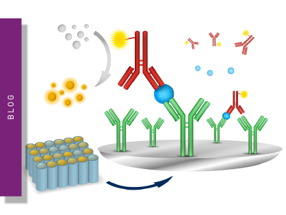
The general principle of how HTRF technology is applied is very similar in different applications. Specific antibodies against each of the two targets (or tags) in the interaction under investigation are labeled with either the donor or the acceptor. If the targets are in proximity, the antibodies will also be close together. By exciting the donor, energy will be transferred to the acceptor, which will emit because of FRET. The final signal is proportional to the amount of binding taking place. Alternatively, donor and acceptor can be covalently bound to the interacting partners.
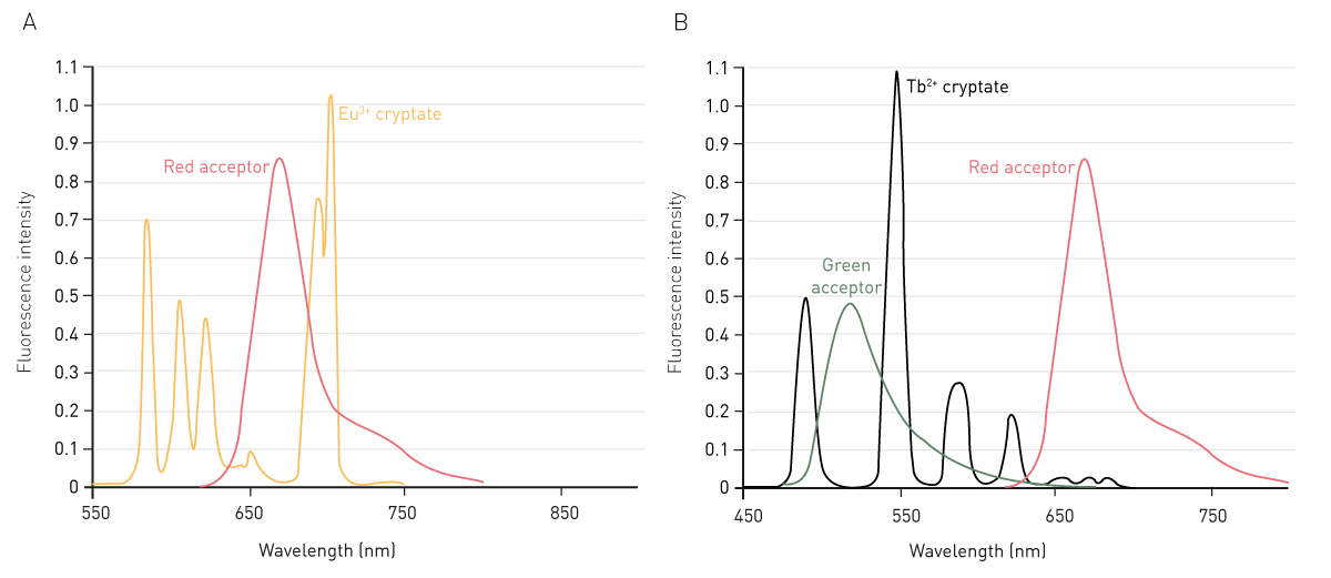
Four specific fluorophores, which form different TR-FRET couples, are used in HTRF. The donor lanthanide fluorophore is either europium (Eu3+) or terbium (Tb2+) cryptate (Fig. 1), whereas the steady-state fluorescence acceptors usually emit in the red range.
As europium and terbium ions are only weakly fluorescent on their own, they need to be embedded in a light-collecting cage to emit light. HTRF technology makes use of a cryptate for this purpose. Like a chelate in other TRF and TR-FRET technologies, a cryptate allows both collection and transfer of energy. The acceptor fluorophore then releases this energy as light at a specific wavelength.
The lanthanide cryptate complex is not subject to photo-bleaching like several conventional fluorophores and is extremely stable under a wide range of chemical conditions.
The terbium cryptate has 10 to 20 times increased quantum yield and a higher molar extinction coefficient compared to europium, features that improve screening performance.1 Both europium- and terbium-based assays can be read on the same HTRF plate reader.
XL665, a pigment purified from red algae, was the first-generation donor developed for HTRF technology. The second-generation d2 is like XL665 but much smaller. It is said to limit steric hindrance problems sometimes suspected in TR-FRET systems.
Both acceptors emit in the red range. Their emission is less likely to be negatively affected by intrinsic medium or compound autofluorescence. Both red acceptors can be coupled with either a europium or terbium cryptate. Terbium donors can be additionally combined with green-emitting acceptors (e.g. fluorescein), allowing for potential multiplexing (Fig. 2).
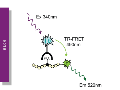
Cryptate donors are typically excited at 337 nm. Acceptor and donor emit at two different wavelengths: 620 nm and 665 nm, respectively. Hence, they can be optically distinguished from each other. Emission at 620 nm is the true emission of the donor and is used as an internal reference, while emission at 665 nm is the acceptor and is used as an indicator of the energy transfer (binding event) taking place (Fig. 3).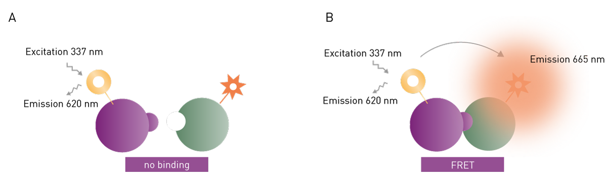
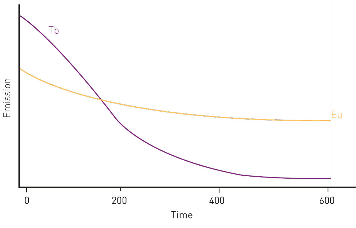
A time delay of 60 microseconds between excitation and fluorescence detection eliminates the non-specific short-lived background emission. Emission is subsequently usually detected over a time period of 400 microseconds (Fig. 4).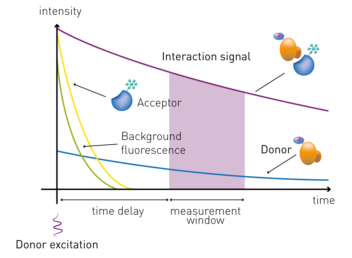
A ratiometric measurement of the intensities of the two emission channels integrated over the 400 microseconds time span is then performed. This normalizes the signal, corrects for well-to-well variability or liquid handling errors, and eliminates interference from the medium and quenching from assay components.
TR-FRET (Time-Resolved Förster Resonance Energy Transfer) is the general detection principle underlying HTRF. HTRF® is a specific implementation of TR-FRET technology developed and trademarked by Revvity, using proprietary lanthanide cryptate donors and validated reagent kits.
In practice, the key distinctions are:
TR-FRET is the broader methodology; HTRF is a specific, commercially validated TR-FRET assay platform
HTRF uses cryptate-based donors (europium or terbium cryptate), which offer greater stability than chelate-based TR-FRET systems
HTRF assays are supplied as ready-to-use kits with defined reagents and validated protocols
Other TR-FRET platforms include LANCE (PerkinElmer/Revvity), LanthaScreen (Thermo Fisher) and THUNDER
Both europium and terbium cryptate-based HTRF assays, as well as other TR-FRET platforms such as LANCE, can be run on the same HTRF-compatible microplate reader.
HTRF is widely used because of its sensitivity and versatility. The combination of time-resolved detection and ratiometric measurements significantly reduces background and increases the detection window between negative and positive signals. Moreover, the lanthanide emission is stable in time, compatible with different reagents and experimental conditions, and does not bleach. Besides its sensitivity, another major advantage is that it is a homogeneous assay. Detection of the bound FRET donor-acceptor complex does not require physical separation from the unbound components to reduce the background. Consequently, HTRF technology does not necessitate in-between separation or washing steps and can be executed with a simple add-and-read protocol (Fig. 5). This minimizes handling steps and makes it more convenient and less time-consuming than other methods such as most types of ELISA or Western Blot. Hence, it is particularly suited for automation-supported screening purposes. This is one of the reasons for its widespread adoption in drug discovery and high-throughput screening.
Key advantages of HTRF assays at a glance:
No wash steps - homogeneous add-and-read protocol reduces handling time and errors
Reduced false positives/negatives - ratiometric detection corrects for compound interference and well-to-well variability
High sensitivity - long-lived lanthanide emission and time-gating eliminate short-lived background fluorescence
Scalable - compatible with 96, 384 and 1536-well microplates for assay miniaturisation
Stable signal - lanthanide cryptates do not photobleach, giving reproducible results over time
Automation-ready - simple protocol supports integration into robotic liquid handling systems
Versatile - applicable to binding assays, enzymatic assays, cell-based assays and biomarker detection
However, limitations also exist. For instance, signal quenching is generated by external interactions with the intramolecular excitation process or fluorescence of library compounds or biological proteins. [1]
HTRF assays require a microplate reader with time-resolved fluorescence (TRF) detection, dual-channel emission capability, and compatibility with 384 and 1536-well formats for high-throughput workflows.
BMG LABTECH’s HTRF-certified readers - the PHERAstar FSX, CLARIOstar Plus and VANTAstar - are purpose-built for HTRF and TR-FRET detection. Key considerations when selecting an HTRF reader include excitation source (laser vs. xenon), simultaneous vs. sequential dual emission, sensitivity, and plate format compatibility.
For a full comparison of reader features and specifications, visit our HTRF microplate reader page.
Signal intensity in TRF detection depends on concentration and not on the overall quantity of a tracer, like in radioactivity. Consequently, HTRF assays can be downscaled while maintaining accuracy and reproducibility. This is further supported using a highly sensitive HTRF plate reader like the PHERAstar FSX.
Assay downscaling or miniaturization is a key step in drug screening. Its goal is the reduction of sample volumes to save on reagents and time while maintaining reliability, robustness, and reproducibility. Although 384- and 1536-well formats are the most commonly used, assays could even be downscaled to 3456-well plates.
An example of assay miniaturisation on BMG LABTECH HTRF readers, including case studies using the PHERAstar FSX in 1536-well format, is discussed in the webinar Miniaturising screening assays: a case study.

The HTRF technology can be applied to analyze binding events in cells or in biochemical assays in 96-, 384-, and 1536-well plate formats for different biological settings such as protein-protein (ligand-receptor) and protein DNA/RNA interactions, GPCR signaling, kinase assays, cytokines, and biomarkers.
Additionally, it is suitable for most cell-based procedures, allowing measurements of the cell lysate in the presence of a cell culture medium.
HTRF technology is widely used in the drug screening community. Advantages include flexibility, reliability, increased assay sensitivity, higher throughput, and fewer false-positive/false-negative results.
The technology is also helpful in the identification of false positives. For more information see AN 359: Identification of false positives in a HTRF® screen for small molecule inhibitors of PD-1/PD-L1.
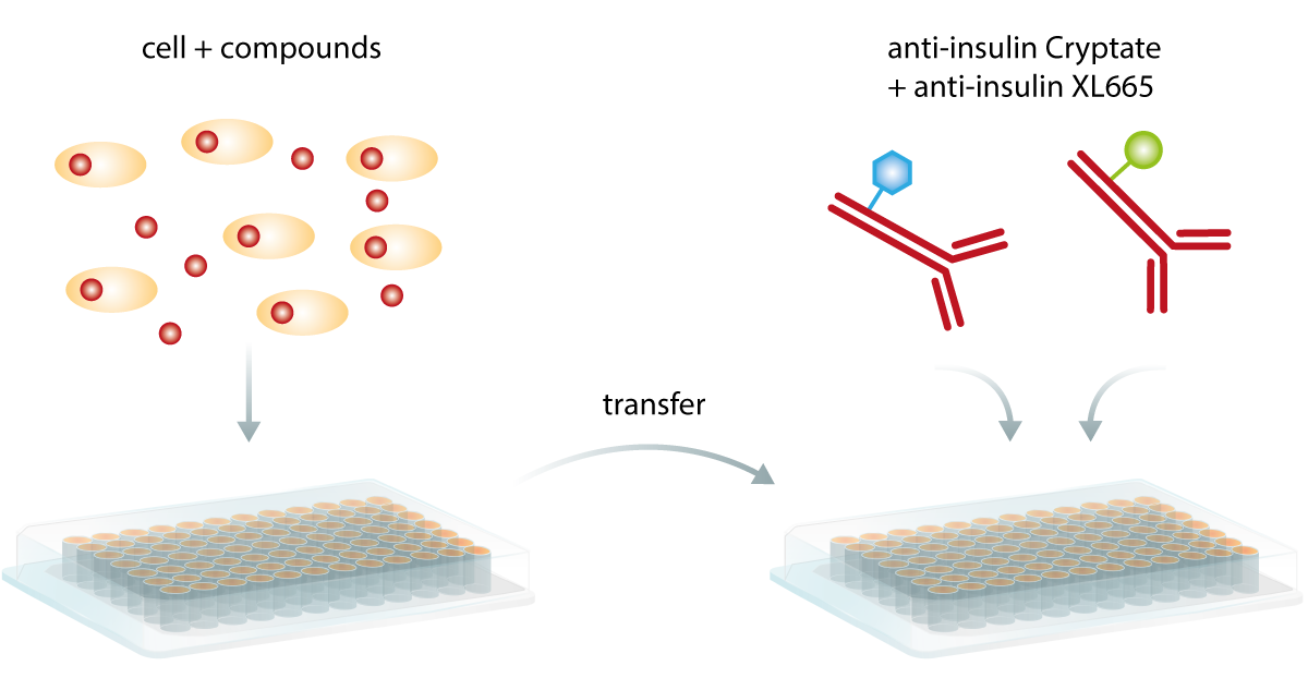
HTRF assays use a homogeneous add-and-read format. Unlike ELISA or Western blot, there are no wash or separation steps. The basic protocol is:
Step 1 - Prepare samples: transfer cells, lysate or supernatant to a microplate (96, 384 or 1536-well)
Step 2 - Add reagents: for instance add the donor-labelled antibody and acceptor-labelled antibody to each well
Step 3 - Incubate: allow antibody binding for the recommended time at room temperature (typically 1–4 hours or overnight)
Step 4 - Read: measure fluorescence on an HTRF-compatible plate reader using TR-FRET mode
Specific incubation times, reagent concentrations and plate formats will vary by kit. Always refer to the Revvity assay kit protocol for exact parameters.
Protein-protein and protein-nucleic acid interactions take place at almost every level in cells. This includes membrane protein communication (like receptor-ligand binding), signal transduction (like G-protein cascades), regulation of gene expression, epigenetics (like histone-modifying activity), etc.
G-Protein Coupled Receptor (GPCR) assays can be divided into two categories, functional and mechanistic assays. Functional assays focus on the quantification of secondary messengers like IP1 or cAMP. As part of the signaling cascade, the second messenger events involving these molecules may be targeted by specific inhibitors to enable their accumulation in the cell. Second messengers can be used as a readout, as their concentration correlates to receptor engagement and ligand binding. This is shown in the application notes “HTRF IP-One assay used for functional screening” and “GPCR activation is measured using Cisbio's cAMP and IP1 HTRF HTplex cell-based assay”.
This mechanistic analysis focuses on receptor organization on the membrane of cells, in particular on receptor oligomerization as a means to mediate signaling.
Enzymatic activity assays, such as kinases, proteases, ubiquitination, acetylation can also be analyzed. Sandwich assays can be used to measure the status change of a substrate, like phosphorylation. Competition assays can be employed to monitor other products generated in the reactions, like ADP.
Downstream signaling products can be used as biomarkers. Their analysis can identify aberrant signaling pathways that possibly play a significant role in inflammatory, metabolic and neurologic diseases. HTRF-based biomarker analysis can be used to test the efficacy of compounds that specifically target these diseases. Examples for biomarker analysis include “Detection of human tau protein aggregation” in neurodegenerative diseases, and “Development of a rapid HTRF insulin assay” for metabolic diseases, in particular diabetes mellitus.
In the drug discovery process, the kinetics of ligand-receptor binding have been traditionally studied only at a late time point. This is mostly because of technical difficulties and the relatively low throughput of these assays.
However, optimizing receptor binding kinetics can be highly beneficial for drug discovery since association and dissociation rates impact drug efficiency and the occurrence of side effects. Moreover, efficacy can be improved, and the duration of action can be prolonged. Finally, binding kinetics seem to play a role in biased agonism.
Therefore, it is desirable to screen drug candidates for their binding kinetics at an early time point, before moving to in vivo models and clinical studies.
The study of binding kinetics can be efficiently performed on microplate readers, mainly relying on TR-FRET kinetic detection for screening purposes and the kinetic study of low-affinity compounds (Fig. 7).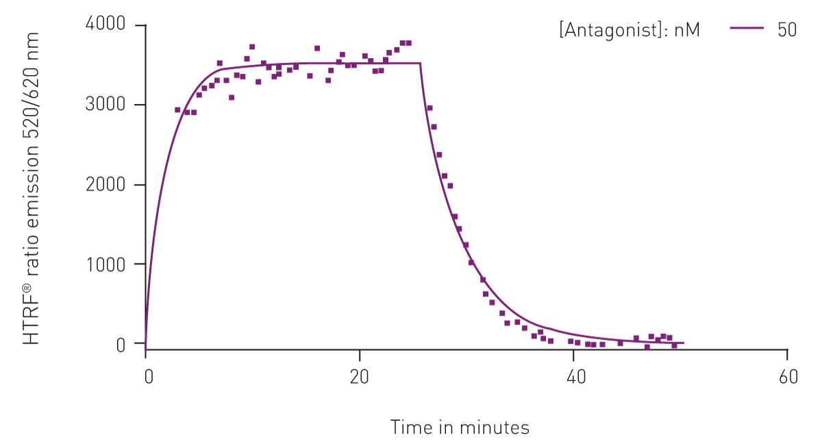
This is discussed in the scientific talk “A TR-FRET approach to measure the kinetics of ligand-receptor binding and its application of fragment screening”, as well as in the application note “Analyze binding kinetics with HTRF”.

The PHERAstar FSX has unique features dedicated to kinetic binding studies that make it superior to any other microplate reader currently on the market. With its high temporal resolution in HTRF detection, and dedicated hardware and software solutions, the PHERAstar FSX easily resolves binding events and calculates association and dissociation rates.

The following application notes demonstrate HTRF measurements performed on BMG LABTECH HTRF microplate readers. Each note includes assay conditions, instrument settings and data demonstrating reader performance.
HTRF Microplate Readers - explore BMG LABTECH’s HTRF-certified instruments
Time-resolved fluorescence explained
HTRF® is a registered trademark of Revvity.
Powerful and most sensitive HTS plate reader
Most flexible Plate Reader for Assay Development
Flexible microplate reader with simplified workflows
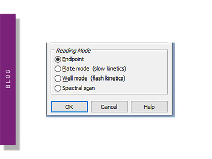
Endpoint and kinetic mode assays are used by scientists to study many processes in the life sciences. Learn about endpoint and kinetic modes on a microplate reader.
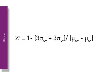
The Z prime value (Z’) is a statistical parameter that can provide practicable information on the quality of an assay. This blog looks at its usage and describes some examples of specific applications.
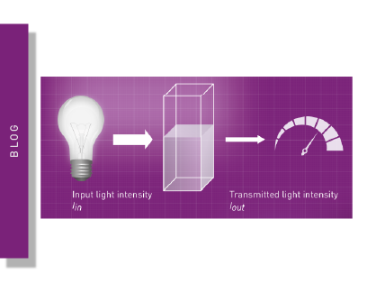
Optical density and absorbance measurements are widely used in the life sciences. This blog looks at practical applications and some of the fundamentals.
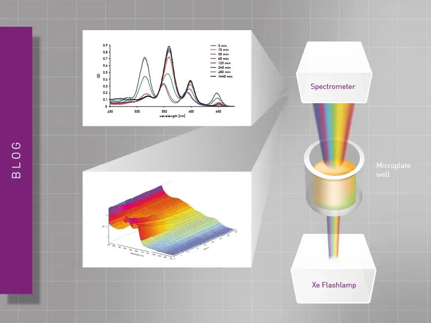
Researchers have different technology options available for absorbance measurements. This blog compares spectrometers and monochromators. What’s the difference?
ELISAs are a popular tool to detect or measure biological molecules in the life sciences. Find out how microplate readers can be used to advance research using immunoassays.
LanthaScreen is a TR-FRET technology which can be used to measure kinase activity, compound binding, and post-translation modification events. Read more about this type of assay here.