





Introduction
Mitochondrial respiration is a central indicator of cellular energy metabolism and an important parameter in studies of cell health, disease mechanisms, and drug responses. Modern oxygen consumption assays provide powerful tools to quantify mitochondrial function in living cells by monitoring changes in extracellular oxygen levels kinetically1.
Depending on the assay design, the measurement may require a controlled atmospheric microenvironment, such as low oxygen levels in the surrounding or physical separation from oxygen in the environment. For this reason, an atmospheric control option or, alternatively, coating the individual wells with oil has been considered an essential prerequisite for performing such assays on a microplate reader.
The Agilent Mito-rOCR Assay provides a simple, oil-free method to quantify extracellular oxygen consumption through a newly engineered sealing component integrated into the assay plate. This microchamber forming seal restricts oxygen exchange between the well and the surrounding air, enabling reliable detection of oxygen depletion without the need for oil.
Assay principle
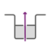
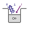
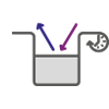
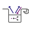
The Mito-rOCR assay measures mitochondrial oxygen consumption by detecting how quickly oxygen in the medium surrounding cells is consumed. The oxygen-sensitive fluorescent sensor becomes less quenched, as oxygen levels drop in the surrounding medium and therefore emits a stronger fluorescence signal with increasing oxygen consumption over time.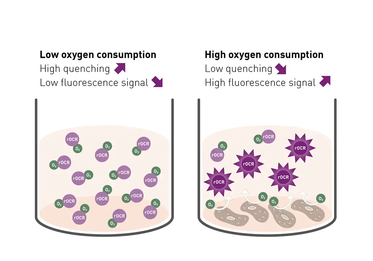
Materials & methods
- HeLa cell line (ACC 57, DSMZ)
- DMEM phenol red-free, high glucose, with 10% FBS, 2 mM glutamine, and 1% pen/strep (all Thermo Fisher)
- Incubator with 37°C, 5% CO2 and humidity control
- Mito-rOCR assay including starter kit (Agilent, #MO-400-4)
- CLARIOstar microplate reader (BMG LABTECH)
Experimental Procedure
HeLa cells were seeded in the 96-well cell culture plate included in the Mito-rOCR assay kit with 15,000 cells/well in 100 µL DMEM supplemented with pen/strep, 10% FBS and glutamine. GOx, Background and Blank wells did not include any cells just cell culture medium. After cells did attach overnight, medium was aspirated and replaced in all wells with 50 µL prewarmed assay medium (DMEM with pen/strep, glutamine without FBS and partly with 15 % Mito-rOCR reagent). After 10 min incubation in the CLARIOstar, preheated to 37°C, additives were added as listed in table 1.
Table 1:
| Type | Additives | Purpose |
| Negative Control | 1μM rotenone and antimycin A (Rot/AA) + Mito-rOCR | Show minimal OCR with present cells |
| Positive Control | GOx+Mito-rOCR | Show maximum OCR (chemically) |
| Background | Mito-rOCR | Show unspecific oxygen decline |
| Blank | - | Show medium auto-fluorescence |
The condensation ring lid of the microplate was then replaced with the rOCR Seal lid, which was fixated with the magnetic top and base plate (also prewarmed to 37°C). The assembled plate was transferred to the CLARIOstar at 37°C and measured for 45 min using the settings displayed in the instrument setting table.
Instrument Settings
|
Time-resolved fluorescence, plate mode kinetic, bottom optic
|
||
|
Optic settings
|
Filters
|
Ex TR |
|
Focal height |
4.8 mm |
|
|
General settings
|
Number of flashes |
50 |
|
Integration start |
30 μs |
|
|
Integration time |
30 μs |
|
|
Gain |
EDR |
|
|
Settling time |
0.2 s |
|
|
Kinetic settings |
Number of cycles |
45 |
|
Cycle time |
60 s |
|
|
Incubation |
37°C |
|
Results & Discussion
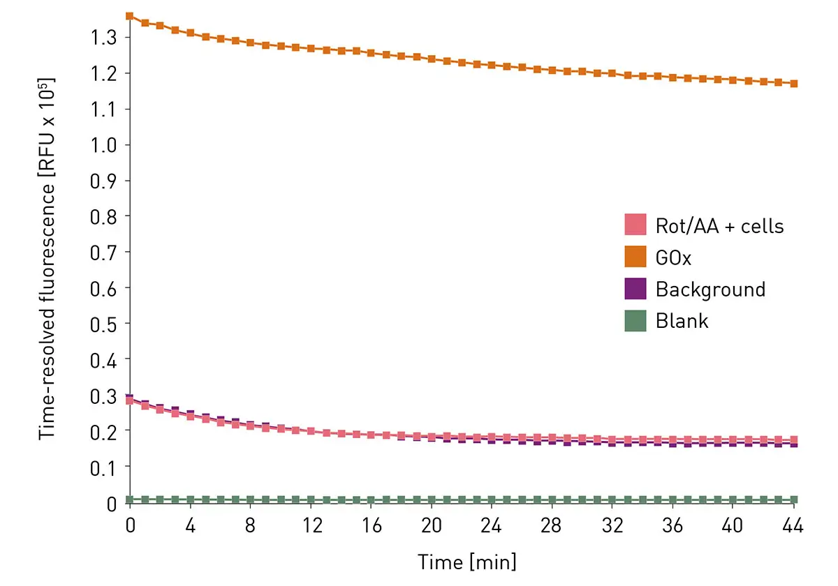
The TRF signal was monitored over 45 min in the CLARIOstar (Figure 2). While the GOx positive control initially exhibited a high signal, both the negative control (cells treated with Rot/AA) and the background wells containing only the Mito-rOCR reagent showed low signal intensities. All three conditions displayed a decrease in signal over time. In contrast, the blank wells with medium exhibited only negligible background fluorescence, which remained constant throughout the measurement.
The kinetic raw data was exported from MARS in .txt format and subsequently imported into Agilent’s Seahorse Analytics software. Within the software, the time-resolved raw intensity is normalized to the background signal, yielding the linearized intensity (Figure 3). The software identifies the linear portion of the kinetic linearized intensity curves and calculates the slopes of the corresponding linear fi ts (OCR; Figure 3, Rot/AA fit in green, GOx fit in blue) to determine the oxygen consumption rate 2.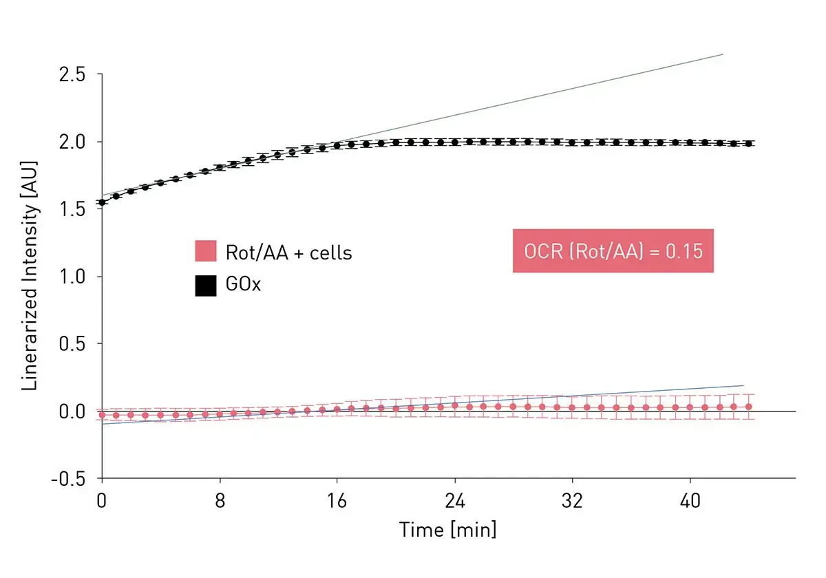
In our setup, the OCR of Rot/AA-treated cells was measured at 0.15 AU/h. This low value is consistent with expectations, as Rot/AA is a well-known inhibitor mixture targeting mitochondrial complexes I and II, thereby suppressing mitochondrial oxygen consumption 2.
Conclusion
This measurement approach demonstrates the full compatibility of the CLARIOstar microplate reader with the Mito-rOCR assay, enabling reliable time measurement of mitochondrial oxygen consumption. The CLARIOstar microplate reader is a powerful platform for time-resolved fluorescence-based OCR measurements, combining sensitive detection with precise temperature control and flexible kinetic acquisition. The combined use of the CLARIOstar and the Mito-rOCR kit offers a simple, oil-free workflow with sensitive detection and effi cient data analysis. Together, it provides a robust and user-friendly solution for studying cellular respiration in live cells.
References
- Divakaruni & Jastroch, A practical guide for the analysis, standardization, and interpretation of oxygen consumption measurements, Nat Metab (2022) 15;4(8):978–994
- Agilent Technologies. User guide: Mito rOCR assay (2024) #5994 7821EN), https://www.agilent.com/cs/library/usermanuals/public/user-guide-mito-rocr-5994-7821 en-agilent.pdf