Introduction
Proteolysis targeting chimeras (PROTACs) are small molecules that have received attention recently as a therapeutic modality and use the cells protein degradation machinery to target the elimination of a specific protein from the cell1. PROTACs achieve this by directly engaging a target protein with an E3 ubiquitin ligase through the action of two binding ligands that are joined by a chemical linker: one specific for the ligase, and the other, the target protein. The formation of this induced ternary complex between the E3 ligase, PROTAC, and target protein is the first mechanistic step required for targeted degradation. A productive ternary complex will result in ubiquitination and ultimately degradation of the protein (Figure 1). The potential for PROTACs as therapeutics is especially relevant for regulating levels of transcription factors and non-enzymatic proteins that are implicated in disease, for which small molecule inhibitors have proven ineffective. While the promise of PROTACs is high, there are still significant challenges for their design and optimization. This includes characterization of the mechanism of action employed by a PROTAC and understanding which steps need to be improved for more efficient degradation2. Here we show how the CLARIOstar with ACU can be used to measure NanoBRET™ signal in live cells to assess the kinetics of ternary complex formation and target protein ubiquitination following PROTAC treatment. Exemplary data is provided for BRD4 and the BET family PROTAC ARV-771, which recruits BET family proteins to the VHL E3 ligase complex.
Assay Principle
BRD4 was tagged with the 11 amino acid peptide, HiBiT, using CRISPR/Cas9 genome editing technology in cell lines stably expressing LgBiT. The high affinity association of HiBiT and LgBiT results in the reconstitution of NanoBiT® luciferase that is fused to the endogenous BRD4 protein. PROTAC-induced ternary complex formation or ubiquitination can then be monitored by expressing a fluorescently labeled HaloTag® fusion to either the E3 ligase recruiter or ubiquitin (Figure 2).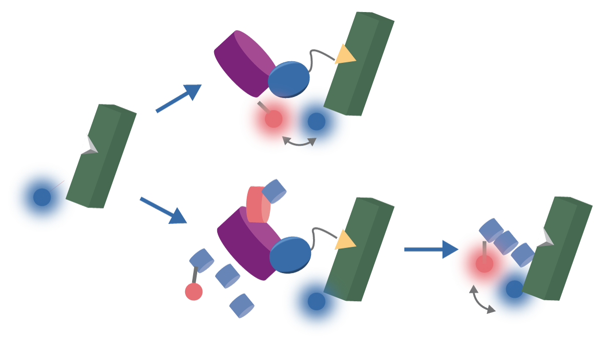
Materials & Methods
-
CLARIOstar with ACU (BMG LABTECH)
-
HaloTag® fusion constructs and NanoBRET™ reagents (Promega)
-
HEK293 cells were obtained from American Type Culture Collection
-
PROTACs and other chemicals and reagents were from commercially available sources
Experimental procedure
For complete experimental details please refer to the NanoBRET™ protocols for ternary complex and ubiquitination. CRISPR/Cas-9 genome-editing was employed on HEK293 cells stably expressing LgBiT to knock in HiBiT to BRD4. Clonal cells were transfected to express a HaloTag®-VHL or HaloTag®-Ubiquitin fusion protein. On the day of the experiment, media was replaced, supplemented with 20 µM Nano-Glo® Vivazine, an extended time-released substrate. After a 1-hour incubation at 37 ° C, 5% CO2, cells were treated with DMSO or the ARV-771 PROTAC. Kinetic NanoBRET™ measurements were collected using a CLARIOstar with ACU employing the following settings.
Instrument settings
| Optic settings | Multichromatic luminescence, plate mode kinetic | |
| Filters | 460-80 / 610 LP | |
| General settings | Settling time | 0.1 s |
| Measurement interval time | 0.5 s | |
| Kinetic settings | Number of cycles | 120 (or 63) |
| Cycle time | 180 s | |
| Incubation | CO2 | 5% |
Results & Discussion
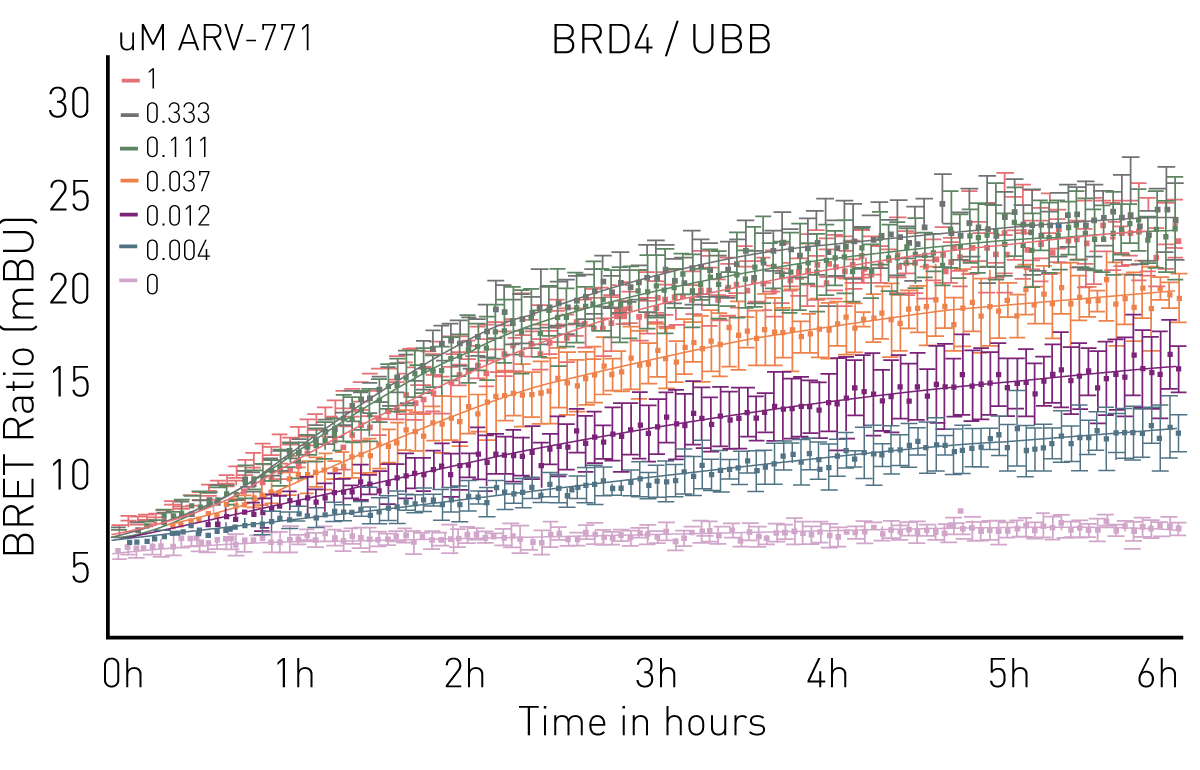
Endogenously tagged HiBiT-BRD4 cells co-expressing either HaloTag®-VHL or HaloTag®-Ubiquitin show rapid and dose-dependent ternary complex formation (Figure 3) and ubiquitination (Figure 4) upon treatment with a concentration series of ARV-771 PROTAC. Ternary complex formation with VHL remained stable at 3 hours, and BRD4 ubiquitination began to reach a plateau at 4 hours. In both assays, NanoBRET™ signal above the baseline was detected down to 4nM ARV-771, demonstrating high sensitivity and assay robustness.

Conclusion
We demonstrate here the application of NanoBRET™ technology and the CLARIOstar with ACU to measure the key steps for targeted protein degradation by PROTACs. The ability to measure the live cell kinetics of ternary complex formation with an E3 ligase as well as the efficiency with which the target protein is ubiquitinated is essential for PROTAC optimization, confirming MoA and understanding degradation efficacy. Both assays can be used in conjunction with endogenously expressed target proteins tagged with HiBiT via CRISPR/Cas9 and show high sensitivity in detecting events at very low concentrations of PROTAC. These tools can further enable an understanding of strategies for chemical optimization, ultimately aiding design and development of highly efficacious PROTAC compounds.
If you are interested in more ways to evaluate PROTACs and other protein degraders, watch this webinar.
References
- Gu, S et al. PROTACs: An Emerging Targeting Technique for Protein Degradation in Drug Discovery. BioEssays. (2018) 40: 1-11.
- Riching, K.M. et al. Quantitative Live-Cell Kinetic Degradation and Mechanistic Profiling of PROTAC Mode of Action. ACS Chem. Biol. (2018) 13: 2758-2770.
- Raina, K et al. PROTAC-induced BET protein degradation as a therapy for castration-resistant prostate cancer. PNAS. (2016) 113: 7124-7129.