PHERAstar FSX
Powerful and most sensitive HTS plate reader


This author profile refers to work created by Dr Andrea Krumm during her tenure at BMG LABTECH - the Microplate Reader Company, where she served as an Applications Specialist contributing scientific content, application notes, and technical expertise. Dr Krumm is no longer employed at BMG LABTECH, but her published materials remain available here for reference and archival purposes. Dr Andrea Krumm is a biotechnology specialist and product manager known for her work in analytical instrumentation and biopharmaceutical analysis. She studied biotechnology and later earned a PhD in cancer biology, focusing on topics such as DNA repair, epigenetics, and tumor biology. After completing her doctorate, she spent several years as an Applications Specialist at BMG LABTECH, where she authored application notes, conducted workshops, and supported scientific customers. In 2020, Dr Krumm joined Tosoh Bioscience GmbH as Product Manager for Analytical Columns.
Calcium is a ubiquitous second messenger, involved in everything from the contractility of the cardiac and skeletal muscles to blood clotting, bone mineralisation, and the functioning of the nervous system. Calcium ions contribute to signal transmission by affecting local electrostatic fields and interacting with proteins to alter their conformations, and many receptors, channels, pumps, transporters, enzymes, and transcription factors play a role in the generation and translation of calcium signals2. For instance, calcium is important in the transmission of nerve impulses via neurotransmitter release but also aids in triggering muscle contraction by communicating with regulatory proteins that allow the interaction of actin and myosin. It even acts as a cofactor for certain enzymes3.
The unique chemistry of calcium ions allows complex molecules to easily bind, even in the presence of large amounts of other ions. This allows the intracellular concentration of Ca2+ to be maintained at the very low levels required for signaling. Even small calcium imbalances can therefore have pathogenic consequences, with links to neurological, endocrine, cardiovascular, pulmonary, digestive, and metabolic diseases, as well as cancers4.
Calcium signaling can be studied in different cells and model organisms to gain a deeper understanding of the mechanisms of a range of physiological processes, including the pathogenesis of diseases such as cancers and neurodegenerative disorders. In this way, studying calcium signaling can help in determining the mechanism of action (MOA) of drugs4 prior to clinical trials. Such tests can assist in predicting potential unwanted drug side effects, and it is critical to have in vitro assays that mimic the in vivo environment as accurately as possible to ensure reliable predictive data.
Cell-based calcium assays allow the study of intracellular calcium levels, which are important indicators for the activation state of ion channels and G-protein coupled receptors (GPCRs), as well as for following the cell damage and apoptosis pathways5. GPCRs, in particular, are common targets for a variety of drugs, as they are implicated in a diverse range of physiological processes and diseases. Although these processes may display different mechanisms and mobilisation of calcium, there are common approaches to monitoring calcium levels.
Calcium flux assays detect intracellular calcium mobilisation in cells and follow the release of Ca2+ into the cytoplasm6. When studying events such as synaptic transmission at the neuromuscular junction, an action potential arrives at the cell membrane, the membrane potential is depolarised, causing voltage-gated L-type calcium channels to open to allow an influx of Ca2+ into the cell. This leads to a release of Ca2+ from the sarcoplasmic reticulum, which also initiates a contraction by activating Ca2+-dependent contractile proteins. One example for a calcium flux assay is highlighted in the application note: Multiplexing the production of two G protein-coupled receptor second messengers using spectrally resolved fluorescent dyes where it was used as a measure for GPCR activation.
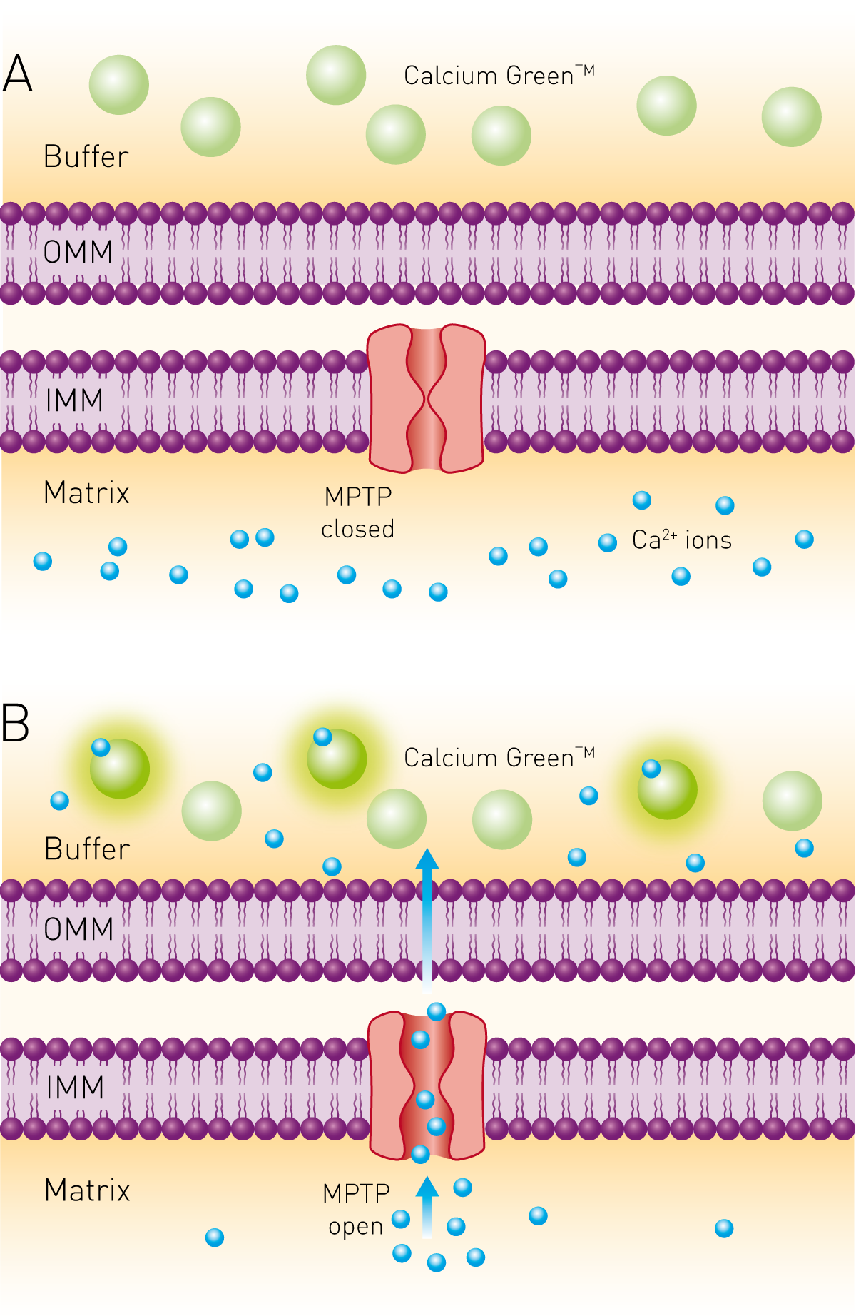
Measuring calcium retention is a good indicator of mitochondrial function. Abnormal mitochondrial function is linked to a whole host of diseases and is the consequence of the heightened opening of the mitochondrial permeability transition pore (MPTP). This leads to an increase in inner mitochondrial membrane permeability, which then permits unregulated movement of ions, including calcium7 (Fig. 1).
The calcium retention capacity of mitochondria can be measured using Calcium Green™-5N – a low-affinity membrane-impermeable dye that increases its fluorescence emission upon calcium binding – to analyse the opening and closing of the MPTP and study the efficacy of MPTP inhibitors. This assay is commonly performed by adding a fixed volume of calcium chloride to a mitochondrial sample at known intervals. Each addition causes a spike in signal that dissipates due to the uptake of calcium into the mitochondrial matrix. Opening of the MPTP collapses the mitochondrial membrane potential, which results in release of the accumulated calcium from the matrix, causing a sudden and sustained increase in fluorescence signal7.
A detailed description of the assay, data acquisition, and analysis is found in the application note “Calcium retention capacity assay evaluates inhibition of mitochondrial permeability transition pore”.
The development of Ca2+ indicators based on fluorescent proteins has assisted greatly in the study of cellular signaling, with different indicators providing different benefits. Coupled with advances in microplate reader technologies, the progressive improvements in these fluorescent probes have significantly increased our understanding of cellular Ca2+ signaling. The ability to incubate within readers with specific conditions, and inject reagents automatically, have allowed traditional cell-based fluorescence assays to be modified to a microplate reader format. It is now even possible to monitor Ca2+ mobilisation through individual channels in intact cells, with millisecond temporal resolution and micrometre spatial resolution8. Common fluorescent dyes include:
Fura-2 is a ratiometric dye commonly used for the measurement of cytoplasmic Ca2+ in living cells. Measurement is based on a shift in the excitation spectrum to shorter wavelengths on Ca2+ binding, with the ratio of emission intensities at 510 nm induced by 340 nm (Ca2+ bound) and 380 nm (no Ca2+) excitation. Ratiometric measurement minimizes the effects of photobleaching or variations in the total amount of dye present, but significant inter-laboratory variability has been reported, and so Fura-2 should only be considered semi-quantitative.
Fluo-4 (an analog of fluo-3) is another dye used for cytoplasmic Ca2+ measurement in living cells and is widely used in high throughput screening applications. Usually, the cell-permeable, non-fluorescent Fluo-4 acetoxymethyl ester is loaded into cells, then converted to the cell-impermeable free Fluo-4 by endogenous cytoplasmic esterases. Fluo-4 fluorescence increases significantly on Ca2+ binding, providing a direct measurement of intracellular calcium levels.
Fluo-8 is an analog of Fluo-3, but offers improved cell permeability at room temperature. It is also significantly brighter than Fluo-3/-4, offering an improved signal-to-noise ratio and allowing it to be used in protocols with no wash step.

The application note “Monitoring intracellular calcium using fluorescent dyes in a mid-throughput assay” details the comparison of three fluorescent Ca2+ assays.
Rhod-2 has one of the strongest and longest-lasting emission signals of any calcium indicator, making it particularly well suited to confocal laser-scanning microscopy and flow cytometry applications in addition to microplate measurements. As with Fluo dyes, they can be easily loaded into cells in the acetoxymethyl ester form. Other versions of the dye, like Rhod-4 are also available. In the following application note, Rhod-4 was used to monitor calcium flux in treated iPSC cardiomyocytes with 100 measurements/s in a live-cell assay using the CLARIOstar with ACU.
There are also calcium biosensors, such as R-GECO (red fluorescent genetically encoded Ca2+ indicator), to study intracellular calcium mobilisation for real-time kinetic readouts. This calcium sensor is based on a viral vector and can detect changes in fluorescence in living cells, as signal intensity increases in response to increases in calcium9.

Colorimetric calcium assays use reagents that undergo a measurable colour change in the presence of an analyte. For instance, o-cresolphthalein reacts with Ca2+ in an alkaline environment to produce a violet-coloured complex, where colour intensity is proportional to calcium concentration in the sample. This quick and easy assay can be used to determine the overall level of Ca2+ and can be performed in a number of different sample types10.
Luminescence assays rely on a chemical or enzymatic reaction that gives off light as a by-product, with a number of assays used to study calcium. Depending on the nature of the assay, this may require a luminescence plate reader equipped with reagent injectors.
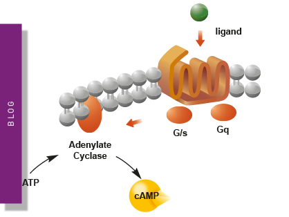
The aequorin luminescence-based assay can be used to analyse Ca2+ levels in vivo, helping to elucidate G-protein mediated Ca2+ signaling, as well as other pathways that are affected by changes in calcium level11. This assay is based on the conversion of apo-aequorin to aequorin with high calcium affinity. Activation of the GPCR will induce intracellular Ca2+ release, which then binds to the aequorin. The aequorin oxidizes a coelenterazine substrate present in the cell suspension to coelenteramide, which emits light12.
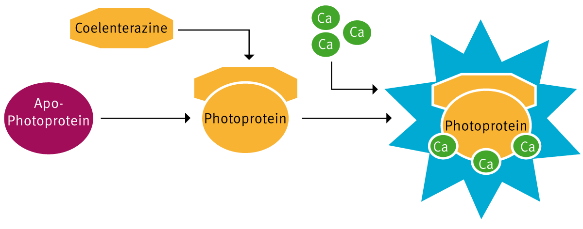
The i-PhotinaR apo-photoprotein can be expressed in cells of interest. In combination with coelenterazine and oxygen, it forms a stable complex, the active photoprotein. Calcium released from intracellular stores upon stimulation of the cells with agonists via GPCRs binds to the photoprotein. The excited photoprotein converts coelenterazine to coelenteramide and emits a flash of blue luminescence (Fig. 3). The biosensor provides a higher light emission than Aequorin and reacts slightly slower, making detection easier.
An intuitive and robust microplate reader is required for all of these calcium assays, as well as to fit the needs of other workflows in the lab. For calcium assays, in particular, a system that is sensitive and can detect results quickly is vital, so that a key moment is never missed. Cell-based fluorescence and luminescence calcium signaling assays perform much better-using bottom reading measurements, as the distance between the cell layer and the detector is reduced and the effect of the cell culture medium minimised.
The CLARIOstar Plus multi-mode plate reader is ideal for calcium assays; it offers ultra-fast data sampling of 100 measurements/second and is equipped with the most sophisticated bottom reading optical system currently available – direct optic bottom reading. Its flexibility enables calcium assays relying on a number of different measurement modes to be run, as well assays for other applications. The platform also incorporates a range of features that make it perfect for live cell-based assays, including an Atmospheric Control Unit, temperature-controlled incubation, multiple shaking options, well scanning, and reagent injectors.

In this interview Timo Strünker discusses the analysis of Calcium signalling and motility in sperm cells.
The application note “Real-time calcium flux measurements in iPSC derived 3D heart tissue” describes how the assay was performed and measured on a CLAIROstar.
In his testimonial, Building a bridge between organoid research and drug discovery, Elad Katz from Navigate Precision Biology discusses the establishment of an organoid platform in 384-well format and how the PHERAstar FSX has been instrumental to detecting calcium oscillations.

Powerful and most sensitive HTS plate reader
Most flexible Plate Reader for Assay Development
Upgradeable single and multi-mode microplate reader series
Flexible microplate reader with simplified workflows
Explore metabolic flux analysis, its significance in research, and how microplate readers enhance measurements for insights.
Explore the vital role of mitochondrial metabolism in cellular function, its measurement techniques, and how microplate readers enhance research in this field.
Learn about applications for bacterial metabolism on a microplate reader.
Life in the depths of the ocean operates under extreme conditions. Find out how proteins from deep-sea luminescent organisms are useful for measurements on microplate readers.
Second messengers play a pivotal role in signal transduction events in cells. But how do you measure these small, transiently lived molecules and how can microplate readers help?
NanoBRET is used to analyse binding events, signaling pathways and receptor trafficking in live cells and has significantly expanded the range and applications of BRET assays.