SPECTROstar Nano
Absorbance plate reader with cuvette port


This author profile refers to work created by Dr Andrea Krumm during her tenure at BMG LABTECH - the Microplate Reader Company, where she served as an Applications Specialist contributing scientific content, application notes, and technical expertise. Dr Krumm is no longer employed at BMG LABTECH, but her published materials remain available here for reference and archival purposes. Dr Andrea Krumm is a biotechnology specialist and product manager known for her work in analytical instrumentation and biopharmaceutical analysis. She studied biotechnology and later earned a PhD in cancer biology, focusing on topics such as DNA repair, epigenetics, and tumor biology. After completing her doctorate, she spent several years as an Applications Specialist at BMG LABTECH, where she authored application notes, conducted workshops, and supported scientific customers. In 2020, Dr Krumm joined Tosoh Bioscience GmbH as Product Manager for Analytical Columns.
Detectors based on biomolecules are increasingly important. This is underlined by the well-known and most ubiquitous biosensor: the glucose meter. Lesser known but undeniably useful are the many biosensors found in biological laboratories. Biosensor technology has seen recent advances, expanding their applications in research and diagnostics.
Researchers value the specific, sensitive and quick detection processes of many biosensors with innumerable applications that often involve microplates.
As key analytical devices in analytical chemistry and life sciences, biosensors play a crucial role in analyzing biological samples and biochemical parameters.
A biosensor is defined as an analytical device that combines a biological component with a physicochemical detector. Bioreceptor interactions between the biological element and the specific analyte trigger a biological response, ensuring selectivity and effectiveness in detection. This biological response leads to signal generation, which is fundamental to biosensor operation.
They are used to detect chemical or biochemical compounds. Let’s first take a look at the components of biosensors.

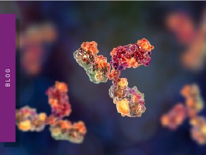
The biological element of a biosensor directly interacts with an analyte of interest. Popular biosensors use antibodies, enzymes, microbes, or nucleic acids for this interaction; these are all examples of biologically derived material.
Each of these biosensors recognizes the analyzed molecules by specifically binding to them or converting them to something else.
Biosensors are designed to detect a target molecule or target molecules with high specificity.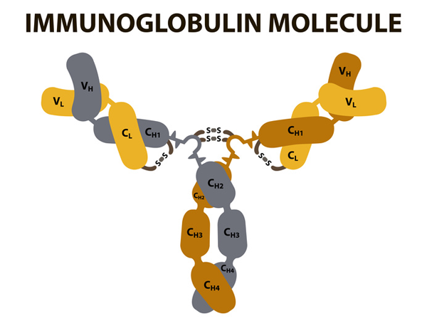
The detector of a biosensor transforms and transduces the signal coming from the interaction of biological elements with analytes into an electrical or optical signal that is used for readout. Optical sensors are a key transduction technique for detecting biomolecular interactions, enabling sensitive and specific detection in biosensor devices.
The so-called biotransducers either measure changes in current, resistance, or charge following a chemical reaction or the change of an optical characteristic such as fluorescence or absorbance. These changes are converted into an electrical signal that can be quantified for analytical purposes.
The prime example of a biosensor, today’s glucose meter, is based on an electrochemical transducer. It quantifies blood sugar with the help of an enzyme (e.g. glucose oxidase) that converts glucose and in the course of that reaction changes the current which is recorded by an amperometric detector. The first potentiometric biosensor was developed in 1969 for urea detection, marking a significant milestone in the evolution of biosensor technology.
Electrochemical sensors are crucial components of modern biosensor platforms, widely used in biomedical diagnostics, environmental monitoring, and food safety due to their sensitivity and versatility. Field effect transistors have also emerged as a modern, highly sensitive method for signal transduction in biosensing devices.
Biosensors using optical transducers record changes of light that occur in the course of the interaction of an analyte and a biological element. One such change is the refractive index which is measured for surface plasmon resonance applications and is a result of molecules binding to a surface. Another example is a color change that reports on enzyme activity which specifically takes place in the presence of the analyte. Furthermore, biosensors that increase their fluorescence or energy transfer between fluorophores in the presence of analyte are often used in life science laboratories. Mostly, the signal change is detected by a microplate reader. Let’s look at some examples of how fluorescent biosensors can be used to measure receptor activation.
In electrochemical detection, electron transfer plays a fundamental role, with redox reactions often involving two electrons, such as those catalyzed by glucose oxidase, which are essential for generating measurable signals. The interface for detection typically involves electrodes, where the choice of materials—such as carbon nanotubes—can significantly enhance device performance by improving sensitivity and electron transfer kinetics. The miniaturization of biosensors to the nano scale, using advanced nanomaterials, enables single molecule detection and further improves detection limits and response times.
One explanation for the increased use of biosensors is their excellent specificity. This feature of the biological element is exploited for the detection of the molecule of interest. Biological interactions such as antibodies binding to their antigens, the conversion of a substrate by an enzyme or the association of two complementary DNA strands are typically very specific as no organism can afford to initiate unnecessary reaction cascades that devour a lot of energy. For instance, microbial biosensors for harmful arsenic are based on a bacterial arsenic exclusion pathway. In the presence of As(III), a transporter is expressed that exports the toxin out of the microorganism. Instead of the transporter system, the biosensor expresses an As(III)-dependent fluorescent reporter gene. As the elaborate As(III) resistance machinery is only started in the presence of arsenic, its recognition is guaranteed to be specific for both the organism and its biosensor.
Another advantage of using biomolecule-based detection is the speed of analysis. The analyte is typically recognized directly and immediately gives a measurable signal. Therefore, biosensors are also commonly embedded in handheld devices for point-of-care testing. For instance, biosensors are found in instruments measuring glucose, drugs of abuse, pregnancy and many more. Biosensors are also cost effective, especially when designed for disposable or single-use applications, making them practical for widespread and resource-limited settings.
Due to the direct response of a biosensor to its analyte, biosensors are often real-time monitors of a molecule or a process connected with it. Such analysis not only gives quantitative information about the presence of a molecule but also temporal information about the presence of an analyte. Continuous monitoring using biosensors is crucial for health management and environmental applications, as it provides real-time data for early detection and ongoing assessment. Furthermore, biosensors can accurately measure analyte concentration across different concentrations, ensuring reliable quantification and performance in various diagnostic and monitoring scenarios.
The applications of biosensors are numerous and diverse. The development and application of biosensing technologies have enabled significant advancements in fields such as disease detection, food safety, and environmental monitoring. The versatility of such biosensors allows them to address a wide range of analytical challenges across these and other sectors. Here are some examples.
The food industry applies biosensors to assess product safety and ensure food safety. These analytical devices play a crucial role in monitoring food quality, traceability, and nutritional value to prevent food contamination and ensure consumer protection.
This includes detection of microbial contaminants, pesticides and toxins or to monitor product quality by detecting specific food components. One example for biomolecule-based assessment of toxins in food is the microbial arsenic biosensor mentioned earlier. This biosensor reports the presence of harmful arsenic by using the genetic regulation of an arsenic-resistance mechanism found in bacteria. In the presence of the hazard, a repressor protein is detached from a gene promoter for the arsenic-resistance system. This derepression activates a reporter gene that expresses green fluorescent protein (GFP) whose signal can be measured.
The glutamate sensor is an example of a biosensor for determining the quality of a food: glutamate enhances flavor and is found in natural products but is often added to food products. The detection of glutamate is often based on the enzyme glutamate oxidase that specifically converts glutamate to alpha-ketoglutarate. Like a glucometer, the glutamate oxidase- catalyzed reaction is detected within the biosensor by a change in current which directly reports on the presence of the flavor-enhancer glutamate.
Biosensors are also used to test environmental samples or samples released into the environment for pollutants. A commonly used device measures the biochemical oxygen demand (BOD) of water or, more specifically, of the microorganisms living in water. The BOD is an important metric to estimate the organic resources of a water sample that can be used by microorganisms to grow. This parameter correlates with the amount of organisms in the water and reflects its level of pollution. It is typically measured in water designated for discharge. The commercially available biosensor measures BOD with a combination of microbes consuming oxygen in the presence of organic compounds and a Clark electrode that determines oxygen concentration.
Recently, novel biosensors were developed that detect residual amounts of pharmaceuticals in the wastewater of municipal treatment plants. This is of particular importance to prevent excessive release of polluted water into the environment and exposure of organisms in water to drugs. Two of the most common drug targets can be monitored by cell-based sensors: cyclooxygenase-1 (COX), which is inhibited by common pain killers, and beta-adrenergic receptor, which is blocked by drugs against hypertension and arrhythmias. The COX-inhibitor-sensing cell line is based on Chinese Hamster Ovary (CHO) cells that carry a fluorescent ratiometric biosensor. A detailed description of the assay principle is found in AN322: Cell-based assay detects residual nonsteroidal anti-inflammatory drugs (NSAIDs) in effluent of municipal wastewater treatment.
The second cell-based biosensor detects β-blockers by means of cAMP production. The second messenger molecule is produced upon stimulation of the drug target, the beta-adrenergic receptor (Fig. 2). Activation of the receptor is prevented, for example, by β-blockers like Metoprolol. However, in response to β-blockers, no cAMP is produced and the divergence can be monitored with a FRET-based cAMP sensor called CEPAC. AN319: Cell-based assay detects residual β-blocker substances in effluent of municipal wastewater treatment plants explains the principle of the CEPAC sensor and how it was used to report on residual drugs in wastewater.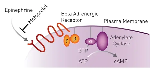 The production of cAMP linked to the activation of the receptor can be monitored with a FRET-based biosensor called CEPAC (Fig. 3). AN319: Cell-based assay detects residual β-blocker substances in effluent of municipal wastewater treatment plants explains the principle of the CEPAC sensor and how it was used to report on residual drugs in wastewater.
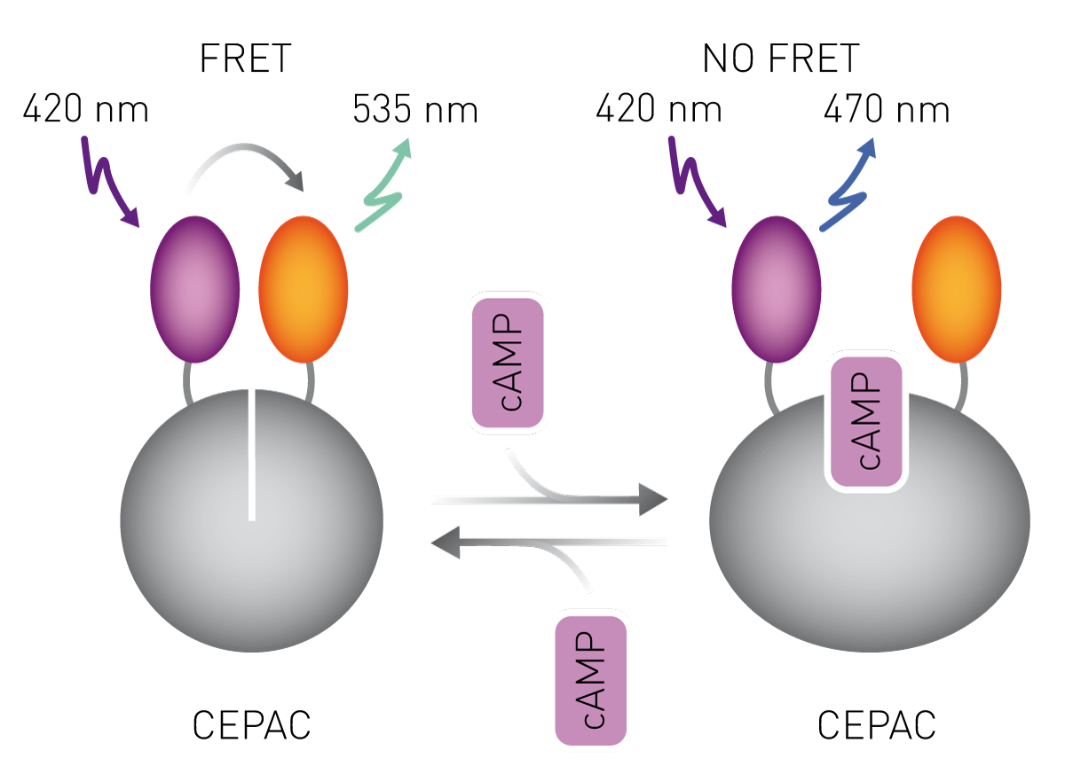
The handheld glucose measurement device was mentioned earlier. It helps diabetics to monitor their glucose levels at home and prevent hyperglycemia. Another emerging biosensor application indicates acute myocardial infarction, a major cause of death. Several systems are commercially available that all detect cardiac troponin, a protein complex indicating heart muscle damage. The biosensor uses an anti-troponin antibody as biological component which binds specifically to cardiac troponin. A secondary antibody subsequently binds to the captured analyte and acts as a mediator in changing the current in the presence of troponin. The biosensor system is integrated into a handheld device and is used by clinicians to diagnose acute myocardial infarcts.
Biosensors are also used for detecting prostate-specific antigen (PSA), which is important for the early diagnosis of prostate cancer and helps inform biopsy decisions.
In addition, biosensors play a significant role in drug discovery by aiding the identification of drug candidates and monitoring treatment efficacy in pharmaceutical research.
Biosensor development continues at pacel. The needs of researchers are diverse. The unique properties of biosensors, such as their biological recognition and molecular binding capabilities, enable innovative applications in healthcare, environmental monitoring, and security.
Biosensors can often be genetically encoded within a cell line of interest and therefore report on changes of a biomolecule in real-time, in the cell of interest, and even in specific cell compartments. Modified fluorescent proteins such as GFP can be linked to a binding domain specific for the analyte. Association of the analyte with the fluorescent biosensor results in a conformational change and subsequently in an increase in fluorescence that can be detected with a microplate reader. Another fluorescence-based biosensor principle uses Förster resonance energy transfer (FRET) between two fluorophores in vicinity with one another. Again, a protein domain binding specifically to the analyte enables detection. The protein is further labelled with two fluorophores which can transfer light from one to another only when they are close together. The proximity is induced by a conformational change upon analyte binding and is read out by measuring both fluorophores in a microplate reader.
An example of a fluorescent biosensor is reduction/oxidation sensitive GFP (roGFP). It contains additional cysteines that form a disulfide bond when oxidized. The oxidized and reduced form of roGFP display different optical characteristics (excitation spectra) which can be measured in sensitive microplate readers. The roGFP sensor is typically expressed by cells and reports in real-time on their redox state as well as on the redox state of specific proteins and enzymes. Mostly, the cells carrying the biosensor are exposed to various treatments and are analyzed in microplate format to enable the required throughput. Learn in the video below how these sensors are used and why sensitive biosensor detection is needed.

In comparison to changes in redox state, the responses occurring upon activation of G-protein coupled receptors (GPCRs) are quicker, but can be readily measured using biosensors as highlighted in the application note: Near-infrared FRET biosensor for multiplexing interrelated signalling of small GTPases. GPCRs are the most abundant class of proteins and more than 30% of FDA-approved drugs target GPCRs. One response to activation of a GPCR coupled to an intracellular Gq subunit is the release of calcium ions (Ca2+) and diacylglycerol (DAG). With the help of two fluorescent biosensors with differing spectral properties, both second messengers were detected in real-time in cells using the CLARIOstar microplate reader. The benefits of biosensors reporting on GPCR are explained in the video below.

In the following example, another second messenger associated with GPCR activation was determined: cAMP. The increased levels of cAMP upon activation with synthetic cannabinoid receptor agonists were monitored with a BRET-based biosensor named CAYMEL. An additional example for a BRET-based biosensor, in this case for the detection of estrogen receptor dimerization, is highlighted in the application note “Novel nanoBRET-Based Biosensor Enables Comprehensive Screening of Estrogenic Endocrine-Disrupting Chemicals (eEDCs) in Live Cells”
Absorbance plate reader with cuvette port
Powerful and most sensitive HTS plate reader
Most flexible Plate Reader for Assay Development
Upgradeable single and multi-mode microplate reader series
Flexible microplate reader with simplified workflows
Gene reporter assays are sensitive and specific tools to study the regulation of gene expression. Learn about the different options available, their uses, and the benefits of running these types of assays on microplate readers.
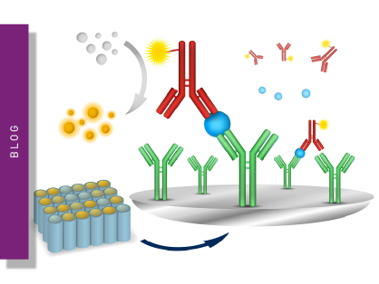
ELISAs are a popular tool to detect or measure biological molecules in the life sciences. Find out how microplate readers can be used to advance research using immunoassays.
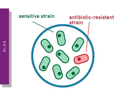
Antimicrobial resistance is a formidable problem across the world. Find out how microplate readers can help tackle resistant microbes.
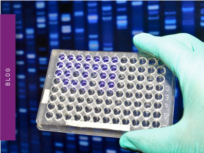
Next generation sequencing (NGS) technologies for DNA or RNA have made tremendous progress in recent years. Find out how microplate readers can advance the quality control of nucleic acids to facilitate NGS.
Light scattering offers distinct advantages for scientists interested in immunology. Find out how the NEPHELOstar Plus is used for high-throughput immunological tests.
The LAL assay ensures that sterile pharmaceutical products and medical devices are safe for human use. This test can be run efficiently and in high throughput on a plate reader.