Introduction
The ABEL® (Analysis by Emitted Light) cell activation kits for whole blood samples and isolated cells are used to analyze the real-time production of free radicals and other reactive species by leukocytes during the respiratory burst. These chemiluminescent assay kits (developed and manufactured by Knight Scientific Limited) involve the use of the light-emitting protein Pholasin®, Adjuvant-KTM (which functions to enhance the light signal during assays with whole blood), and the cell stimulants PMA (phorbol-12-myristate-13-acetate) and fMLP (n-formyl-methionyl-leucyl-phenylalanine).
Pholasin® is a photoprotein isolated and purified by Knight Scientific Ltd (KSL) from cultivated specimens of the bioluminescent mollusc, Pholas dactylus, the Common Piddock. In the assay, Pholasin® emits light in the presence of free radicals and reactive species produced by the NADPH oxidase system of stimulated leukocytes. The assay is ultrasensitive, using 20 μL of blood diluted 1:100 (the equivalent of 0.2 μL whole blood) in a final volume of 200 μL. Light is measured in a BMG LABTECH microplate reader.
Materials & Methods
The materials were purchased from the following manufacturers:
- ABEL® cell activation test kit for whole blood or isolated cells with Pholasin® and Adjuvant-KTM
- Knight Scientific Ltd.
- BMG LABTECH Microplate Reader with reagent injectors.
Blood samples were drawn into 5 mL evacuated vials containing EDTA and analyzed on the day of collection. All other reagents required were provided with the kit and were prepared following the instructions supplied with the kit. The microplate was prepared with the reagents and diluted whole blood samples according to the detailed protocol provided. Plasma was separated from an aliquot of each sample and assayed as the control for the corresponding whole blood sample.
All samples were assayed in triplicate. Prior to the preparation of the microplate, the injectors were primed with the cell stimulants: injector 1 with PMA and injector 2 with fMLP. The temperature was also set to 37°C. Once ready, the plate was inserted into the reader to allow the reagents and samples to reach 37°C before measuring the luminescence. During this time, the instrument was set up with the following parameters:
With PMA:
|
Mode:
|
Plate
|
|
Gain:
|
3300 - 3500 (depending on reader)
|
|
Measurement:
|
Time/well: 1 s
|
|
Positioning:
|
Delay: 0.1 s
|
|
Shaking:
|
Orbital, during measurement
|
|
Cycle time:
|
This parameter is automatically calculated from the number of samples being analyzed, i.e. the number of wells being measured, and is adjusted appropriately for the number of samples being assayed on a specific 96 well plate.
|
|
Number of cycles:
|
This is calculated from the total assay time (s) within plate mode divided by cycle time (s).
|
PMA was injected after 60 seconds, therefore the cycle at which injection was to occur was calculated according to the cycle time of each particular assay.
With fMLP:
|
Mode:
|
Well
|
|
Gain:
|
3300 - 3500 (depending on reader) |
| No. of Intervals: |
200
|
| Measurement time/well: | 2 s/well |
| Positioning delay: |
0.1 s
|
| Injection 2 start time: |
60 s
|
|
Shaking:
|
Orbital, during measurement |
Upon completion of the experiments, the injectors were washed with water, 10 mM NaCl(aq), water, 50% ethanol, and a final rinse with water. Thorough washing was very important for the correct maintenance of the injectors.
Results & Discussion
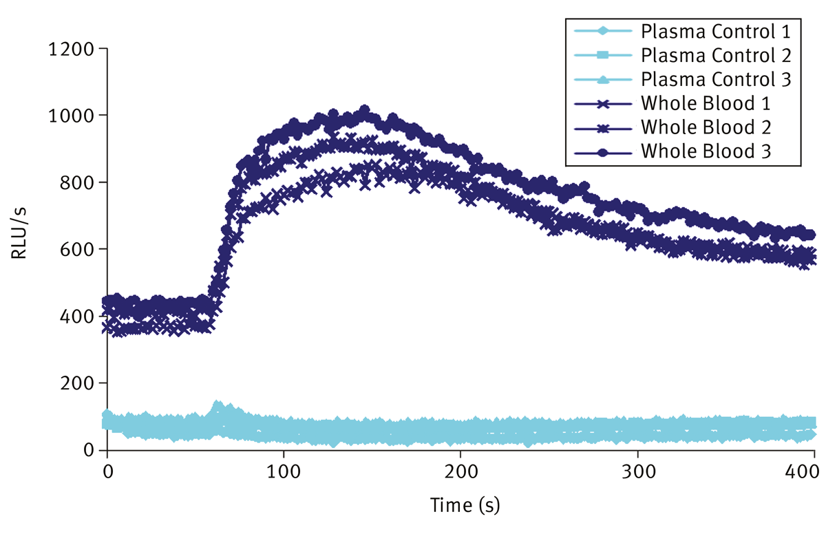
The luminescence was measured as relative light units per second (RLU/sec). The increase in measured emission of light illustrated the presence of free radicals released by activated leukocytes during the respiratory burst following stimulation. With fMLP, which activates the NADPH oxidase by binding to receptors on the plasma membrane, the respiratory burst commenced almost immediately (within approx. 5 s) as can be seen from the increase in the signal peak in Figure 1.
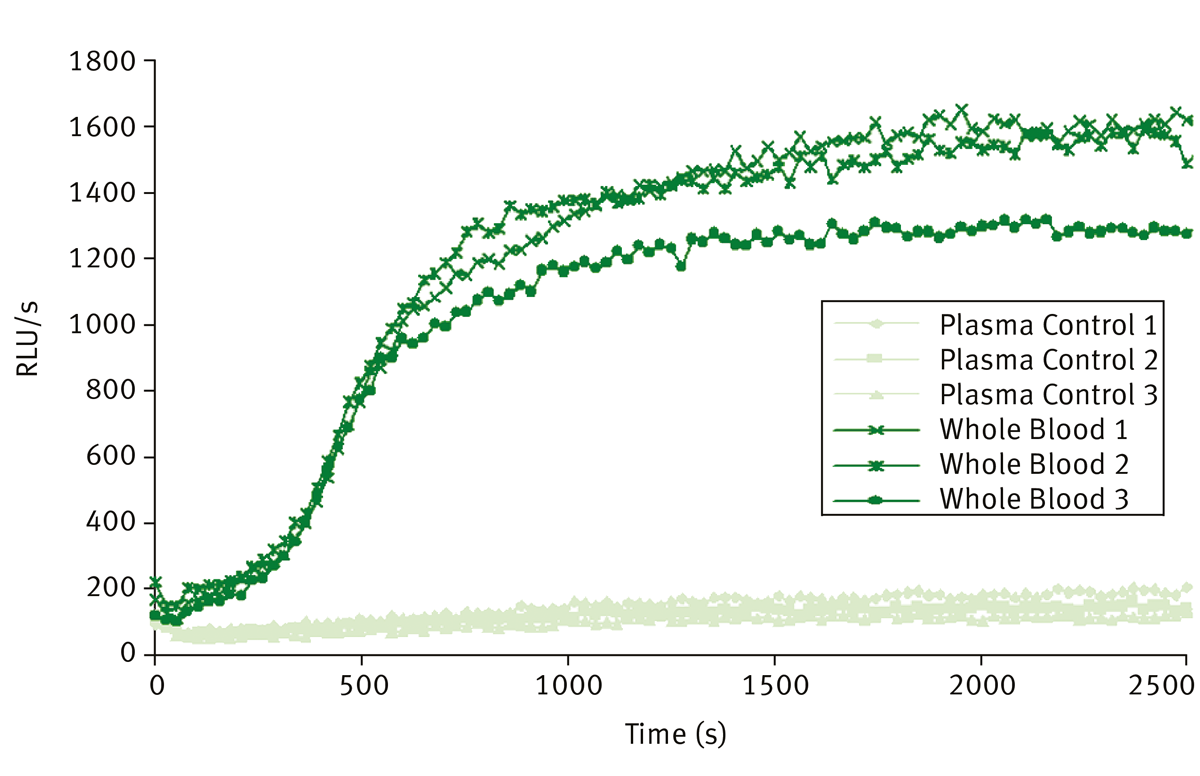
 The response started to tail off after 2-3 minutes. A longer lag time was observed in response to PMA (Figure 2) which enters the cell and activates the NADPH oxidase system via direct activation of protein kinase C. With PMA the NADPH oxidase system of both the plasma membrane and the secondary granules of leukocytes is activated. Figure 2 shows the results of measuring luminescence for a total time of 45 minutes, with PMA injection after the initial minute of measuring time. The signal increased to a maximum level and then became constant. Plasma was used as a control to measure baseline luminescence, as it contains no cells.
Using the BMG LABTECH microplate reader to measure the light emitted in these assays had a number of advantages. The sensitivity of detection was more than adequate to detect free radicals produced from leukocytes in 0.2 μL of blood (containing on average 500 neutrophils).
The automated injectors provided the ability to inject the cell stimulants and measure luminescence simultaneously, which was of greatest advantage when injecting fMLP. Stimulation with fMLP is initiated by binding to receptors resulting in the rapid production of superoxide (2-5 s lag time), which is detected by a rapid increase in light emission from Pholasin®. The ability to maintain the temperature control allowed the assays to be carried out at physiological temperature. Of particular value in the whole blood assay was the ability to measure light while the microplate was being shaken; this feature ensured that the blood cells along with all the reagents were in suspension during the assay. Temperature control and shaking were important factors for obtaining good reproducibility and ensuring that any differences in results observed were due to inherent differences in the activity of the leukocytes.
ABEL is a registered trademark of Knight Scientific Ltd.