Introduction
Changes in the intracellular concentration of Ca2+ ions is the basis for numerous cellular responses; from receptor signalling to mediating contractile function. Thus accurate techniques to monitor intracellular [Ca2+] are in high demand for both academic and industrial research. Traditionally, monitoring intracellular Ca2+ requires live-cell fluorescence imaging. Advances in microplate reader technology, including the ability to incubate at 37°C and 5% CO2 and inject reagents automatically, have allowed the adaptation of the traditional fluorescence-based assays to a microplate format. This greatly increases the throughput and automation of such assays.
This application note compares the suitability of 3 commercially available fluorescent Ca2+ dyes; Fura-2AM, Fluo-8AM and Cal-520AM used to monitor histamine-stimulated Ca2+ mobilisation in human umbilical vein endothelial cells in the CLARIOstar® plate reader equipped with atmospheric control unit.
Assay Principle
Materials & Methods
- 96-well microplate (black clear bottom, Greiner Bio-one)
- CLARIOstar with atmospheric control unit (ACU), BMG LABTECH
- Fura-2AM and Fluo-8 (TefLabs, UK), Cal-520AM (Stratech, UK), histamine hydrochloride (Sigma-Aldrich, UK)
Cell Culture
Human umbilical vein endothelial cells (HUVEC) were isolated from fresh umbilical cords and cultured in black 96-well plates coated with gelatin (1 mg/ml) in M199 +20% FCS.
Calcium-dye loading and cell stimulation
Cells were loaded with 2 µM calcium dye (Fura-2AM, Fluo-8AM or Cal-520AM) in medium for 30 minutes at 37°C. Loading solution was washed off using Krebs buffer solution and then cells were incubated in 190 µL Krebs in the CLARIOstar (set to 5% CO2 and 37°C) for a further 10 min to allow complete deesterifi cation. Histamine (20X working solution, 200 µM) was loaded into the on-board reagent injection system, and added to desired wells at 10 µL to achieve a final concentration of 10 µM. Background fluorescence was obtained in wells containing unloaded cells in Krebs buffer.
Data analysis
Fura-2AM: Background fluorescence at 340 and 380 nm (ex) was subtracted from raw fluorescent intensity at the corresponding wavelengths. Background corrected fluorescence at 340 nm (ex) was divided by that at 380 nm (ex) to obtain Ratio 340:380.
Fluo-8AM and Cal-520AM: Background fluorescence at 488 nm (ex) was subtracted from raw fluorescent intensity. The average baseline (pre-injection, FO) fluorescence was taken and all data points expressed relative to this value (F/FO).
Instrument Settings
| Optic settings | Fluorescence intensity, well mode kinetic |
|||
| Monochromator settings | ||||
| Excitation | dichroic | Emission | ||
| Fura-2M | 335-10 380-10 |
420.5 | 520-30 |
|
| Fluo-8AM | 480-10 | 497.5 | 525-30 | |
| Cal-520AM | ||||
| Gain | 2000 | |||
| Focus height | 4.3 mm | |||
| Optic | Bottom optic |
|||
| General settings | Number of flashes: | 50 per interval | ||
| Settling time: | 0.5 s | |||
| Kinetic settings | Number of intervals: | 120 | ||
| Interval time: | 1 s | |||
| Injections | 10 µl in interval 11 | |||
| Incubation | 37°C | |||
| Atmospheric control | 5% CO2 | |||
Results & Discussion
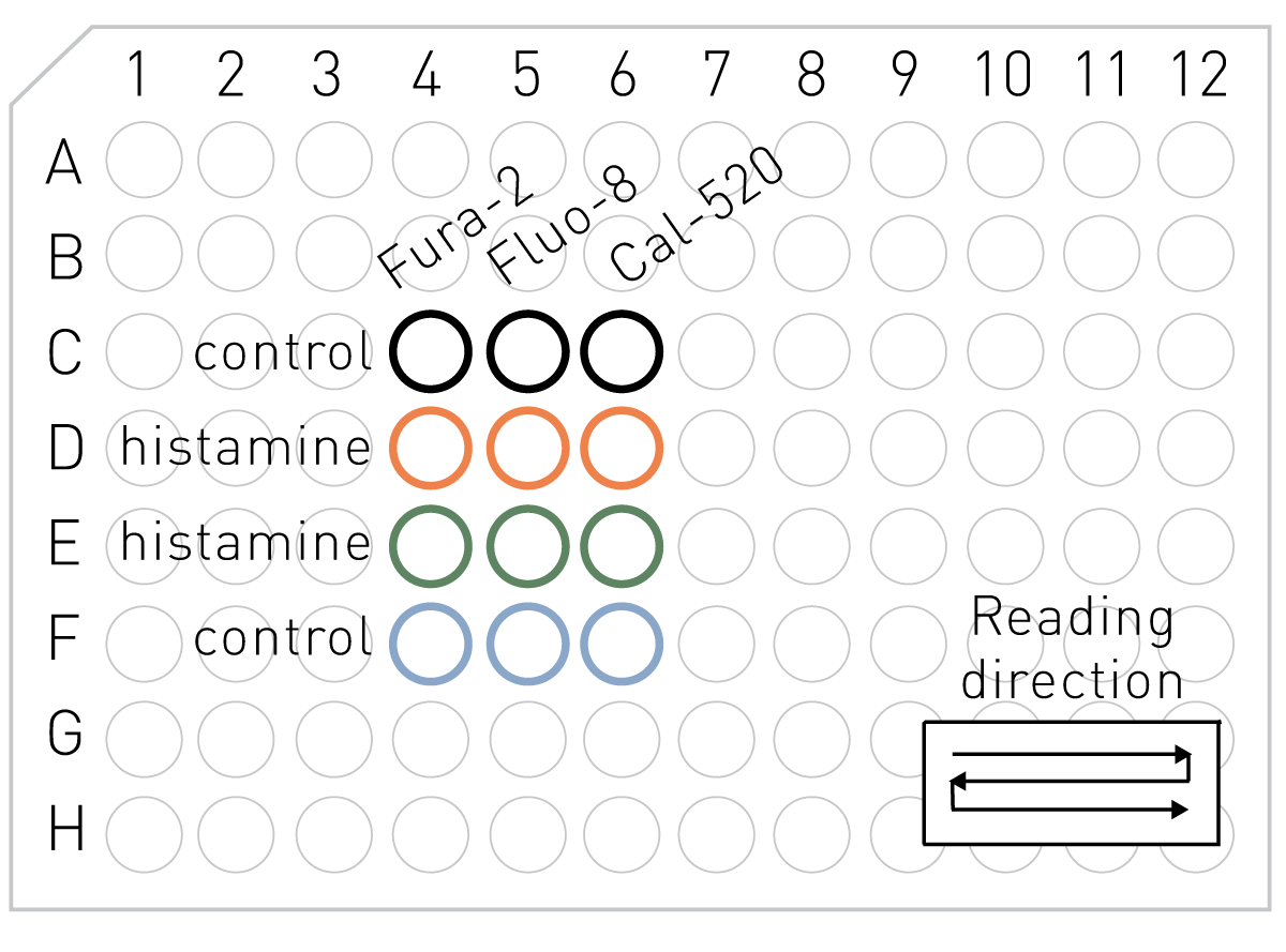
To investigate which Ca2+ dye performs best in a 96-well plate format assay, cells were loaded with either Fura-2AM, Fluo-8AM or Cal-520AM and treated with control (ddH2O) or histamine (10µM) and intracellular Ca2+ levels monitored as outlined in Fig 1. Figure 2 illustrates the results from duplicate wells, expressed as background corrected fluorescence intensity at 520nm. The wellmode kinetic function of the CLARIOstar enables rapid cycle times, and the recording of rapid calcium release after stimulation. Independent of dye used, stimulation with histamine results in a peak/plateau pattern reminiscent of similar measurements using epifluorecent imaging. Cal-520 demonstrated the highest F/F0 upon histamine stimulation, which was comparable to the R340/380 obtained with Fura-2.
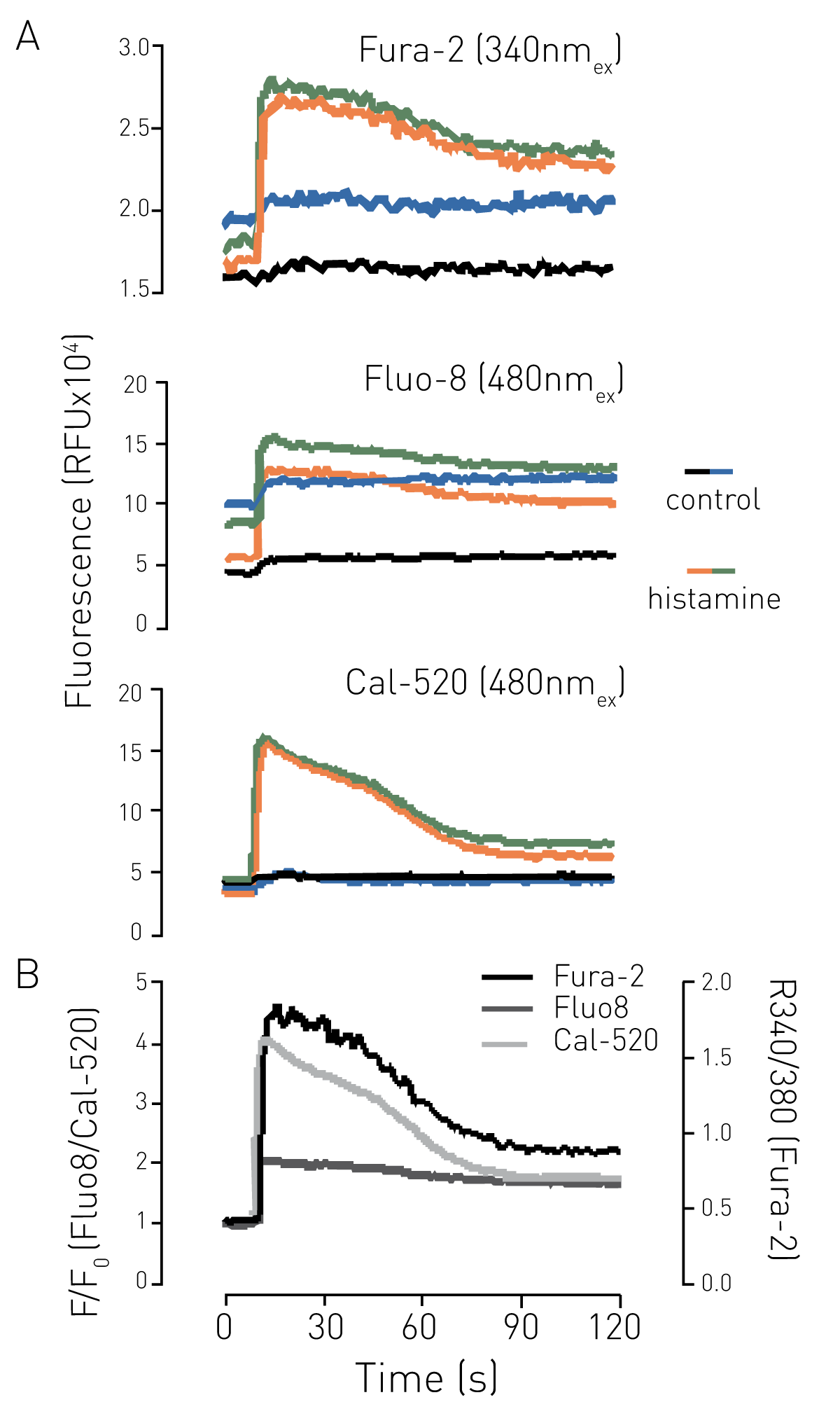
When using the wellmode option, each well is read in turn, and there is a time difference between measuring the first and last well. Therefore, a factor to consider when choosing a suitable dye is the extent of leakage from the cytosol of the dye, particularly when monitoring intracellular Ca2+ levels, which are ~105X lower than extracellular concentrations. Dye leakage would be detected as an increase in baseline fluorescence over time, as shown in Figure 3. Fluorescence was only monitored periodically to avoid photo-bleaching effects. While Fluo-8 showed the strongest baseline shift over time, with Fura-2 also showing a gradual increase, no substantial baseline increase was detected with Cal-520 over the time course investigated.

Conclusion
Fluorescent dyes have long been used to monitor intracellular Ca2+ levels in living cells by fluorescent imaging. Here we have adapted such protocols for use in a 96-well plate format in the CLARIOstar plate reader. Dye leakage can contribute significantly to the accuracy of such measurements, and here we have shown that Cal-520AM appears as good as Fura-2 at reporting histamine-stimulated Ca2+ mobilisation in endothelial cells in a 96-well plate format, with minimal leakage over a 30 minute time frame. Such attributes make this dye useful for mid/high-throughput analysis of intracellular Ca2+ levels in living cells.