PHERAstar FSX
Powerful and most sensitive HTS plate reader


Barry Whyte is Application Scientist and Science Writer at BMG LABTECH in the United States. He has PhD and Bachelor of Science (BSc) degrees in biochemistry from the University of Bristol in the United Kingdom and more than 20 years of experience in the life sciences and science communications. Over the years, Barry has worked on three continents and traveled widely. He enjoys building on his international work experience and learning new ways to help scientists advance their research.
The monocyte activation test is a cell-based, in vitro assay to test for pyrogens that is an alternative to traditional animal testing methods like the rabbit pyrogen test. 1-3 Pyrogens are a broad range of substances that may contaminate pharmaceuticals, biologics and medical devices. The detection of pyrogens is essential to prevent potentially life-threatening immune reactions from happening. In this context, the monocyte activation test plays an important role in many quality control and bioanalysis processes for the manufacture and batch testing of drugs and medical devices due to its ability to test for endotoxin and non-endotoxin pyrogens. For example, the monocyte activation test is often used to ensure the production and safety of vaccines, antibiotics and plasma-derived drugs. 3
Monocytes are a type of leukocyte and are part of the immune system. They can differentiate into macrophages and monocyte-derived dendritic cells. These cells either eliminate the pathogen or alert other blood cells to help destroy the invading infectious organism and prevent infection. When monocytes are activated by pyrogens, they produce cytokines (e.g. interleukins or tumor necrosis factor-α or TNF- α). The detection of these molecules serves as a reliable indirect test for the occurrence of fever. Different types of immunological assays including Enzyme-Linked Immunosorbent Assays (ELISAs) and other types of immunoassays offer sensitive, quantitative measurements of cytokines. Each of these assays provides different options for throughput. For example, cytokine production can be detected using gene reporter assays which offer a rapid readout, wide dynamic range and simplified protocol. We will take a look at the different assay options for monocyte activation tests later in this blog. The monocyte activation test is a particularly sensitive test, with a low limit of detection that is crucial for product-specific validation.
This type of analysis is essential for batch release testing of parenteral products, replacing the rabbit pyrogen test with a more reproducible assay for pyrogen testing.
In this blog, we look at the monocyte activation test and examine how microplate readers support this test for pyrogens.
Pyrogens trigger fever in the body through the stimulation of monocytes (Fig.1). In brief, pyrogens are recognized by different monocyte receptors. These activated receptors trigger intracellular signaling pathways within the monocyte cells. Different signaling pathways lead to effectors that promote transcription of pro-inflammatory cytokine genes. Monocytes activated in this way secrete cytokines (in this case endogenous pyrogens) that travel through the bloodstream to the brain where they can stimulate the production of prostaglandins [Exogenous pyrogens arise from outside the body and induce fever reactions after parenteral administration. In contrast, endogenous pyrogens such as interleukins or tumor necrosis factor alpha (TNFα) are produced by the body itself as a reaction to contact with exogenous pyrogens]. The binding of prostaglandin to cell receptors in the hypothalamus leads to an increase in the body’s thermoregulatory set point. This interaction sets in motion the body’s heat generating and heat conservation mechanisms that are a feature of the fever response. In some cases, these changes are life threatening.
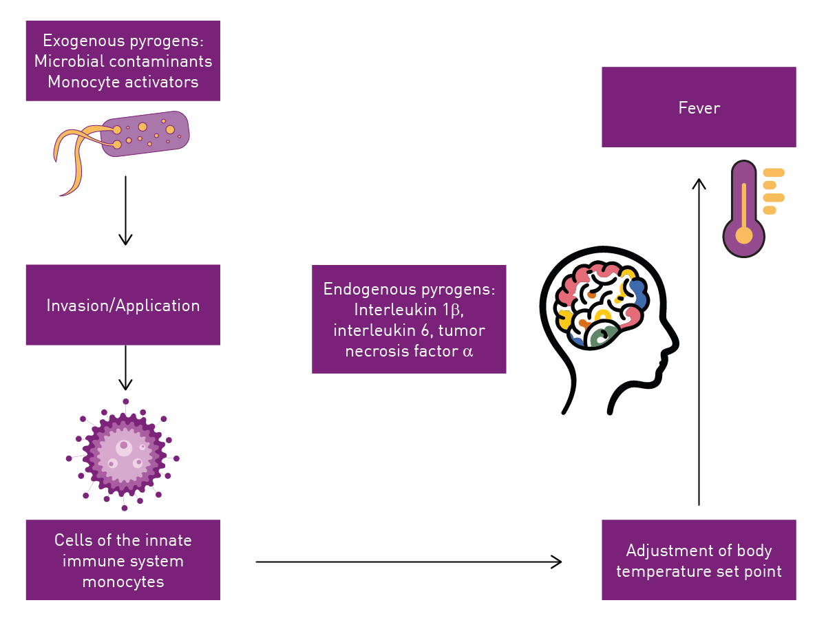
It is therefore important to determine the cytokine concentration for measuring the immune response and accurately assessing pyrogen levels. You can read more about the different steps in immune signaling that ultimately affect temperature control in the blog Pyrogens and pyrogen testing.
Monocytes are a good option as a detection test for pyrogens due to their role in the immune response in humans. The test essentially leverages the role that monocytes play in innate immunity. As mentioned in the previous section, when pyrogens activate these key cells of the innate immune system, they secrete pro-inflammatory cytokines. These readily generated cytokines can be detected in different in vitro assays at high sensitivity under conditions suitable for a reliable, fast testing method.
Different monocyte activation test kits are available commercially, which can be categorized into two main types based on their cell sources: those utilizing cell lines and those relying on donor-pooled peripheral blood mononuclear cells (PBMCs). Both cell sources effectively detect endotoxins and non-endotoxin pyrogens.
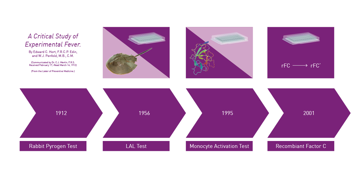
Different tests for pyrogens have been developed over time (Fig. 2) each with unique attributes.4,5 The monocyte activation test is not only an alternative to complete pyrogen tests like the rabbit pyrogen test (i.e. offering detection of both endotoxin and non-endotoxin pyrogens) but also for other bacterial endotoxin tests (BET tests). Its ability to detect bacterial endotoxins makes it an option as an alternative to the traditionally used Limulus Amebocyte Lysate (LAL) test (amebocyte lysate test), for example, or more recently developed assays like the recombinant factor C (rFC) test. For products where bacterial endotoxins are a concern, it is also crucial to assess risks related to non-endotoxin pyrogenic substances during both the production process and upon batch release which makes the monocyte activation test an attractive test option to cover both groups of substances. Since the monocyte activation test significantly contributes to ensuring patient safety by detecting both endotoxin and non-endotoxin pyrogens, it reduces health risks associated with contaminated medications.
 Criteria for monocyte activation tests
The preparation and analysis of pyrogen samples according to established pharmacopoeia standards often involve creating a standard curve. This process includes diluting pyrogen samples to achieve a range of concentrations, which are essential for ensuring accurate measurements in pyrogen testing. Establishing a standard curve allows for the conversion of measured cytokine concentrations into pyrogen equivalent units, providing a precise assessment of pyrogen presence in samples. This type of reference curve is important in pharmaceutical preparation and analysis.
A thorough risk assessment is critical when evaluating pyrogens, especially during product validation and batch release processes. This ensures the absence of pyrogenic substances from the final manufacturing steps and products by employing tests like the monocyte activation test. These tests detect the broadest range of pyrogens, including those from Gram-positive and Gram-negative bacteria, as well as yeasts, molds, and viruses.
As indicated, the main counterpart of the monocyte activation test in the pyrogen testing family is the rabbit pyrogen test. However, the rabbit pyrogen test sometimes lacks reproducibility and accuracy. It is also relatively expensive and raises ethical concerns due to the use of live animals. As an in vitro alternative, the monocyte activation test offers several benefits over traditional methods (Fig. 3). The monocyte activation test provides improved accuracy, good sensitivity and reduced use of lab animals aligning with current regulatory recommendations and advancements in the field.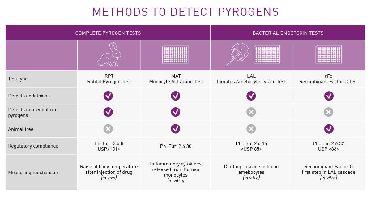
Regulatory bodies and guidelines, such as the European Pharmacopeia, have advocated for the adoption of in vitro alternatives like the monocyte activation test in place of traditional animal-based tests. Hundreds of thousands of rabbits are used worldwide each year for pyrogen testing and alternatives are needed. The European Pharmacopeia General Chapter has for some time suggested replacing the rabbit pyrogen test with the monocyte activation test as a validated approach to ensure products are labeled ‘pyrogen free’. In June, 2024, the European Pharmacopeia Commission removed the rabbit pyrogen test from its monographs and mandated the adoption of non-animal methods like the monocyte activation test. The European Pharmacopeia Commission abolished the use of the traditional rabbit pyrogen test from 1 July 2025. 6 The test will be completely replaced by non-animal methods.
While progress is being made to reduce the number of animals used in testing, animal use remains considerable worldwide despite the availability of alternative tests. Further developments in regulatory requirements coupled with ongoing innovation in testing methods should help reduce unwanted animal tests.
As mentioned earlier, monocytes activated by pyrogens produce cytokines that can be detected by either immunological assays or gene reporter assays each with different throughput options on a microplate reader. Monocytes from different sources can be used for these assays. These include monocytes isolated from whole blood (fresh or cryopreserved), isolated primary monocytes (peripheral blood mononuclear cells or PBMCs) (fresh or cryopreserved) and monocytic cell lines. Cytokine production can also be detected using gene reporter assays which offer a rapid read out, wide dynamic range and simplified protocol. However, they require monocytes to be genetically modified to express the appropriate reporter gene.
Gene reporter techniques rely on the use of a reporter gene typically placed downstream of a regulatory sequence. If the regulatory sequence is activated or repressed the reporter gene is expressed at different levels and the product can be measured quantitatively using different detection techniques. You can read more about these types of assays in the blog Gene reporter assays. LumiMAT™ is a gene reporter assay available for the measurement of endotoxin and non-endotoxin pyrogens.
The LumiMAT™ assay
Traditional monocyte activation tests have issues with lot-to-lot variability and often depend on labor-intensive ELISA assays. LumiMAT™ is a monocyte activation test that uses luminescence to detect pyrogens. The LumiMAT™ assay uses the monocytic cell line NOMO-1 in which the luciferase reporter gene has been introduced to express luciferase protein in response to transcription factor NF-κB when these cells are activated by pyrogens.
Conventional monocyte activation tests use ELISA methods and peripheral blood mononuclear cells. While they offer good reactivity, variations can arise due to differences between donors and there may also be constraints due to donor shortage. These ELISA-based methods are time consuming and typically take around 1.5 days to perform. In contrast, LumiMAT™ makes use of a convenient reporter assay in a stable reporter cell line. This offers a stable supply of cells, good reactivity and less lot-to-lot variability. Significantly, the LumiMAT™ assay allows rapid testing in around 5 hours. LumiMAT™ therefore offers a faster readout, higher sensitivity, wider dynamic range and a simplified protocol compared to other monocyte activation tests. You can read more about the LumiMAT™ assay in the application note Fast, highly sensitive pyrogen detection with the LumiMAT™ assay performed on BMG LABTECH readers.
Microplate reader features like incubation and shaking provide benefits for monocyte activation tests. The cell-based assays that are the central element of monocyte activation tests require a temperature of 37°C and, in the case of LumiMAT™, incubation at 5% CO2. Only a temperature incubation option makes it possible to monitor the signal development of the assay over time. All BMG LABTECH readers offer accurate temperature regulation up to 45°C (optionally up to 65°C).
The Atmospheric Control Unit from BMG LABTECH provides researchers with a system that enables control of both the oxygen and carbon dioxide concentrations in an independent manner. Consistent stirring options also deliver benefits for assays where this is critical. This accessory is especially relevant when it comes to application of cell-based assays as it transforms the reader into an incubator. The ACU allows the development of the luminescent signal derived from the reporter cell line in LumiMAT assays to be monitored over time in the microplate reader, instead of only observing the endpoint signal after the predefined incubation time. The VANTAstar®, the CLARIOstar® Plus, and the Omega series can be combined with the Atmospheric Control Unit.
All BMG LABTECH microplate readers have exceptionally fast reading capabilities. In addition, the Omega series, VANTAstar, CLARIOstar Plus, and PHERAstar® FSX are multi-mode microplate readers capable of detecting absorbance, fluorescence intensity and luminescence assays. This broad detection capability enables them to support all available pyrogen assays—except for the rabbit pyrogen test. The Omega series, CLARIOstar Plus and PHERAstar® FSX come with on-board injectors that can offer the very best options for detection at the time of injection. The VANTAstar can be equipped with a modular injection unit. The available shaking options support users in the proper mixing of assay reagent before the kinetic monitoring starts.
It is also possible to detect cytokines using fast immunological methods that involve semi-homogeneous protocols. The application note Highly sensitive ELISAs in 90-minutes: SimpleStep ELISA® kits and the SPECTROstar® Nano describes a sandwich ELISA assay technology that makes use of a semi-homogeneous assay format (Fig.4). This provides a faster way to determine cytokines and other analytes compared with conventional ELISA assays and, at the same time, delivers outstanding sensitivity, specificity and reproducibility. The sample preparation involved in assessing cytokine release is crucial for accurate measurement and immune response activation. A single-wash protocol greatly reduces assay time. In the study, the SimpleStep ELISA kit was used to measure the levels of human interleukin-6, an inflammatory cytokine capable of inducing maturation of B-cells and fever.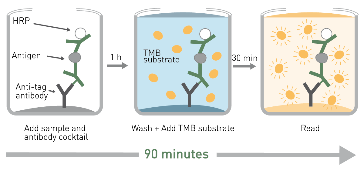
The application note Multiplex analysis of inflammatory cytokines from primary human macrophages using a FLUOstar® Omega includes a description of the detection of different inflammatory cytokines using luciferase reporter gene assays (luminescence). Transcription factor NF-κB was detected using a luciferase reporter assay after the transfection of the primary human macrophages with an adenovirus. The FLUOstar Omega provided a versatile, automated platform for these types of measurements.
Different ELISA readouts are possible for the various cytokines involved in pyrogen responses, for example interleukin-6, interleukin-1β or TNF-α. For conventional monocyte activation tests, absorbance, fluorescence intensity and luminescence measurements are all methods of choice to determine pyrogen levels accurately. In most cases, absorbance or fluorescence are typically used for detection. Although less common, luminescence is an option for some types of ELISA assays.
Conventional monocyte activation tests benefit from the speed, accuracy, sensitive detection, and cost-effective savings on materials and resources that microplate readers can deliver. Microplate readers also contribute to reproducible results in monocyte activation tests, ensuring reliable outcomes in detecting pyrogenic contaminants.
The application note Multiplex analysis of inflammatory cytokines from primary human macrophages using a FLUOstar® Omega also describes the detection of different inflammatory cytokines using ELISA assays (absorbance). In the study, different cytokines were measured using peripheral blood mononuclear cells in 96- and 384-well microplate formats. TNF-α concentrations were determined in both microplate formats if the ratio of the cells and culture volume were kept constant. Interleukin-6 concentrations were also measured under similar conditions (Fig.5). ELISA measurements were used to assess interleukin-10 and IP-10 levels. Transcription factor NF-κB was detected using a luciferase reporter assay after the transfection of the primary human macrophages with an adenovirus. The FLUOstar Omega provided a versatile, automated platform for these types of measurements.
The monocyte activation test is an effective assay for ensuring that products are pyrogen-free before they are released, which is consistent with regulatory directives to replace traditional testing methods with this more sensitive approach. Ensuring the absence of pyrogens in parenteral pharmaceuticals, biologics and medical devices prevents serious health complications, including inflammatory responses and possible organ failure. The use of reliable testing methods like the monocyte activation test to detect both endotoxin and non-endotoxin pyrogens in these products mitigates the risks of contamination during the manufacturing process. In this context, maintaining consistency and comparability of results across different tests and manufacturing sites is crucial, as variability introduced by different batches can affect assay results. A stable pyrogen-free supply of products from various manufacturing sites is essential.
Traditional pyrogen testing methods often require labor-intensive manual preparation leading to inefficiencies, increased costs, and inaccurate results. As we have seen in this blog, recent refinements to the monocyte activation test are reducing inefficiencies by implementing automation-compatible testing methods, decreasing costs through the implementation of sustainable testing methods, and improving testing performance by generating improved sensitivity of testing methods.
In many cases, workflows have been reduced from days to hours with appropriate quality control by adopting monocyte activation tests. Modifications to existing protocols for monocyte activation tests are extending applications to a wider range of interventions, for example by allowing direct detection of pyrogens on the surfaces of a wider range of medical devices.
As innovation proceeds, monocyte activation tests will become increasingly relevant to more products arising from biotechnology, drug discovery, microbiology, immunology and many other areas of assessment including quality control and bioanalysis. Effective solutions for pyrogen detection will be crucial in ensuring the quality and safety of these products.
Further developments in in vitro alternative tests will help provide more accurate, reliable, and humane testing methods, reducing the use of lab animals and improving testing efficiency. Regulatory agencies are advocating for these alternatives, emphasizing their benefits. This should help raise the visibility of what methods like the monocyte activation test can bring to patient safety across the globe.
Powerful and most sensitive HTS plate reader
Most flexible Plate Reader for Assay Development
Flexible microplate reader with simplified workflows
Upgradeable single and multi-mode microplate reader series
Absorbance plate reader with cuvette port
Microplate nephelometer for light-scattering and turbidity measurements
Learn about the bacterial endotoxin test (BET test) and its role in ensuring the safety of pharmaceuticals, biologics and medical devices.
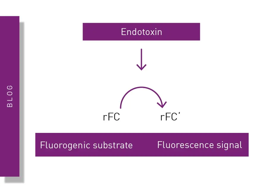
The recombinant factor C (rFC) test is used to detect bacterial endotoxins that can cause fever and adverse events when introduced into the body. Learn about the rFC test and the advantages it offers for endotoxin detection in the life sciences.
Pyrogen tests are vital to ensure the safety of different health interventions including pharmaceuticals, medical devices and an array of biological products. This blog looks at the different types of pyrogens as well as some of the widely used pyrogen tests.
Gene reporter assays are sensitive and specific tools to study the regulation of gene expression. Learn about the different options available, their uses, and the benefits of running these types of assays on microplate readers.
You want to make sure that your cell culture is free of mycoplasma? BMG LABTECH microplate readers are the perfect measurement platform to read MycoAlert® mycoplasma detection kits.