SPECTROstar Nano
Absorbance plate reader with cuvette port


This author profile refers to work created by Dr Andrea Krumm during her tenure at BMG LABTECH - the Microplate Reader Company, where she served as an Applications Specialist contributing scientific content, application notes, and technical expertise. Dr Krumm is no longer employed at BMG LABTECH, but her published materials remain available here for reference and archival purposes. Dr Andrea Krumm is a biotechnology specialist and product manager known for her work in analytical instrumentation and biopharmaceutical analysis. She studied biotechnology and later earned a PhD in cancer biology, focusing on topics such as DNA repair, epigenetics, and tumor biology. After completing her doctorate, she spent several years as an Applications Specialist at BMG LABTECH, where she authored application notes, conducted workshops, and supported scientific customers. In 2020, Dr Krumm joined Tosoh Bioscience GmbH as Product Manager for Analytical Columns.
The OD600 provides information on the growth of microorganisms such as bacteria and yeast. More specifically, microbial growth correlates with the number of organisms in a population and reports how it changes over time. Information about microbial growth is needed in a variety of biological and medical research areas:

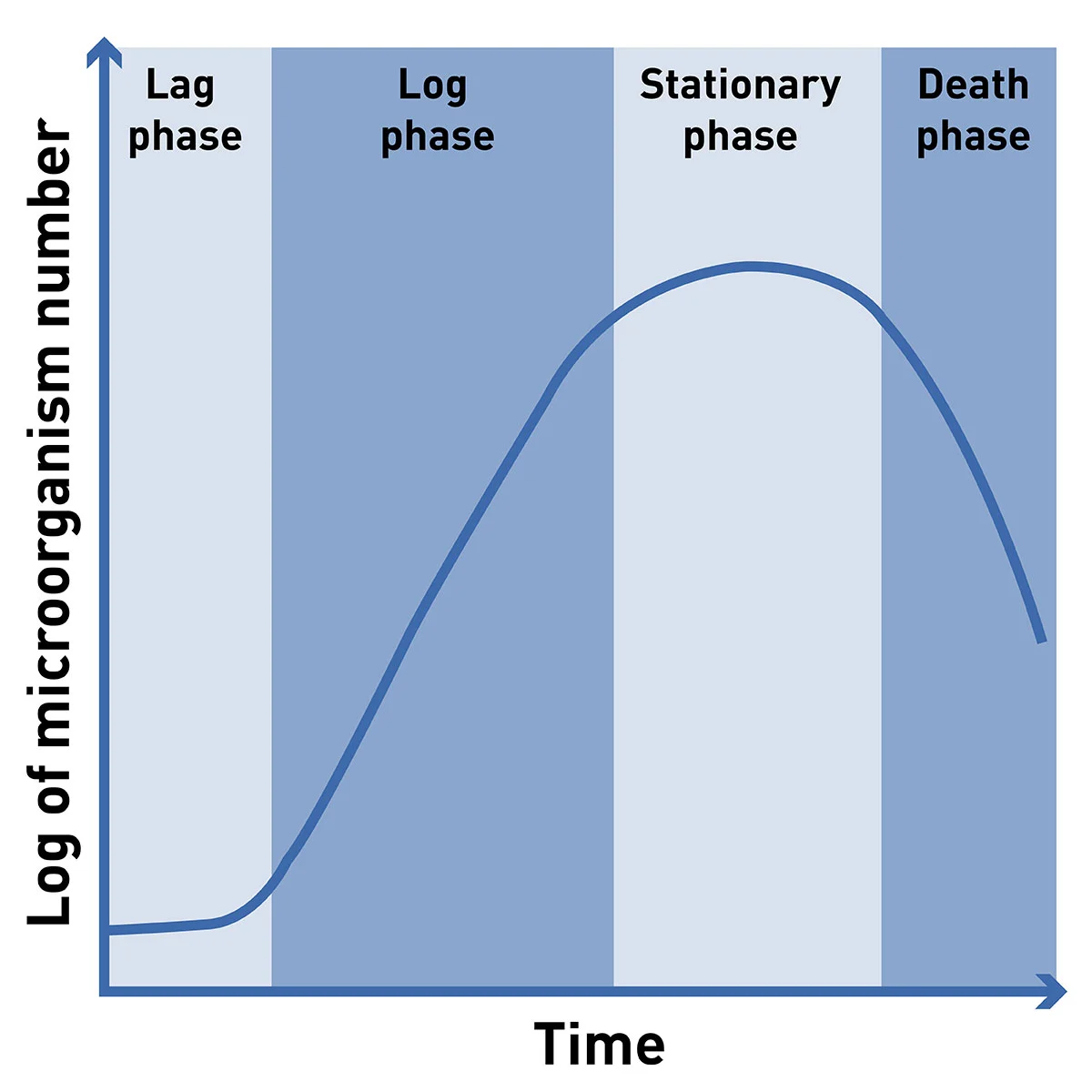
Monitoring the change of a microbial population over time results in a characteristic curve (Figure 1) called a growth curve. It consists of four growth phases:
Lag-phase
After starting the culture by placing a few microorganisms into the culture medium (inoculation), the organism needs to adapt to its new environment. During this time no division takes place and the organism density stays the same.
Log-phase
Once the microorganism has adapted to its new surrounding it starts to reproduce. Each cell doubles in a specific time leading to exponential microbial growth. When the logarithm of growth is plotted against time, this growth phase produces a line. The Log-Phase is the time when producing microorganisms is most effective. It further serves to calculate growth parameters such as the doubling time and growth rate.
Stationary phase
The reproduction of microorganisms is limited by nutrients present in the culture medium. Once the carbon source or other essential components are used up, the organisms stop dividing. This stage of microbial growth curves is reflected in a stable microorganism density at a high level.
Death phase
As the environmental conditions of a microbial culture get worse due to the deprivation of nutrients, salts, amino acids, or the increase of harmful waste products such as acids or ethanol, the population starts to die. This gets visible by an overall decrease in microorganism density.
The most common way to assess microbial growth in solution is the measurement of the optical density at 600 nm or short OD600. The method is based on absorbance detection mode and basically determines which portion of the light passes through a sample, more specifically through a suspension of microorganisms. How does the transmitted light relate to the growth of microbes? Particles in solution scatter light and the more particles (microorganisms) can be found in a solution, the more light is scattered by them. Therefore, a replicating population of bacteria or yeast increases light scattering and measured absorbance values. At the same time, this means that the absorbance mode is only exploited to determine the extent of light scattering instead of measuring the physical absorbance of light energy by absorbing molecules. This is an important point with practical consequences: OD600 measures light scattering and not absorbance!
As explained, the OD600 measurement is a light-scattering and not an absorbance-based method. Accordingly, the Beer-Lambert law which states a linear relation of absorbance and the concentration of an absorbing molecule is theoretically not applicable for OD600 measurements. In practice, however, it has been shown that despite the absence of absorbance, the Beer-Lambert can be used for low-density cultures. In other words, the OD600 value can be directly related to the number of microorganisms in very low-density suspensions. For higher density cultures, the OD600 measurement results in a rather parabolic curve. The number of organisms in such a culture can only be calculated after calibration of OD600 values to a count of organisms1.
If you are looking for approaches to get the most out of your OD600 measurements, take a look at our HowTo Note “How to optimise OD600 measurements”.
Cuvette or microplate?
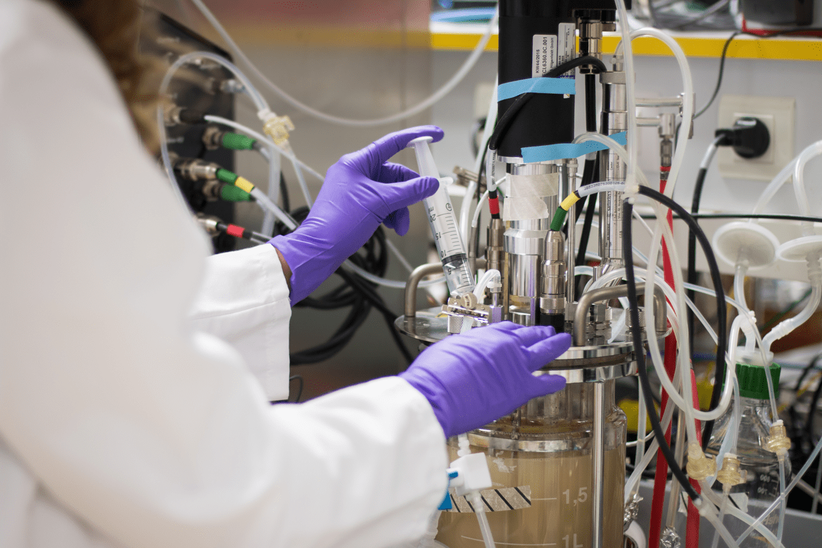
Cuvette measurements are recommended to monitor production processes with huge volumes. At regular intervals (e.g. 1 hour), samples are taken to determine the density of microorganisms. The method requires approximately 1.5 ml of microorganism suspension and the time to draw samples (Figure 2).
 Alternatively, microplates can be used to continuously monitor microbial growth. It requires only 200 µl of suspension and automatically measures the progress in growth at a chosen interval. As there are typically 96 wells in a microplate it is perfectly suited to measure multiple samples in replicates and to compare different media conditions or strains. For mid-to high-throughput applications, processes can be automated. An example is shown in the video below. Here, a Clariostar Plus was integrated with a PAA S-LAB Pro automated handler and a LiCONiCs incubator for growth and detection in high-throughput of cells under physiological conditions.
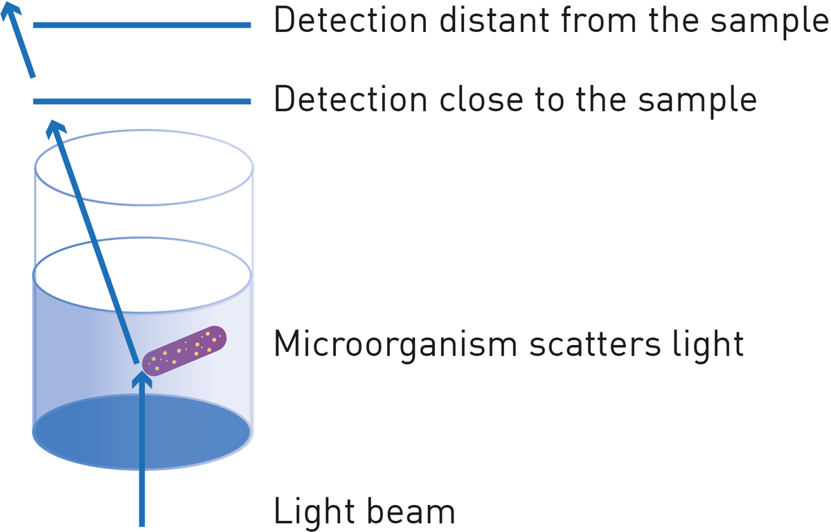
The measurement of light scattering by particles using the absorbance mode may give different results from one instrument to another. The reason is that different instruments use different light beams and the detector is positioned at different distances from the sample. Think of a light beam being scattered by a microorganism: a nearby detector still captures the light, while a detector positioned further away does not (Figure 3). Therefore, choosing one instrument series for microbial growth measurements by OD600 is recommended. Clearly, the decision for cuvettes or microplates narrows the choice as either a cuvette spectrophotometer or a microplate reader is required. Instruments that perform both, cuvette and microplate measurements, such as the SPECTROstar Nano are an alternative that equips you for all (future) needs.
When is calibration needed?
If the OD600 values should be used to determine the amount/concentration of organisms in solution (e.g. microorganisms/ml) or if a correction of the non-linear nature of the OD600-scattering measurement is needed. For the measurement, microorganism suspensions with known microorganism concentrations are prepared and OD600 is measured at the instrument of choice using the same volume as used in later experiments. The measured values can then be used to generate a calibration curve. OD600 values of unknown samples can later be related to this curve and the concentration can be back-calculated. As the light-scattering changes with the size and shape of particles, it is recommended to repeat calibration for each organism of interest. Likewise, calibration needs to be performed when changing the analysis instrument or the sample volume.
The measurement of microbial growth by OD600 in microplates is increasingly used in combination with detection modes such as luminescence or fluorescence. The measurement of microbial growth serves as normalization for another functional assay. An article published in Nucleic Acids Research used the CLARIOstar multi-mode plate reader to measure translation in Staphylococcus aureus (2). A beta-galactosidase reporter was measured using a fluorescence readout and the signal indicating enzyme activity was normalized to OD600. Another example monitored L. monocytogenes gene expression using luminescence and normalized it to OD600 values (3).
Nephelometry
The nephelometric method to measure microbial growth is also based on the light scattering of microorganisms. Contrary to OD600 measurements where the loss of transmission due to scattering is measured, nephelometry directly detects the scattered light. An example of monitoring the growth of Candida albicans by nephelometry in a 96-well plate is given in the application note Nephelometric monitoring growth of Candida albicans using BMG LABTECH’s NEPHELOstar® Plus. Compared to the absorbance-based method, nephelometry is more sensitive and detects lower amounts of particles, therefore being suitable for suspensions with very low microorganism numbers.
Stable fluorophore expression
A fluorophore such as GFP or RFP stably expressed by the microorganism of interest is capable of reporting on microbial growth. Fluorescence intensity is linear to the fluorophore concentration. Thus, it reports an increase in fluorescent microorganisms. The application note Expression of a stable green fluorescent protein mutant in group B streptococcus. Growth, detection, and monitoring, with the CLARIOstar®, compare microbial growth determination using stably expressed GFP to OD600 and explain a method of circumventing autofluorescence using polarized fluorescence emission. OD600 is difficult to use for the detection of mycoplasma contamination, as mycoplasma is very small, slowly proliferating, and usually to be detected in combined cultures with eukaryotic cells. Other mycoplasma contamination tests are effective here.
Various methods report on the growth of microorganisms. If you would like to read about the solutions BMG LABTECH offers for your microbial research, discover our dedicated microbiology brochure.
Absorbance plate reader with cuvette port
Powerful and most sensitive HTS plate reader
Most flexible Plate Reader for Assay Development
Upgradeable single and multi-mode microplate reader series
Flexible microplate reader with simplified workflows
Learn about the bacterial endotoxin test (BET test) and its role in ensuring the safety of pharmaceuticals, biologics and medical devices.
The monocyte activation test is a cell-based assay used to detect pyrogens. Learn more about the monocyte activation test and how microplate readers can support these crucial tests for quality control and bioanalysis.
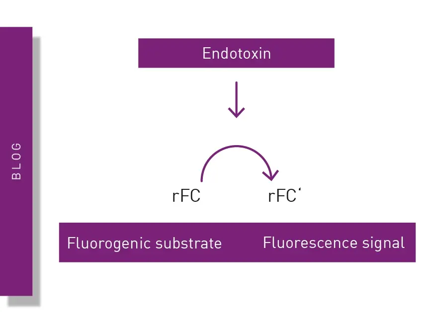
The recombinant factor C (rFC) test is used to detect bacterial endotoxins that can cause fever and adverse events when introduced into the body. Learn about the rFC test and the advantages it offers for endotoxin detection in the life sciences.
Pyrogen tests are vital to ensure the safety of different health interventions including pharmaceuticals, medical devices and an array of biological products. This blog looks at the different types of pyrogens as well as some of the widely used pyrogen tests.
Learn about applications for bacterial metabolism on a microplate reader.
Gene reporter assays are sensitive and specific tools to study the regulation of gene expression. Learn about the different options available, their uses, and the benefits of running these types of assays on microplate readers.