Introduction
With about 1000 members, G protein-coupled receptors (GPCRs) represent the largest superfamily of proteins that serve as receptors localized to cell surface membranes. They share conserved elements and employ similar hetero-trimeric proteins to initiate production of second messengers to participate in a wide variety of physiological responses. Due to their physiological importance, GPCRs are the target of about 1/3 of approved drugs1.
Despite all we have learned about GPCR’s and their signalling there is still much to learn, for example, the complex interactions between receptors and the signalling cascades they initiate1. To further our understanding of these important responses ION Biosciences has produced several tools to detect GPCR stimulated second messengers. They have used a variety of fluorophores that span the visible spectrum as a part of these tools thus enabling the detection of multiple signalling events simultaneously.
Here we show how the detection of calcium and thallium flux can be detected simultaneously. Using the CLARIOstar Plus with LVF monochromator allowed easy adjustment of the detection wavelengths to optimize the multiplex detection.
Assay principle
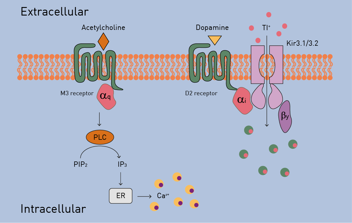
Figure 1 shows two second messenger production events occurring after stimulation of the M3 and D2 receptors by acetylcholine and dopamine, respectively. Release of intracellular calcium can be detected by Fluo-Gold because of Gq signalling following M3 activation. Simultaneously, stimulation of the D2 receptor initiates potassium flux through a G protein-coupled inwardly rectifying potassium channel (GIRK). This can be detected using surrogate thallium ions and Thallos.
Materials & methods
- PDL coated 384-well plate, clear bottom, black (Corning® Falcon®, #CLS353219)
- Thallos AM and Fluo-Gold AM (ION Biosciences, #1381A, #1045E)
- CLARIOstar Plus (BMG LABTECH)
- Cells, other chemicals and reagents were obtained from commercial sources
Experimental Procedure
HEK293 cells were transduced with D2 receptor, Kir3.1 and Kir3.2 for 24 hours and cultivated in a PDL coated microplate. Afterwards, cell culture media were removed, and cells were loaded with Thallos AM and Fluo-Gold AM dyes for 1 hour prior measurement. Cells were treated with 100 µM acetyl-choline and/or 10 µM dopamine 20 seconds after the start of the measurement using the on-board reagent injectors on the CLARIOstar Plus.
Instrument settings
|
Fluorescence Intensity (multichromatic), Bottom Reading, Kinetic (well mode)
|
|||
|
Optic settings
|
|||
|
Thallos AM (= optimized)
|
|||
|
Preset
|
Excitation |
Dichroic
|
Emission
|
|
GFP
|
470-15 (465-15) |
491.2 (485.2)
|
515-20 (507-18)
|
|
Fluo-Gold AM (= optimized) |
|||
|
Alexa Fluor 532
|
520-20 (525-17)
|
542.5 (546.2) |
570-30 (574-30)
|
|
Gain and Focus
|
Adjusted
|
||
|
Kinetic settings |
|||
|
Number of intervals
|
320 |
||
|
Interval time
|
1 second |
||
|
Flashes per chromatic
|
20 |
||
|
Injector settings
|
|||
|
Volume
|
10 µl |
||
|
Pump speed
|
50 µl/second |
||
|
Injection start time
|
20 seconds |
||
|
General settings
|
|||
|
Target Temperature
|
37 °C |
||
Results & Discussion
Initial studies showed that all dyes tested from ION Biosciences worked very well individually (data not shown), but we wanted to test multiplexing capacity. Figure 2 shows that the default settings available on the CLARIOstar Plus for the relevant fluorophores for Thallos AM and Fluo-Gold AM work well but exhibit some bleed-through (Fig. 2A). A small adjustment of the detection wavelengths, easily achieved because of the LVF monochromator, decreases the bleed-through (Fig. 2B).
Additional validation tests showed the specifi city of detection of the Ca2+ and Tl+ signaling pathways (data not shown). Figure 3 shows the simultaneous detection of acetylcholine stimulated Ca2+ release via Fluo-Gold and dopamine-mediated activation of a GIRK channel via Thallos fluorescence. Both dyes were suitable for the multiplex approach. 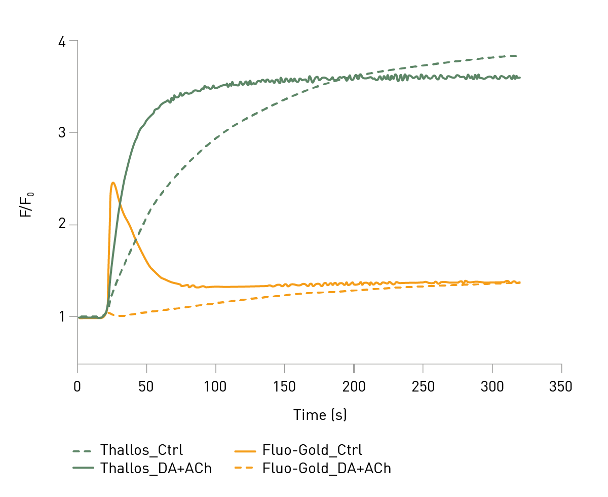
Conclusion
We show the successful multiplexing of two fluorescent dyes to enable the simultaneous real time detection of activation of distinct GPCR signaling pathways. These results provide proof of principle for the application of these and similar dyes from ION Biosciences to further our understanding of complex GPCR signaling events.
References
- Zhang, M., et al. G protein-coupled receptors (GPCRs): advances in structure, mechanisms and drug discovery. Sig Transduct Target Ther (2024) 9: 88