Introduction
Ferroptosis is a unique form of cell death that is characterized by iron-dependent oxidative damage to membrane phospholipids and is involved in important pathological mechanisms of neurodegenerative disease. Excessive reactive oxygen species (ROS) also target sensitive fatty acids and promote lipid peroxidation, which then impairs the integrity of lipid membranes and induces suicide signaling cascades.1 ROS can be produced by the mitochondria during hypoxia – reoxygenation. The activation of the mitochondrial sodium/ calcium exchanger NCLX during acute hypoxia drives superoxide production at complex III.2 Inhibition of the of Na+ import through NCLX is a promising approach to block this pathway and inhibit lipid peroxidation.
Here, the CLARIOstar Plus microplate reader was used to measure the inhibition of lipid peroxidation in a fluorescence–based lipid peroxidation assay while changing oxygen levels with the Atmospheric Control Unit (ACU).
Assay principle
The lipid peroxidation assay was conducted in cell cultures subjected to normoxia or ischemia-reperfusion by labeling cells with Bodipy 581/591 C11, a lipophilic fluorescent probe used to determine lipid peroxide levels (fig. 1) 3.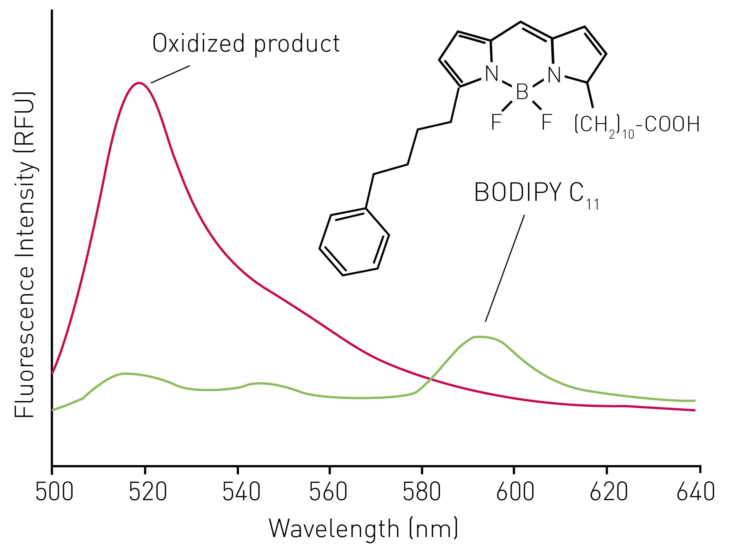
- 96-well, black, clear-bottom plates (Thermo Fisher Scientific)
- SK-N-DZ human neuroblastoma cells
- Bodipy 581/591 C11 (#D3861, Invitrogen)
- DMEM FluoroBrite™ (#A1896701, Gibco)
- DMEM, no glucose, no glutamine, no phenol red (#A1443001, Gibco)
- CLARIOstar Plus microplate reader with an Atmospheric Control Unit (BMG LABTECH)
Experimental procedure
SN-N-DZ cells were seeded 24 hours prior to the lipid peroxidation assay in 96-well black, clear-bottom plates. Cells were exposed to Bodipy 581/591 C11 at a concentration of 2 µM for 30 min.
In a preliminary experiment, cells were treated with a ferroptosis inducer and also inhibitor to validate the suitability of the measurement protocol for lipid peroxidation assay.
For the main experiment (fig. 2), the culture medium was replaced with DMEM FluoroBrite™ (incl. 0.5% FBS) for the normoxia control (plate 1) and DMEM without glucose, glutamine or phenol red for the ischemia reperfusion plate (plate 2). Initial measurements of the lipid peroxidation assay were taken simultaneously from both plates at 18% oxygen. Then, the normoxia plate was kept in a standard cell incubator while the ischemia-reperfusion plate stayed inside the CLARIOstar Plus, and oxygen levels were lowered to 1% for 2 h. Thereafter, oxygen levels were reverted back to 18% to mimic reperfusion, and the culture medium was changed to FluoroBrite™ (incl. 0.5% FBS).
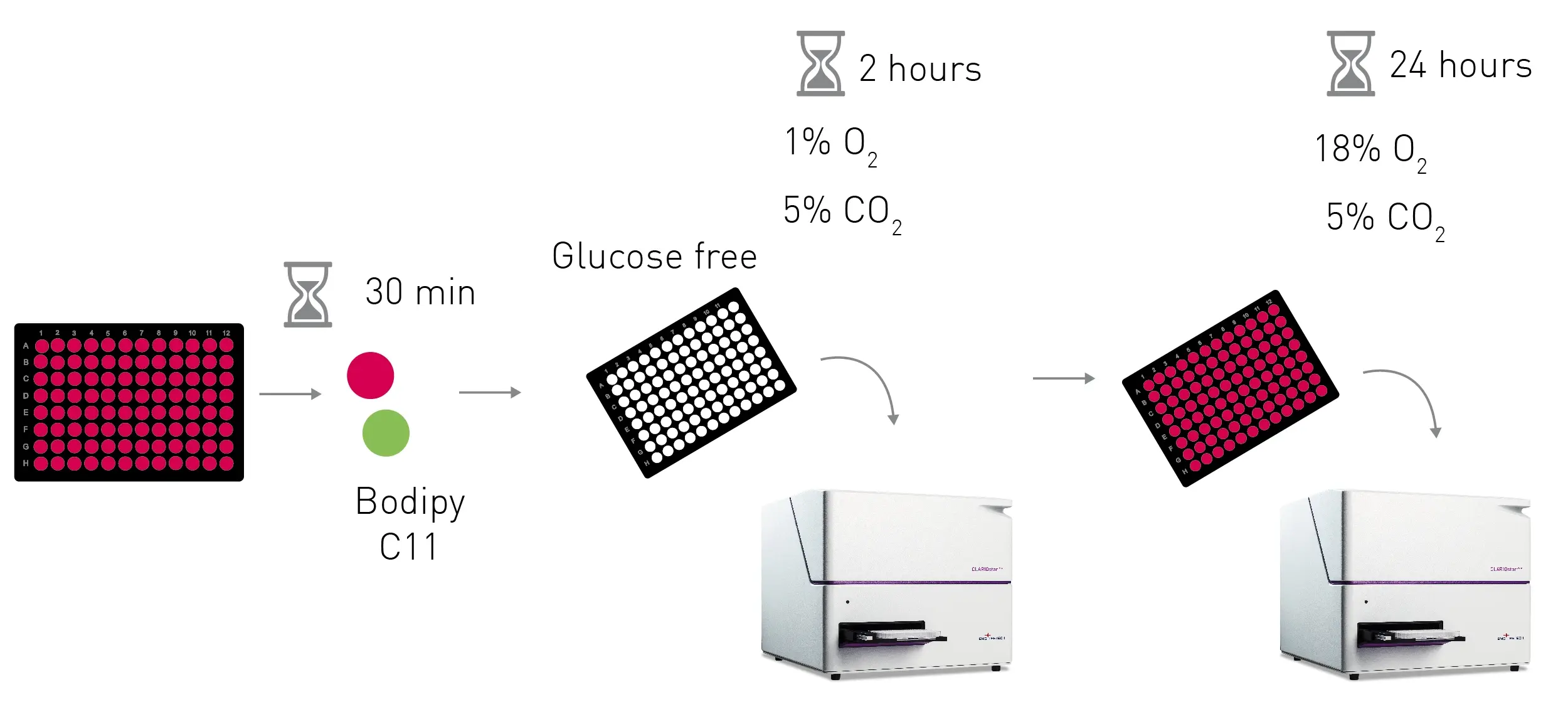
Concurrently, treatment with NCLX or ferroptosis inhibitors was added and both plates were incubated in normoxia for 24 h until they were measured again. Lipid peroxidation was determined by calculating the ratio between the maximum fl uorescence at 520-530 nm and 590-600 nm emitted by the Bodipy 581/591 C11 probe. In this lipid peroxidation assay, a higher ratio corresponds to elevated levels of lipid peroxides.
Instrument settings
|
Optic settings
|
Fluorescence Spectrum, Endpoint
|
|
|
General settings
|
Number of flashes per well | 20 |
| Settling time | 0.5 sec | |
|
Endpoint settings
|
Excitation wavelength (nm) | 485-10 |
| Emission wavelength (nm) | 509-8 to 640-8 | |
|
Incubation
|
37ºC, 1%-18% O2, 5% CO2 |
|
Results & Discussion
The ferroptosis inducer RSL3 was used as a positive control, while the ferroptosis inhibitor Ferrostatin – 1 was used as a negative control to assess the protocol for the lipid peroxidation assay (fig. 3).
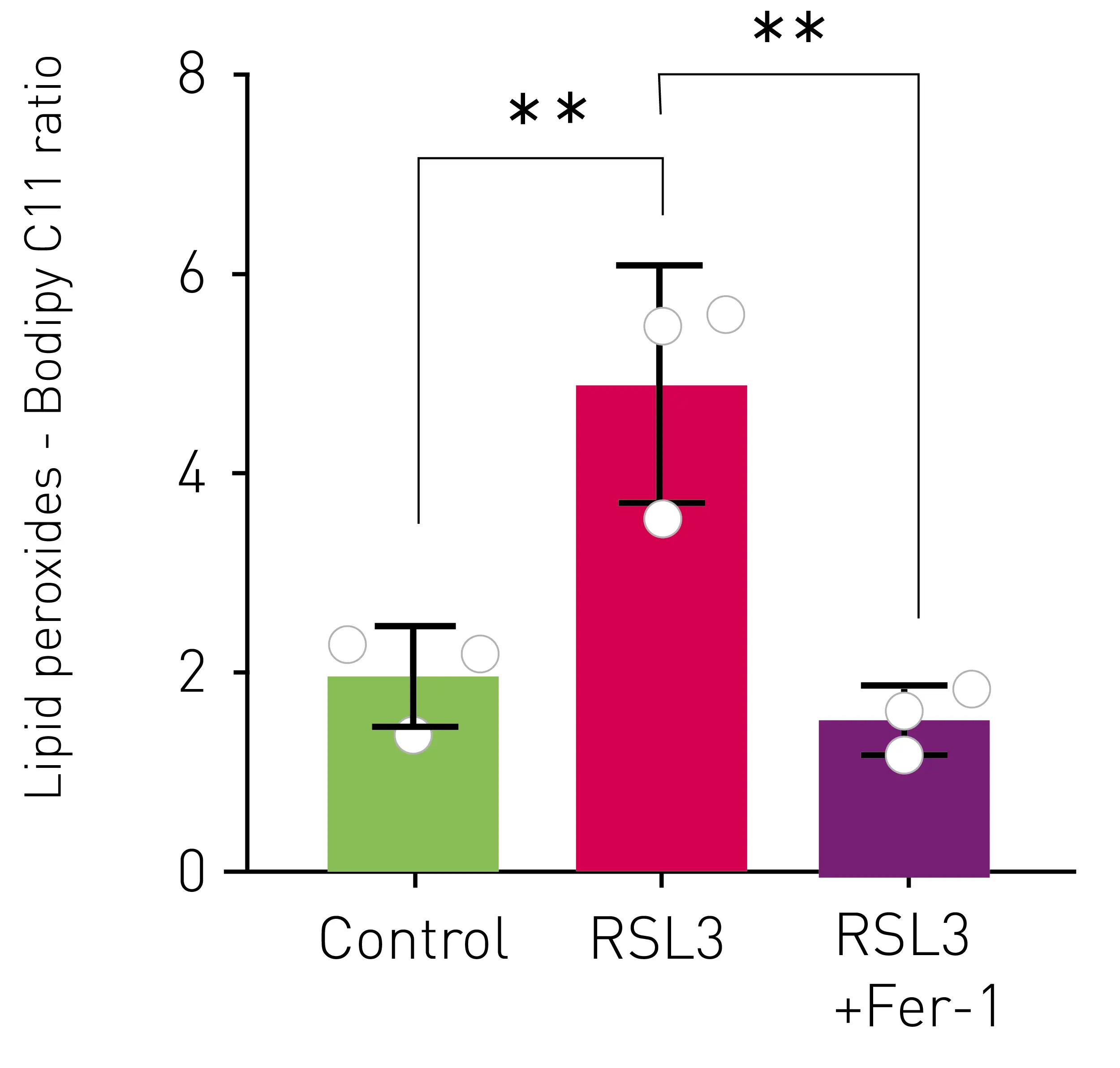 The results shown in figure 3 confi rm that the lipid peroxidation assay protocol successfully detected the lipid peroxides produced by the ferroptosis inducer. Hence, the same protocol was used to quantify lipid peroxide levels in the normoxia and ischemia-reperfusion models.
Figure 4 illustrates the increase in Bodipy 581/591 C-11 ratio observed for cells subjected to both normoxia and ischemia-reperfusion with and without treatment in the lipid peroxidation assay. The horizontal line marks the initial levels of lipid peroxides observed in normoxia.
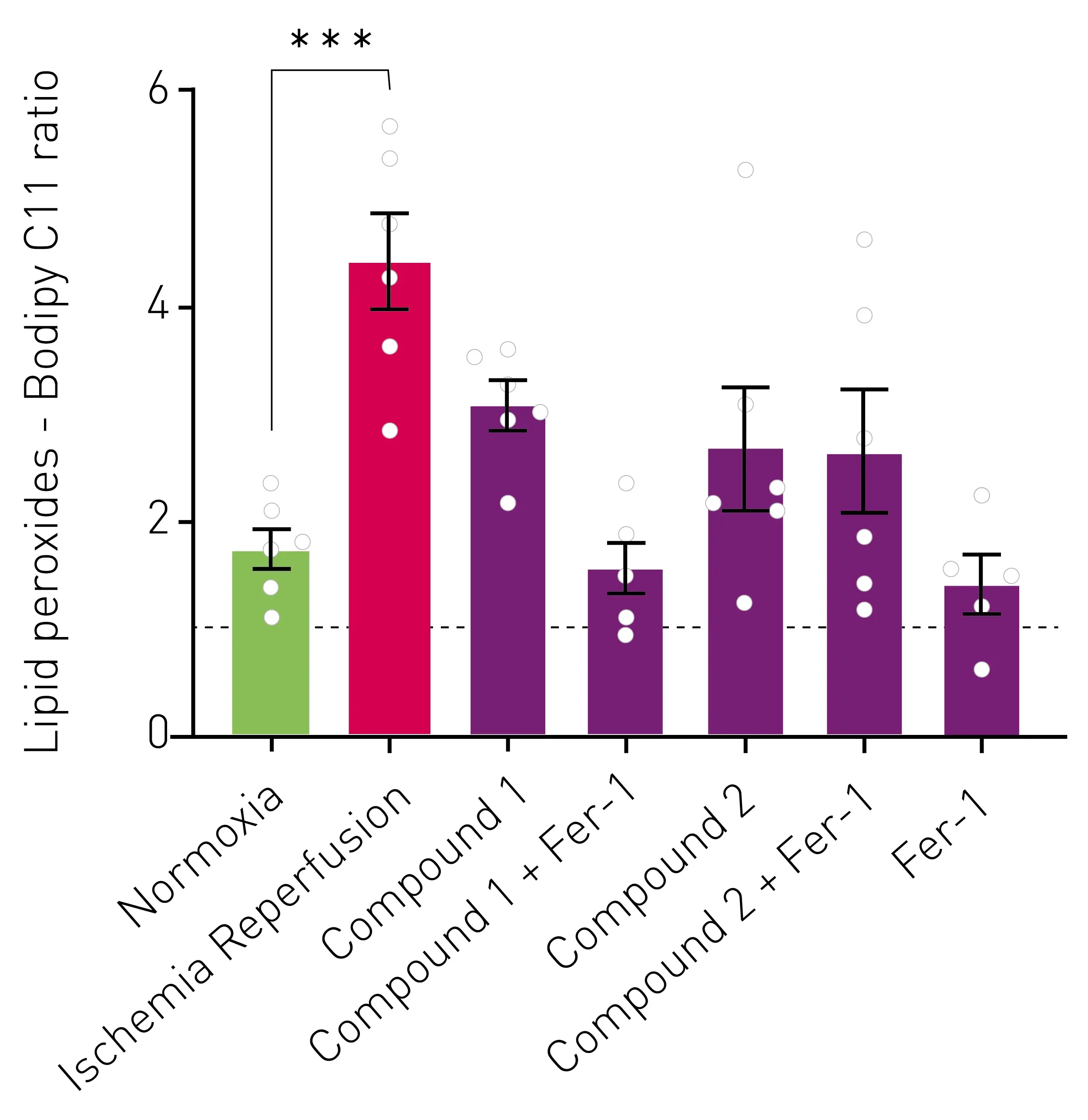
Cells treated with NCLX and/or ferroptosis inhibitors exhibit a smaller increase in the Bodipy 581/591 C-11 ratio that determines lipid peroxides in the lipid peroxidation assay. Importantly, this increase is not signifi cantly different from the rise observed in normoxic cells. Furthermore, only the cells subjected to ischemia-reperfusion without any treatment show a signifi cant increase in lipid peroxide levels.
Conclusion
The CLARIOstar Plus microplate reader with ACU enables to manipulate oxygen levels to replicate the conditions of ischemia and reperfusion in cell models. Lipid peroxides can be successfully determined by measuring a lipid peroxidation assay based on Bodipy 581/591 C-11 fluorescence in the CLARIOstar Plus.
There is a signifi cant increase on lipid peroxidation levels observed after 2 hours of ischemia and 24 h of reperfusion in SK-N-DZ neuroblastoma cells. Nevertheless, the administration of an NCLX and/or ferroptosis inhibitor during reperfusion seems to reduce production of lipid peroxides in this process. This reduction may ultimately contribute to diminished cell death caused by ferroptosis.
References
-
Dixon SJ, Lemberg KM, Lamprecht MR, et al. Ferroptosis: an iron-dependent form of nonapoptotic cell death. Cell. 2012;149(5):1060-1072.
-
Hernansanz-Agustín P, Choya-Foces C, Carregal Romero S, et al. Na+ controls hypoxic signalling by the mitochondrial respiratory chain. Nature. 2020;586(7828):287-291.
-
Drummen GP, van Liebergen LC, Op den Kamp JA, Post JA. C11-BODIPY (581/591), an oxidation-sensitive fluorescent lipid peroxidation probe: (micro)spectroscopic characterization and validation of methodology. Free Radic Biol Med. 2002;33(4):473-490.