Introduction
Since its introduction in the 1970s, the ELISA (enzyme-linked immunosorbent assay) remains a very popular technique to assess the presence or quantity of an analyte.1,2 Despite the high utility of this approach and the continued advances to improve sensitivity, most ELISA-type assays require washing steps. These steps are time-consuming and tedious or require equipment dedicated to the task.
Immunoassays that use SPARCL from Lumigen provide no-wash detection. We have previously shown the performance of the Omega microplate reader in the detection of this luminescent assay.3 Here we show the CLARIOstar is also excellent for detection of SPARCL immunoassays. Furthermore, we show that the SPARCL assay can be performed in a 384-well format for improved throughput.
Assay Principle
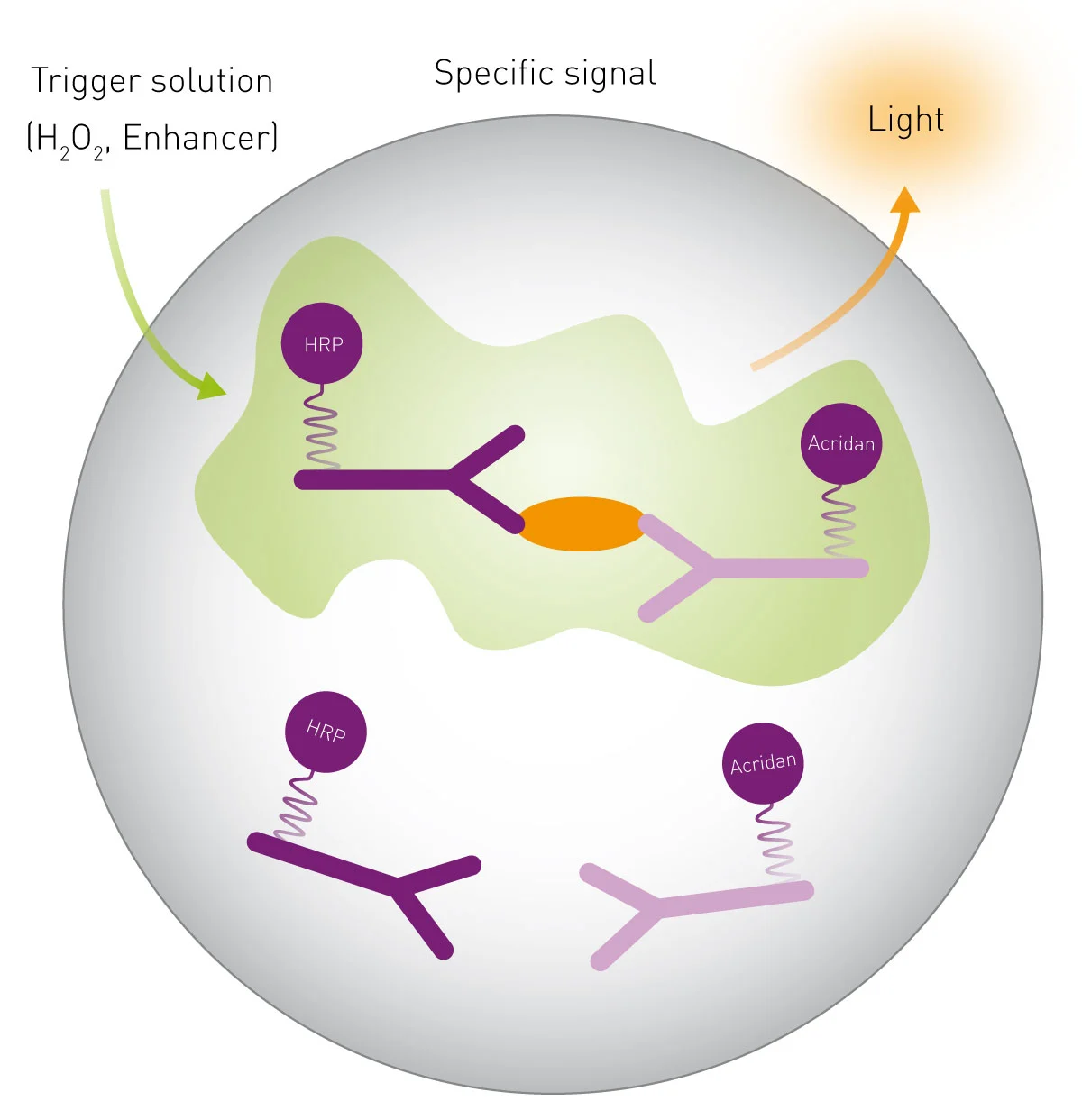
As the SPARCL acronym implies (spatial proximity analyte reagent capture luminescence) it detects an analyte in solution and creates a light signal (Figure 1). It is an immunoassay that employs 2 antibodies: one bound to acridan the other bound to HRP (horseradish peroxidase). The binding of these antibodies to an analyte brings the substrate and enzyme into proximity such that when a trigger solution (H2O2) is added a light signal is produced. The intensity of the flash of light is proportional to the amount of analyte present making it a tool for quantifying the analyte.
Materials & Methods
- SPARCL Generic Human IgG Pharmacokinetic Assay Kit (Lumigen)
- CLARIOstar microplate reader (BMG LABTECH)
- White, 96-well plates (Greiner)
- White, 384-well plates (Corning)
Standard curves were prepared using 1:2 serial dilutions. For tests using 96-well plates, human IgG dilutions were prepared in rat serum. The maximum concentration of IgG was 2,875 ng/mL. To show assay performance in 384-well plates human IgG was diluted in PBS with 0.1% BSA. The maximum concentration was 1,000 ng/mL.
Assays using 96 well plates were performed as described in the instructions from Lumigen.4
Assays using 384-well plates were performed as described4 with the following modifications: antibody mixture (20 µl) was added to wells followed by 10 µl of varying concentrations of IgG for a standard curve. Following incubation 2 µl of background reducing reagent was added.
Plates were read using CLARIOstar test protocols with the following settings which incorporate injection of trigger solution.
Instrument settings
| Detection Mode: | Luminescence, top reading |
| Detection Method: | Well mode, Kinetic |
|
No. of intervals: |
100 (96 well), 50 (384 well) |
|
Interval time(s): |
0.02 |
|
Gain: |
3500 (96 well), 3300 (384 well) |
|
Focal height (mm): |
11.0 |
|
Emission filter: |
No filter |
| Injection needle: | H3 |
| Injection start time: |
0 s |
| Injection volume (µl): | 75 (96 well), 30 (384 well) |
| Pump speed: | 300 µl/s |
| Aperture spoon: | For 384 well only |
Results & Discussion
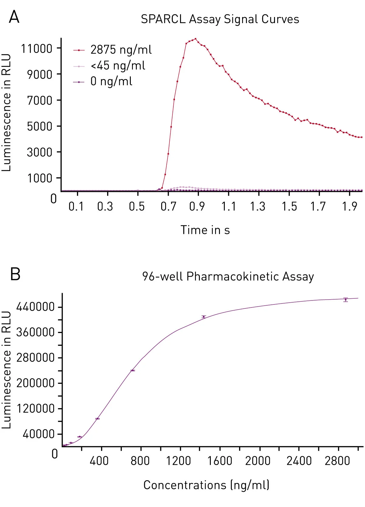
The SPARCL assay produces a flash luminescent signal that has a fairly reproducible signature. The signal detected in a 96-well plate using the CLARIOstar corresponds well with what has been previously observed (Figure 2A).
Using MARS data analysis software, a sum of luminescent signals over the 2 second time period of data collection was obtained. From this, we can show that the response is robust with signal to blank calculated to be 587 for the high concentration standard. Furthermore, regression analysis shows that the data fits to a 4-parameter fit as expected (Figure 2B) from which calculation of unknown concentrations could be possible.
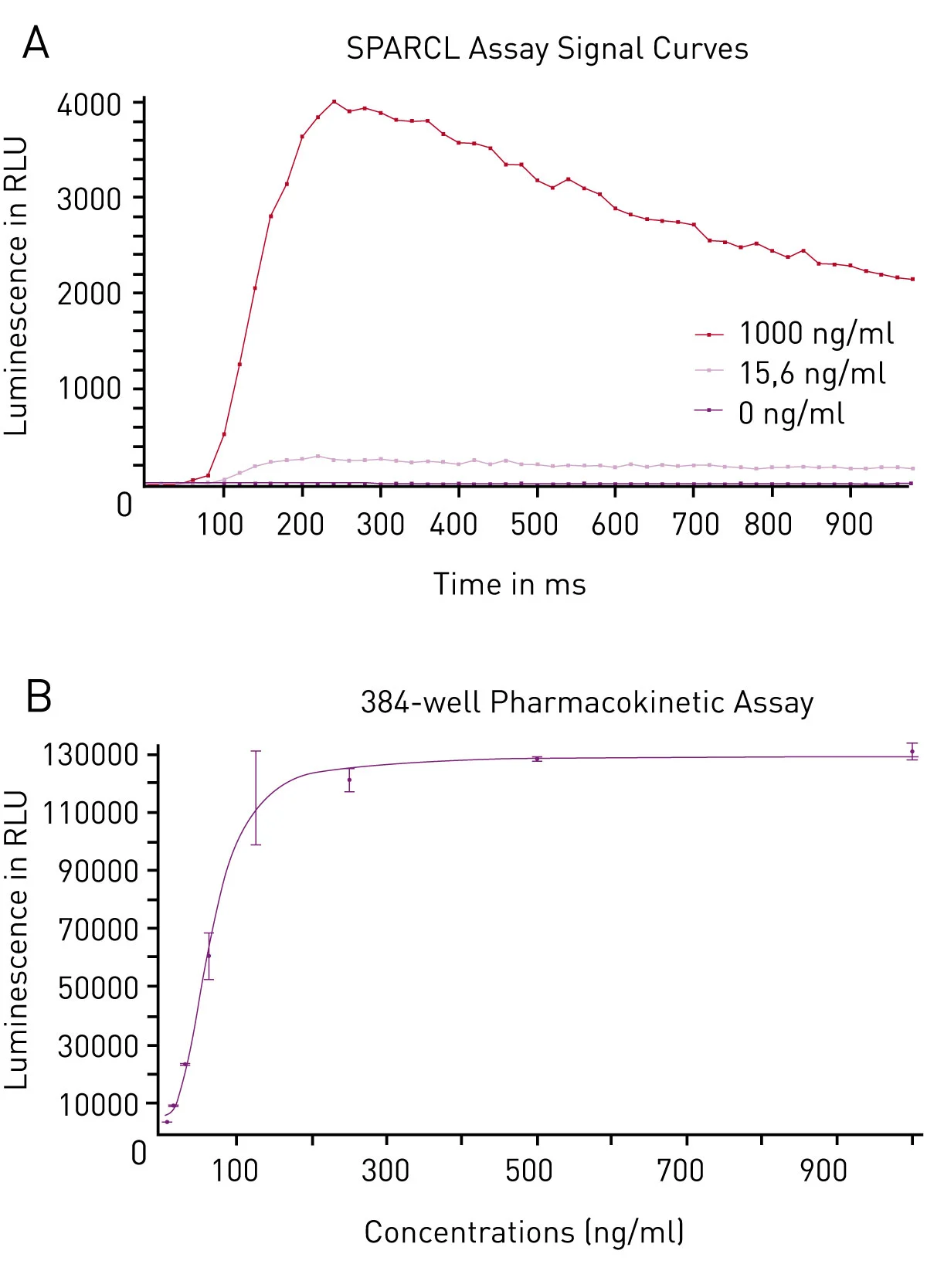
We were further able to confirm the ability to perform the SPARCL assay in a 384-well plate format. The data collected in the 384-well tests exhibited a similar response to injection of trigger solution (Figure 3A).
Once again, upon the completion of data analysis, we could confirm that the assay performed in 384-well plates was similarly robust based on signal to blank values (=576 for the high standard). In addition, the expected 4-parameter fit curve can be used to correlate concentration to signal (Figure 3B).
Conclusion
Lumigen’s SPARCL assay is highly compatible with the BMG LABTECH CLARIOstar microplate reader. We obtained robust responses in this pharmacokinetic assay which detects human IgG. For those who seek higher throughput, we find the 384-well plate option also performs very well.
References
- Engvall, E. and Perlman, P. (1971). Enzyme-linked immunosorbent assay (ELISA) quantitative assay of immunoglobulin G. Immunochemistry 8, 871–874.
- Van Weeman, B.K. and Schuurs, A.W.H.M. (1971). Immunoassay using antigen-enzyme conjugates. FEBS Letters 15, 232–236.
- Xie, W., Cameron, M. and Peters, C. (2015). Using SPARCL technology to Develop Immunoassays for Biomarker Detection and Pharmacokinetic Studies.
For more information about SPARCL contact Lumigen at SPARCL@beckman.com or visit www.lumigen.com/detection-technologies/sparcl
Lumigen and SPARCL are trademarks of Lumigen, Inc.