Introduction
Protein ubiquitination is an essential part of the regulation of a wide variety of biological processes. Although ubiquitination is commonly associated with proteasomal degradation, roles in immune signaling and cell cycle regulation have also been identified. The diversity of regulation is made possible by the fact that mono- and distinct forms of poly-ubiquitination have separate regulatory roles.
As expected for such a vital process, dysregulation of ubiquitination is associated with numerous diseases including cancer and auto-immune disorders. Therefore, research efforts have sought ways to correct dysregulation and more recently have sought to harness the protein degradation abilities of ubiquitination with protein targeting chimeric molecules (PROTACs). These efforts have been hindered by a paucity of tools to look at all stages of the protein ubiquitination cycle.
Here we describe a simple high throughput screening (HTS) assay called UbiReal1. Using fluorescence polarization (FP), fluorescently labeled ubiquitin (Ub) can be tracked to monitor the sequential conjugation and deconjugation states of Ub. BMG LABTECH’s CLARIOstar microplate reader is well known for its excellent FP performance which is showcased in this application.
Assay Principle
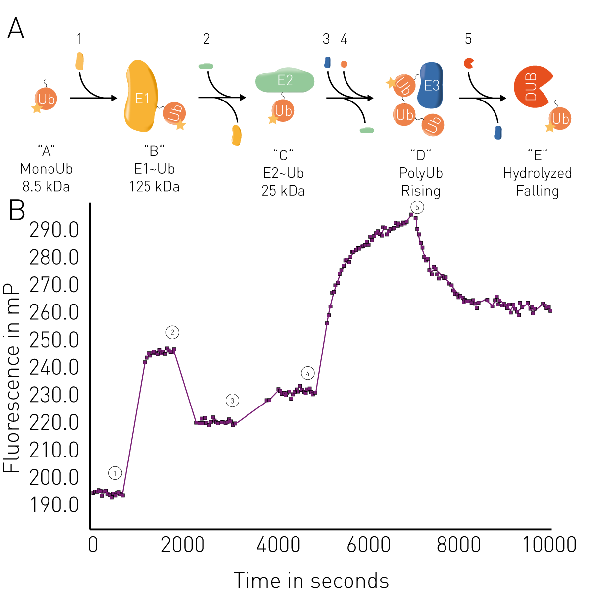
In humans, ubiquitination is controlled by hundreds of proteins that are broadly categorized as writers, readers, and erasers. The writers are made up of E1 Ub-activating enzymes, E2 Ub-conjugating enzymes, and E3 Ub ligases that act in sequence to transfer Ub (Figure 1A, steps 1-4). Ub signals are then interpreted by Ub binding proteins termed readers that direct appropriate cellular outcomes. Following signaling, eraser proteins termed deubiquitinases (DUBs) remove and recycle the Ub modification (Figure 1A, step 5).
FP can be used to monitor a molecule’s rotation in solution with the understanding that larger molecules rotate more slowly and thus demonstrate a higher FP value. Previous results had shown that mono-Ub can be distinguished from poly-Ub chains based on changes in FP signal2. This led to the investigation of whether FP monitoring of fluorescent Ub could distinguish between the various states seen in the ubiquitination cascade. Representative data in Figure 1B shows: 1) conjugation of labeled Ub to E1 caused a large shift in FP, 2) formation of the E2~Ub conjugate decreased FP slightly, 3) transition to an E3~Ub conjugate exhibits a detectable increase of FP from 2, 4) conjugation of additional unlabeled Ub is observed as a time-dependent increase in FP, and 5) removal of Ub by DUBs is evident as a decrease in FP.
Materials & Methods
- Greiner, black, 384-well, small volume, HiBase microplates
- CLARIOstar® microplate reader
- Proteins were obtained or expressed and purified as described in Franklin & Pruneda1
- Chemicals were obtained from commercial sources
Experimental Procedure
Samples were prepared using a master solution resulting in a final volume of 20 µL with a buffer composition of 25 mM sodium phosphate (pH 7.4), 150 mM sodium chloride, 10 mM MgCl2, and 100 nM TAMRA-Ub.
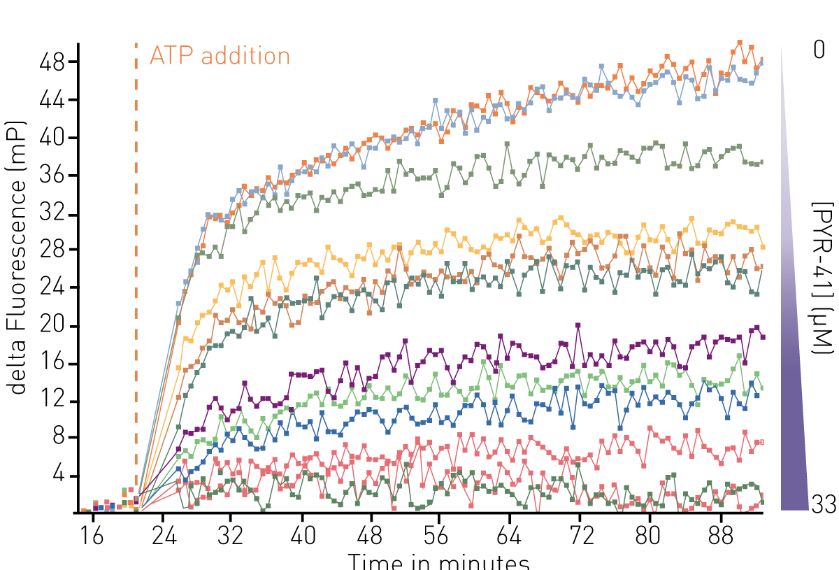
For E1 Inhibition assays, 0.5 µL of the inhibitor PYR-41 at various concentrations in DMSO, or DMSO alone as a control, were added to the master solution which also contained 125 nM E1. FP was monitored for 10 cycles using the settings indicated below, at which point the test was manually paused and 1 µL of ATP was added to initiate the reaction ([final]= 5 mM) and monitoring continued for 70 minutes.
Instrument Settings
| Optic settings | Fluorescence polarization, plate mode kinetic |
| Filters: Ex: 540/Di: LP566/Em:590 | |
| General settings |
Number of flashes: 20 well per cycle |
| Settling time: 0.1 s | |
| Kinetic settings | Number of cycles: 187 or 120 |
| Cycle time: 40 or 41 s |
Data analysis
MARS data analysis software was used to calculate FP values and Z’, an indicator of assay quality, using equations available in the software. In addition, the average was calculated, and baseline correction was performed using the signal observed for 10 cycles prior to ATP addition.
Results & Discussion
Figure 1B shows that all stages of the ubiquitination cycle can be monitored by the UbiReal approach. However, it is anticipated that most investigations, including drug discovery, will focus on isolated steps in the pathway. Therefore, we sought to assess the performance of UbiReal in a more focused manner.
Figure 2 shows results that focus on an assay to analyze the conjugation of TAMRA-Ub onto E1. Furthermore, it shows how this assay could be used to assess the effect of chemical inhibition. Specifically, showing the concentration-dependent effect of the known chemical inhibitor PYR-413. Assay quality data (Z’=0.59) indicate that this assay is suitable for HTS.
 In addition, the UbiReal approach was used to study the activities of E2s, E3s, and DUBs in more focused assays (data not shown here). All are found to be suitable for HTS based on Z’ assessment of assay quality1.
Conclusion
The UbiReal method addresses the need for a universal HTS assay to assess Ub conjugating and deconjugating activity. The assay has many attractive features (convenient, quantitative, robust, and scalable) that will make it useful for mechanistic work and as an HTS for small molecules. Excellent FP detection and data analysis from BMG LABTECH nicely complement this useful assay.
References
- Franklin, T.G., and Pruneda, J.N. A High-Throughput Assay for Monitoring Ubiquitination in Real-Time. Front. Chem. (2019) 7: 1-11.
- Ye, Y. et al. Polyubiquitin binding and cross-reactivity in the USP domain deubiquitinase USP21. EMBO Rep.(2011) 12: 350-357
- Yang, Y. et al. Inhibitors of ubiquitin-activating enzyme (E1), a new class of potential cancer therapeutics. Cancer Res. (2007) 67: 9472-9481.