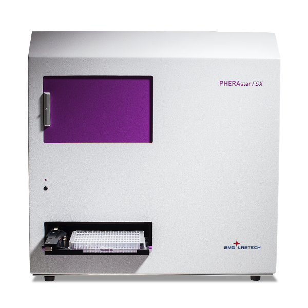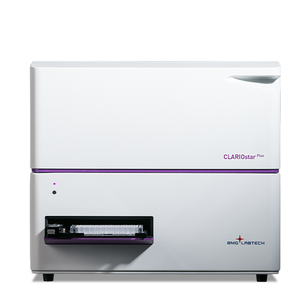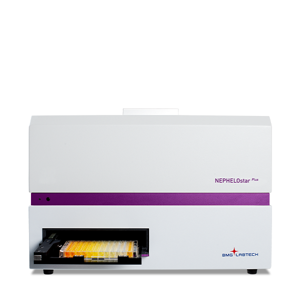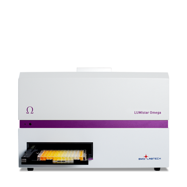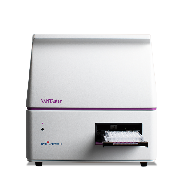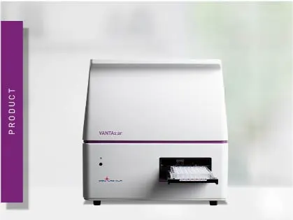
VANTAstar
Flexible microplate reader with simplified workflows





Barry Whyte is Application Scientist and Science Writer at BMG LABTECH in the United States. He has PhD and Bachelor of Science (BSc) degrees in biochemistry from the University of Bristol in the United Kingdom and more than 20 years of experience in the life sciences and science communications. Over the years, Barry has worked on three continents and traveled widely. He enjoys building on his international work experience and learning new ways to help scientists advance their research.

Prions are infectious, misfolded proteins that cause neurodegenerative diseases such as bovine spongiform encephalopathy (BSE), Creutzfeldt-Jakob disease (CJD) and chronic wasting disease (CWD) (Fig. 1). These debilitating disorders affect humans (CJD), cattle (BSE), cervids, and other wild animals such as deer and elk (CWD).1-4 Prion diseases, also known as transmissible spongiform encephalopathies, result from misfolded proteins that accumulate as aggregated molecules primarily in the cells of the nervous system including the brains of infected animals.
Prions propagate after regularly folded prion proteins (PrPC) are converted into misfolded, aggregation-prone PrPSc proteins that build up and yield amyloids (Fig. 2; see also note on nomenclature in figure legend). These misfolded proteins and their aggregates damage nerve and other cells and, over time, lead to a decline in brain function and ultimately death.
The Real-Time Quaking-Induced Conversion (RT-QuIC) assay is a rapid, sensitive, quantitative test that detects prions by amplifying the protein misfolding activity and tracking amyloid formation by fluorescence using a chemical dye. These measurements can be made in different ways but are typically performed on microplate readers for reasons that we will see later.
RT-QuIC assays are widely used by researchers in academic labs and industry studying prion diseases and other neurodegenerative disorders.5 Since they were first introduced, RT-QuIC assays have greatly accelerated experiments and analysis of prions compared with earlier more costly and lengthy bioassays where infected animals were studied over months, and where analysis often depended on post-mortem brain tissue.
In research using samples from cerebrospinal fluid, RT-QuIC assays showed high accuracy with 90.3% sensitivity and 98.5% specificity for prion diseases.6 By detecting prion seeding activity from less invasive samples, including nasal brushings, research has also shown that RT-QuIC assays can support earlier detection of prions which may be useful for diseases like CJD.7
This blog describes the origin of RT-QuIC assays, how the method works, where microplate readers fit into the workflow, and provides examples of applications ranging from prion and neurological disease research to emerging research in diagnostic development as well as AI-enabled drug discovery.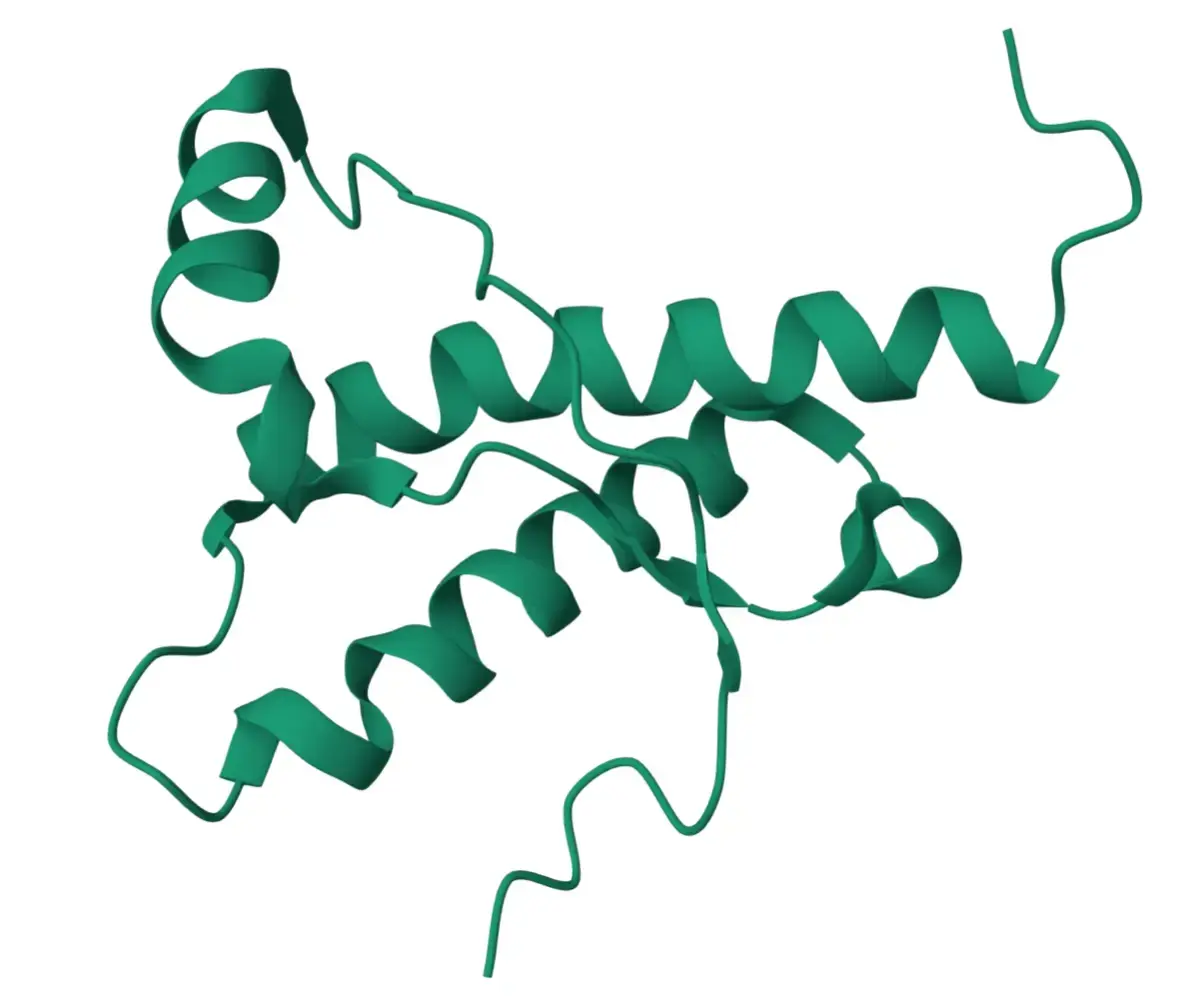
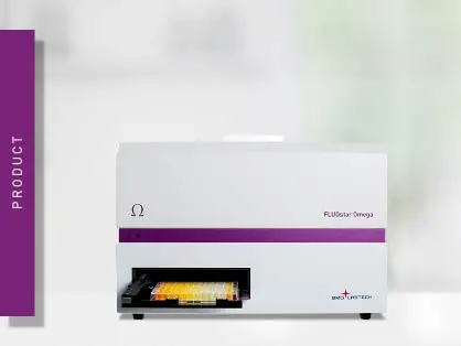
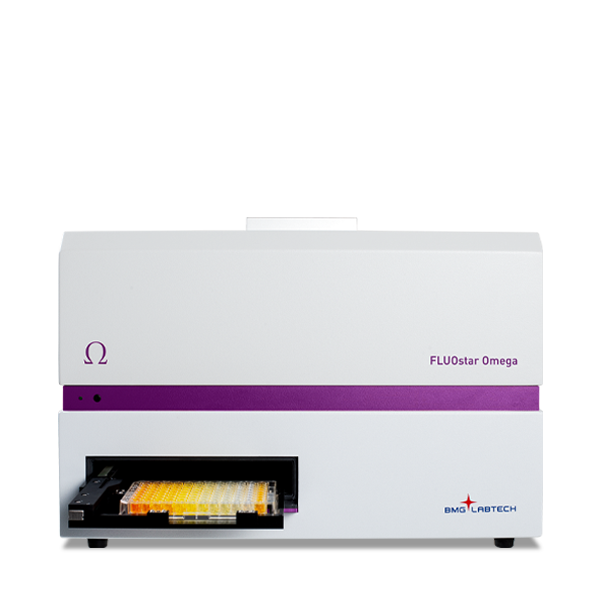
The RT-QuIC assay was first described in 2010 as a new test for ultrasensitive human prion detection, offering a highly sensitive, rapid and quantitative way to assess prions.8,9 As a new method, it offered considerably higher throughput than conventional assays. The approach described in the original paper, which was developed at Rocky Mountain Laboratories in the United States, was performed on a FLUOstar® Omega microplate reader from BMG LABTECH which offered the robust qualities and precision needed to support the vigorous intermittent shaking required for the analysis.8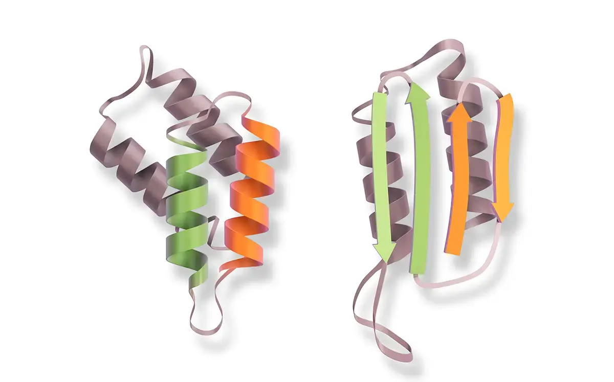
The FLUOstar Omega microplate reader from BMG LABTECH quickly went on to be an international standard for prion analysis. Since then, RT-QuIC has been adopted as an assay of choice for researchers looking not only to study prion diseases like BSE, CJD and CWD but also for other neurological diseases like Alzheimer’s and Parkinson’s that arise in part due to the misfolding, aggregation and accumulation of other proteins (e.g. alpha-synuclein, tau), in the brain that also give rise to amyloid.
The RT-QuIC assay in use today combines a shaking-induced conversion step with an amyloid seeding assay that is used to estimate the relative amount of prion seeding. This combination allows the RT-QuIC assay to amplify and link protein misfolding activity (the prion seeding part of the assay) to the formation of amyloid which can be detected at high sensitivity through fluorescence measurements and use of chemical dyes. In this sense, the RT-QuIC assay is an indirect but sensitive measurement of protein misfolding events and a direct assay of protein aggregation and amyloid accumulation.
Prions typically replicate by a series of sequential, amplifying events. Misfolded PrPSc acts as a point of recruitment of normal PrPC cellular prion proteins. This interaction converts the recruited normal protein into the abnormal conformation setting in place a chain reaction where the protein-protein aggregates grow to produce a larger seed of misfolded proteins (Fig. 3).
As the aggregates grow, they can fragment to produce new nucleation sites. The seeds are eventually transformed into fibers of amyloid-rich assemblies that comprise significant amounts of beta-sheet conformations of the misfolded proteins.
RT-QuIC assays typically involve prion-seeded conversion of the alpha helix-rich form of bacterially expressed recombinant PrPC to a beta sheet-rich amyloid fibrillar form, with recombinant prion protein used as the assay substrate. A commonly used truncated substrate spans amino acids 90-231, helping support prion-specific detection and studies of prion strains.
Most RT-QuIC assays use thioflavin T (ThT) as a fluorescent dye and fluorescence reporter of amyloid, with the ThT signal serving as the fluorescence readout to monitor amyloid formation in real time, but alternative dyes or detection methods can be used for specialized assays or experiments (for more information on ThT see the companion blog (The Thioflavin T assay for monitoring amyloid formation). Thioflavin T binds quantitatively to beta-sheet rich structures found in aggregated proteins and generates increased fluorescence. Fibril formation is recorded by detecting an increase in fluorescence emission associated with the binding of dye to amyloid fibrils, the structures linked to prion disease that form after seeding. When ThT is added to prion proteins, it becomes incorporated as polymerization occurs which causes an increase in fluorescence over time (Fig. 3).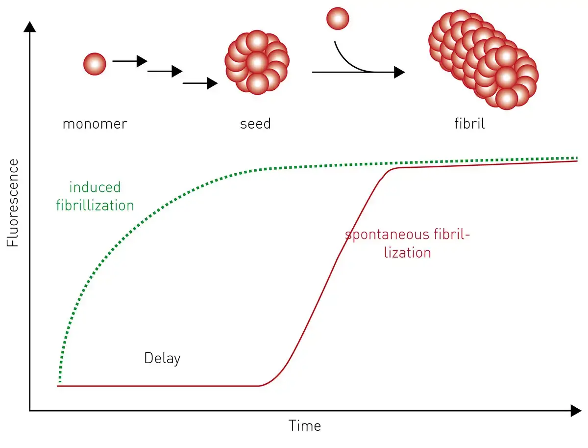
Other dyes such as Congo Red are available that bind to amyloid but show reduced specificity. As part of the RT-QuIC assay’s cyclic amplification principle, cyclic shaking accelerates fragmentation of the growing fibrils and produces a rapid amplification of the original prion seed. In practice, RT-QuIC assays generate characteristic amplification curves that comprise a baseline phase, an exponential increase in fluorescence, and a plateau phase. The lag time, maximum fluorescence, and reaction kinetics can provide quantitative or semi-quantitative measurements of prion seeding activity. The different steps in the RT-QuIC assay are summarized schematically in figure 4.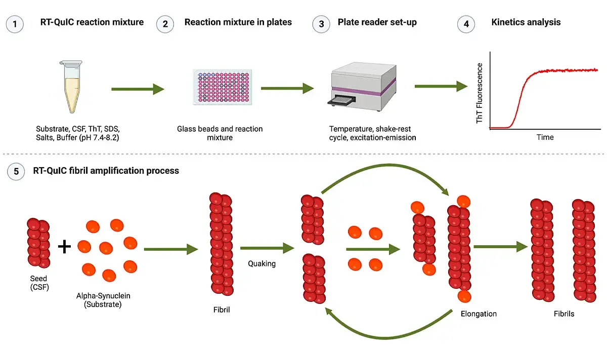
In practice, the RT-QuIC assay measures serial dilutions of samples, which allow the seeding dose to be estimated statistically. In a prion assay, very small amounts of infectious protein are added to normal prion recombinant protein to seed or cause protein misfolding as observed in different neurological diseases. The assay is quantified by measuring serial dilutions of the samples and determining at which point seeding activity is lost (SD50), which is the end point dilution.
In addition to RT-QuIC assays other detection options are available to study different aspects of the molecular interaction of prions. For example, the bioluminescent prion assay is an option to study the dimerization of the prion protein and researchers have also used this approach to see how this type of interaction contributes to pathogenesis.10
Microplate readers offer a robust platform for performing prion assays and measuring protein misfolding and aggregation. The ability to combine sensitive, high-performance kinetic fluorescence measurements with periodic cycles of rigorous shaking and consistent temperature control makes microplate assays one of the most widely adopted and practicable systems for effective RT-QuIC assays. Fluorescence bottom reading measurements can be taken at regular intervals with shaking for up to days at a time.
In addition to robustness and reliability, the high-throughput capabilities and versatility of microplate readers make them well suited for prion analysis whether it’s for basic research, diagnostics or drug discovery (Table 1).
Table 1: Summary of advantages of using microplate readers for RT-QuIC assays.
|
Feature |
Benefit |
| High throughput | Able to process tens to hundreds of samples rapidly saving time and resources |
| Sensitivity & specificity | Capable of detecting trace amounts of prion seeds and low amounts of amyloid improving detection and accuracy |
| Real-time fluorescent monitoring | Track amyloid formation as it happens, providing kinetic data and not just endpoint results |
| Quantitative readouts | Provide fluorescence intensity measurements that permit comparisons of aggregation kinetics and treatment effects across many conditions (multiplexing options) |
You can learn more about the benefits of RT-QuIC assays for amyloid amplification and how these analyses are supported by the capabilities of microplate readers in the scientific talk “Optimizing sensitivity and specificity in the RT- QuIC assay” by Dr. Davin Henderson from Colorado State University.
RT-QuIC assays find many uses in prion studies and for emerging and established investigations of other neurological diseases that involve protein misfolding and amyloid accumulation. A few examples give a sense of the breadth of applications.
In the application note Real-time quaking-induced conversion assay for prion seeding, RT-QuIC assays were used to monitor prion seeding using fluorescence intensity measurements on the FLUOstar Omega microplate reader (Fig. 5). This kind of RT-QuIC analysis also supports biomarker work in human prion disease.
Data are typically generated as kinetic fluorescent plots. RT-QuIC assays were measured every 15 minutes for 20-68 hours while alternately shaking for a minute and resting for a minute. The shaking and resting periods needed to activate the assay were controlled using the MARS software.
The robustness of the FLUOstar Omega reader was demonstrated in this application note by its ability to measure continuously for days at a time without a break in shaking or temperature fluctuations. A Spearman-Kärber analysis was used to estimate the seeding dose or dilution at which 50% of the wells became ThT positive per gram of tested tissue.
This application note highlights not only the stability of BMG LABTECH microplate readers for long-term shaking but also demonstrates how the features of bottom reading, double orbital shaking, temperature control, and analysis using the MARS software contribute to measuring the fluorescence signal of these assays.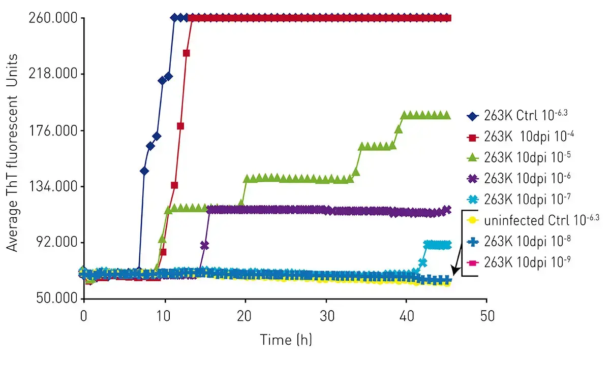
As mentioned earlier, research has demonstrated that RT-QuIC methods can be used with cerebrospinal fluid samples for early detection of prions. In some cases, RT-QuIC assays have been used to measure prion seeding activity in skin samples with 94% sensitivity.6
Researchers are also looking at ways to use RT-QuIC assays for other uses such as disease surveillance in populations of prion-infected animals in the wild.
In the paper “RT-QuIC detection of CWD prion seeding activity in white-tailed deer muscle tissues” scientists measured prion-seeding activity in tissue samples from wild and farmed deer that may have been exposed to CWD.11 Plates were shaken on BMG LABTECH FLUOstar Omega microplate readers at 700 rpm (57 s double orbital shaking followed by 83 s resting). Fluorescence measurements were recorded after 21 shake/rest cycles at three different temperatures (45 °C, 50 °C, and 55 °C; the highest temperature setting was for investigating whether decomposing tissues would converge at specific dilutions).
Measurements of this type allow scientists to make side-by-side comparisons of rates of amyloid formation in different tissues of animal populations (Fig.6). While these methods are still under development, these types of data could in time be useful for decision making for interventions related to the safety of the food chain.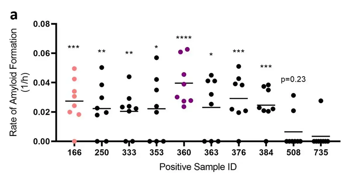 Increasingly, scientists are integrating artificial intelligence-based methods into their discovery-related research. The paper “Discovery of potent inhibitors of α-synuclein aggregation using structure-based iterative learning” 12 describes a machine learning approach to identify small molecule inhibitors of alpha-synuclein aggregation, a process thought to be involved in Parkinson’s disease and other synucleinopathies.
In this study, researchers used RT-QuIC to test the ability of selected small molecules to slow the aggregation of alpha-synuclein induced by brain tissue samples from patients. The machine learning method involved chemical libraries containing millions of compounds that were screened computationally to identify small molecules that bind to the amyloid aggregates and block their proliferation. A small number of top-ranking compounds were then tested experimentally in RT-QuIC assays to select the most potent inhibitors of aggregation. The information gained from these experimental assays was fed back into the machine learning model in an iterative manner to identify highly potent compounds.
The alpha-synuclein (αSyn) RT-QuIC protocol involved measuring ThT fluorescence at 45-minute intervals through the bottom of the microplate on an Omega series reader at 37°C. The results revealed that this approach could identify compounds two orders of magnitude more potent than previously reported compounds.
Demand for RT-QuIC assays is poised to increase as researchers tackle the many unmet medical needs in neurological disease linked to protein misfolding and protein aggregation.
In the paper “A same day α-synuclein RT-QuIC seed amplification assay for synucleinopathy biospecimens” 13, researchers reported the development of a faster α-synuclein RT-QuIC assay that is as analytically sensitive as previous assays. Influential factors in optimizing the assay speed and performance included temperature, salts, the use of beads, shaking speed, and the presence of detergents. The reported assay can be completed in ≤12 h for samples from different tissues (in this case brain, skin, and intestinal mucosa). Samples from cerebrospinal fluid took a few hours longer. This study was performed on an FLUOstar Omega reader. Such increases in speed that offer same-day analysis improve the practicality, cost-effectiveness, and throughput of measurements of pathological forms of α-synuclein for fundamental research, clinical diagnosis, and therapeutics development for diseases like Parkinson’s and other synucleinopathies. The limited number of biospecimens used in the study provide an initial proof-of-principle for future applications. However, the researchers emphasized that testing on much larger and more complex sample sets is required to better establish the assay’s utility in further applications.
The olfactory mucosa has also been explored as a minimally invasive sample in research studies of human prion disease.7 These advances may support earlier diagnosis of disease and strengthen studies looking to use surrogate biomarkers such as tau protein and phosphorylated tau for the direct evaluation of disease-related prions.14
Beyond diagnostics and surveillance14,15, RT-QuIC is also relevant across other protein misfolding diseases. Emerging evidence suggests prion-like propagation by other protein species in disorders such as multiple system atrophy.
In clinical trials, translatable biomarkers are needed that track the progression and severity of the neurodegenerative diseases impacted by protein misfolding and aggregation, and assay refinement may also improve differentiation across molecular subtypes and different neuropathologies, including genetic human prion diseases and fatal familial insomnia.
Progress in artificial intelligence, machine learning and modeling techniques will enhance studies of protein folding and misfolding which should also bring further advances to the drug discovery space.
More advanced detection technologies and innovation in specific RT-QuIC assays should translate into further benefits for researchers.
BMG LABTECH readers, including the FLUOstar Omega and VANTAstar®, continue to lead the way for prion detection and activity measurements, offer a robust platform for shaking for extended periods of time (over multiple days), while periodically reading the fluorescent signal as ThT is bound to amyloid.
You can read more about prions, the link between misfolded proteins and neurological diseases, the use of microplate readers and the different offerings from BMG LABTECH in the following blog articles:
Flexible microplate reader with simplified workflows
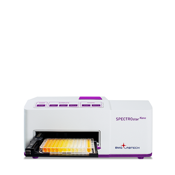
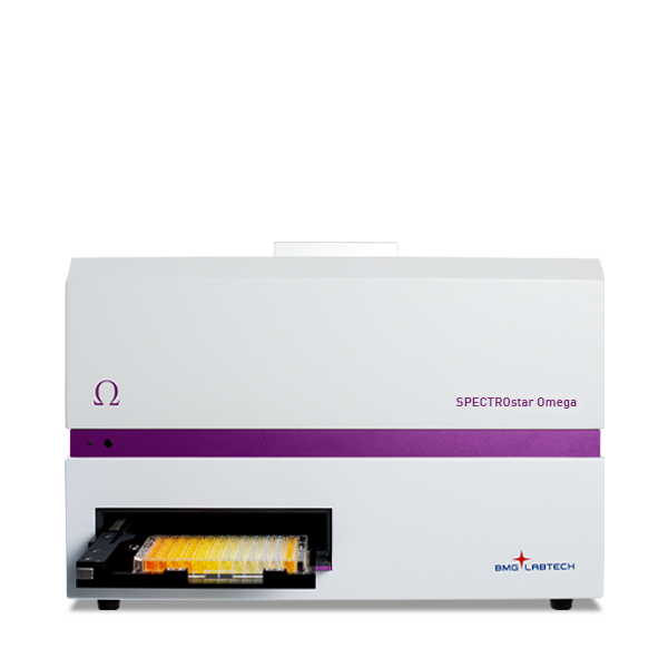
Upgradeable single and multi-mode microplate reader series
The fluorescent dye Thioflavin T (ThT) is widely used to study amyloid proteins. Explore the uses of microplate readers to support ThT assays for the study of protein aggregates.
Neurodegenerative disease ultimately leads to the death of neurons as neuronal functions deteriorate. Find out how microplate readers can be used to study neuronal cell death and its link to neurodegenerative disease.
The disruption of mitochondrial function is linked to neurodegenerative diseases. Find out how microplate readers advance research into mitochondrial dysfunction and neurodegenerative diseases.
The way proteins misfold and aggregate is linked to many neurodegenerative diseases. Find out how microplate readers can help advance research into protein misfolding.
Alpha-synuclein is a key protein involved in neurodegenerative diseases like Parkinson’s. Find out how microplate readers can help advance alpha-synuclein research.