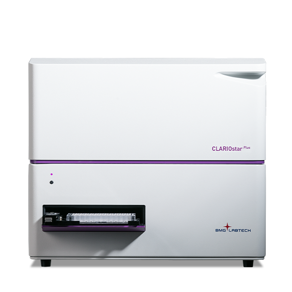
CLARIOstar Plus
Most flexible Plate Reader for Assay Development





Barry Whyte is Application Scientist and Science Writer at BMG LABTECH in the United States. He has PhD and Bachelor of Science (BSc) degrees in biochemistry from the University of Bristol in the United Kingdom and more than 20 years of experience in the life sciences and science communications. Over the years, Barry has worked on three continents and traveled widely. He enjoys building on his international work experience and learning new ways to help scientists advance their research.

Thioflavin T (ThT) is a small, fluorescent molecule (a benzothiazole dye) that binds quantitatively to sites in the grooves created by beta-sheet-rich structures found in some aggregated proteins (Fig.1). ThT typically generates increased fluorescence when it binds to these sites on cross beta-sheet-rich proteins. This type of interaction occurs for proteins like amyloids that are thought to play a crucial role in many diseases.
ThT assays find widespread use for many areas of research linked to neurodegeneration including mitochondrial dysfunction, neuronal cell death and prion research where it is often the fluorophore of choice for detailed, robust analysis of amyloids. Alzheimer’s disease is linked with the presence of two proteins in the brain: beta-amyloid and tau protein. Parkinson’s disease involves the accumulation of the alpha-synuclein protein in the brain.
The utility of ThT to study aggregated proteins means that it can also be used for other applications not related specifically to neurodegeneration. Emerging opportunities are arising to investigate the impact of protein misfolding and aggregation on other disease areas and in basic research where amyloids play a role.
This article describes the ThT assay, gives a few examples of applications, and highlights the technological features of a microplate reader that help deliver optimal performance for ThT assays.
For more information on the use of ThT in prion assays see the companion blog Real-time quaking-induced conversion (RT-QuIC) assays.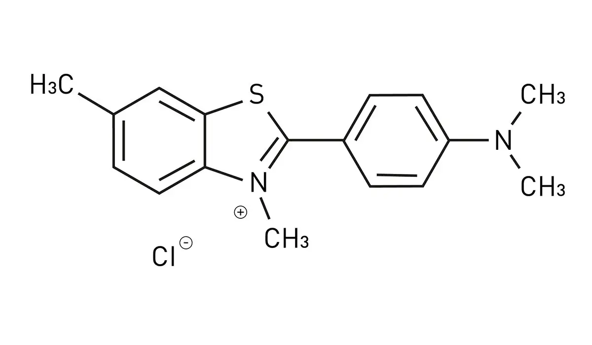
Thioflavin T (ThT) was first synthesized in 1959 and found initial use as a histological stain for amyloid protein aggregates in tissues. One of the first applications for detecting and quantifying amyloid fibrils in vitro was reported thirty years later when a team of researchers in Japan described its use to quantitate amyloid fibrils in mouse tissue.1
In their paper, the spectral changes that take place upon ThT binding to amyloid fibrils and the increase in the fluorescence emission that occurs at specific wavelengths due to protein binding by ThT were described. A few years later, LeVine demonstrated the use of ThT to monitor synthetic beta-amyloid aggregation of fibril peptides in solution.2
The early spectral observations described by Takeda and colleagues and the work of LeVine were vital for the development and proliferation of the quantitative, real-time kinetic assays for ThT in use in laboratories around the world today.
When ThT binds to aggregated amyloid fibrils, it displays a dramatic shift of the fluorescence excitation maximum (from 385 nm to 450 nm) and the emission maximum (from 445 nm to 482 nm), with binding reported in the sub-micromolar Kd range. This ThT fluorescence originates from the dye bound to amyloid fibrils, and the shifted emission spectrum is a useful readout of fibril formation.1,2
The several orders of magnitude increase in ThT fluorescence intensity upon binding of the fluorophore to amyloid fibrils makes it an unusually sensitive and efficient reporter, enabling real-time, in-solution observation of the fibrillization process, although signal is also affected by experimental conditions such as neutral pH and ionic strength.
ThT concentration should also be controlled carefully, as ThT has a critical micellar concentration of about 4 μM.
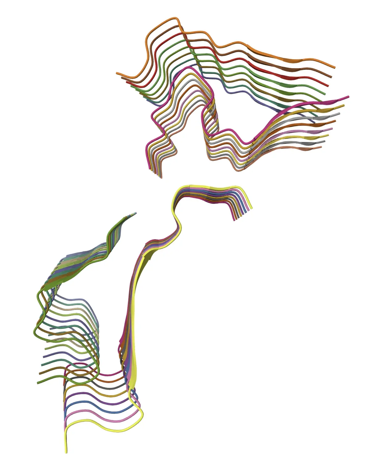
Beta-amyloid 40 fibrils are a good example of an oligomeric protein rich in grooves created by beta-sheet-rich structures that can bind ThT.3 In the three-dimensional structure of a beta-amyloid 40 fibril shown in figure 2, the regularity of the peptide molecules stacking into highly ordered beta-sheet structures is evident. Beta-amyloid 40 fibrils have the characteristic cross-beta sheet architecture. The beta strands run approximately perpendicular to the fibril axis, while each β sheet layer aligns parallel to the fibril axis and helps generate repetitive grooves and channels on the fibril surface.
This architecture helps explain the ThT binding site: surface ridges and channels created by specific amino acids, including hydrophobic residues, support ThT binding, and these structural features are central to how amyloid structures are recognized. In practice, ThT molecules act as a molecular rotor, so restricted rotation upon binding increases fluorescence, and the exact binding mode depends on the local amyloid fibril structure.
Precursor amyloid fibril structures such as oligomers and protofibrils can produce variable signals because oligomeric species and other aggregate species often show low fluorescence emission, and the ThT assay cannot reliably distinguish between different aggregate species.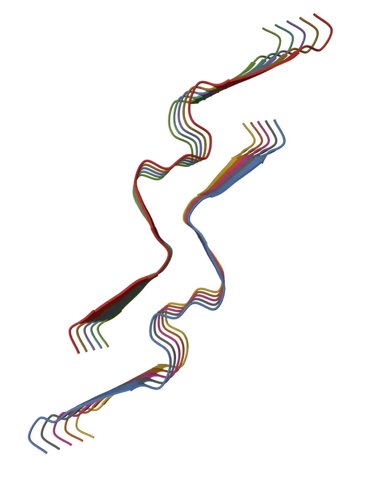
The grooves are crucial for ThT binding since they offer repetitive sites for interaction (this is shown schematically in Fig .3 for alpha-synuclein fibrils; alpha-synuclein is a small, 140-amino-acid protein found in nerve cells that can form amyloid fibrils and is linked to Alzheimer’s disease). When ThT binds to an amyloid structure, its rotational freedom becomes restricted which produces the large increase in fluorescence. 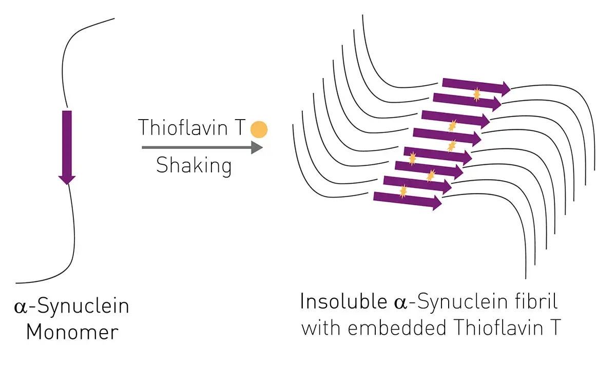
Regular globular proteins generally produce little or no ThT fluorescence because they lack the repetitive binding sites that allow strong interactions with ThT. Some misaggregated or misfolded proteins can bind ThT but misfolding alone is not always a prerequisite for ThT binding and increased fluorescence. As before, amyloid fibrils exhibit strong ThT binding because the fibrils contain the ordered cross-beta-sheet architecture that provides repetitive binding environments for ThT. Precursor amyloid fibril structures such as oligomers and protofibrils can produce ThT fluorescence but this is less consistent than for fully formed fibrils.4-6
Microplate readers offer several key advantages for the performance of ThT assays (Table 1). In addition to throughput, sensitivity, robustness and reliability, they offer versatility for different applications.
Table 1. Summary of benefits offered by microplate readers for metabolic flux measurements.
|
Feature |
Benefit |
| High throughput | Process tens to hundreds of samples simultaneously supporting applications like compound screening, studies of protein variants, or disease-related samples |
| Sensitivity | Detect trace amounts of mature amyloid fibrils improving detection and accuracy |
| Real-time fluorescent kinetic measurements | Track amyloid formation as it happens, allowing analysis of lag phase, fibril growth rate, and aggregation kinetics |
| Temperature control | Maintain defined assay temperatures which are important for protein aggregation studies and their fluorescence measurements |
| Shaking options | Provide a robust system for shaking options often essential for ThT-related assays |
| Reproducibility | Automated shaking and temperature control reduce variability between wells and experiments |
| Quantitative readouts | Provide fluorescence intensity measurements that permit comparisons of aggregation kinetics and treatment effects across many conditions (multiplexing options) |
The main mode of measurement for ThT assays is fluorescence intensity detection but as we will see later options exist for fluorescence polarization measurements with alternative fluorophores that may be applicable for some specific uses. In screening workflows, however, ThT fluorescence can be biased by exogenous compounds, including small molecules, so confirmatory data analysis is important.
ThT assays remain the method of choice for looking at amyloid fibrillization and aggregation.
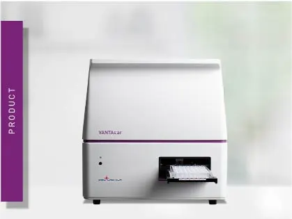
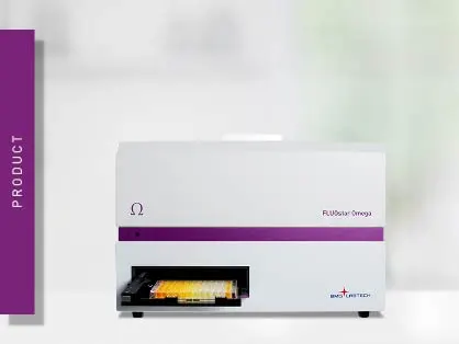
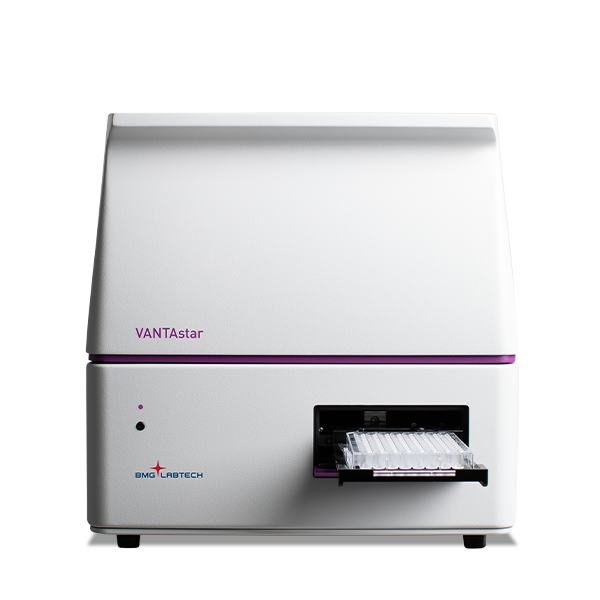
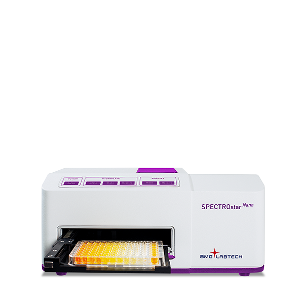
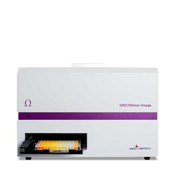
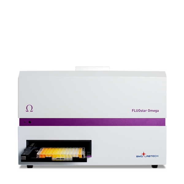
The FLUOstar® Omega microplate reader is the ideal choice for researchers looking to focus on ThT assays as an application for their work. Their advanced functions for shaking and temperature control are combined into a robust system suitable for fast, higher throughput assays for the monitoring of amyloid aggregation. Fluorescence bottom reading measurements can be taken at regular intervals with shaking for up to days at a time. Both the VANTAstar® and CLARIOstar Plus also offer features suitable for ThT assays.
ThT assays with fluorescence intensity measurements to look at amyloid aggregation can be measured in real time. Here we provide a few examples of ThT assays to show how microplate readers can be used in practice.
In the BMG LABTECH application note “Following Abeta fibrillization/aggregation in real-time using a FLUOstar Omega microplate reader” the accumulation of misfolded amyloid protein aggregates linked to Alzheimer’s disease is measured using ThT.
In this assay and application, the fibrillization process can be followed over time. Amyloid fibril formation proceeds through a nucleation-dependent polymerization reaction, so before aggregation or fibrillization can start a critical amount of initial aggregation seeds needs to be present or spontaneously formed (Fig. 4). Once enough seeds have formed a massive and steep increase of fibrillization can be monitored by following the increase of ThT fluorescence due to incorporation into newly formed fibrils. After some time, a plateau is reached indicating the end of the reaction. 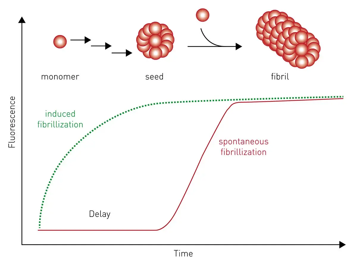
Figure 5 shows some signal curves for samples and controls where ThT incorporation into newly formed amyloid-beta fibrils is measured. The results were obtained with the FLUOstar Omega microplate reader and the long-term shaking capability of the microplate reader was used to monitor amyloid-beta aggregation. The lag times derived from signal curves proved to be a useful measure for the fibrillization process.
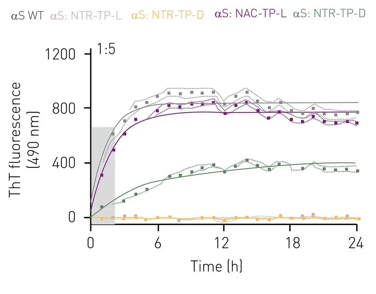
The application note Peptidic inhibitors of α-Synuclein preventing Parkinson’s disease-associated fibrilization and cytotoxicity describes experiments where ThT assays were used in inhibitor studies of alpha-synuclein aggregation in fibril nucleation and elongation assays. The FLUOstar Omega microplate readers used in the study are remarkably well equipped to withstand the harsh conditions required to run the ThT assays. Figure 6 shows kinetic ThT fluorescence measurements for fibril elongation assays in the presence of different inhibitors which were screened for their ability to impact the fibrillization process.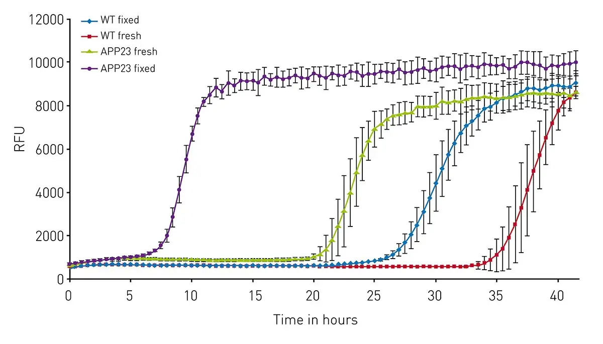 Fluorescence measurements with ThT while widely used are not the only options available to researchers to study fibril formation. The application note Novel aggregation-specific fluorogen monitors prefibrillar protein aggregation by fluorescence polarisation (FP) describes the use of plane polarized light and a novel aggregation-specific fluorogen [bis(triphenylphosphonium) tetraphenylethene or TPE-TPP] to monitor prefibrillar species of amyloid proteins. As mentioned earlier ThT works best with later-stage or fully formed fibrils. TPE-TPP-based fluorescence polarization measurements were made on a CLARIOstar microplate reader offering temperature control to 65° C, heavy duty shaking, and high sensitivity detection.
ThT readouts are sometimes validated with orthogonal structural methods such as electron microscopy or atomic force microscopy..
In the previous section, we saw how ThT assays can be used to study neurodegenerative diseases like Alzheimer’s disease. In principle ThT can be used to study any disease process linked to amyloid accumulation and protein aggregation.
Scientists are beginning to explore in more detail how such events contribute more broadly to other diseases including those linked to areas like metabolism. In this context, ThT assays can be used as part of studies to improve existing interventions for diseases like diabetes. The protein insulin is responsible for maintaining normal glucose levels in part by activating the insulin receptor and is the primary treatment for diabetes. However, insulin is prone to unfolding and forming cross-beta fibers (Fig. 7). Fibrillization is known to complicate insulin storage and therapeutic applications. The molecular details of the insulin fibrillization process remain to be established, which hinders efforts to prevent the fibrillation process.
In the paper “Structural basis of insulin fibrillation” researchers characterized insulin mutants that displayed reduced fibrillization while maintaining native insulin receptor signaling activity. As part of the study, insulin point mutants resistant to fibrillization but retaining signaling activity were produced. ThT assays were used as a measurement tool to confirm that the generated point mutants had lost the ability to form fibrils.7 ThT assays were performed on a BMG LABTECH FLUOstar Omega microplate reader at 37°C. The fluorescence signals were scanned every hour with excitation filters at 430/10 nm (center wavelength/bandwidth) and emission filter at 475/30 nm (center wavelength/bandwidth). This is just one example of new application options for ThT assays.
The future use of ThT assays will extend beyond measuring amyloid formation towards understanding the mechanism of different diseases, identifying drug targets, and developing more sensitive approaches to detect protein fibril aggregation. In neurodegenerative disease ThT assays will continue to be an assay of choice to support research on amyloid-beta in Alzheimer’s disease, alpha-synuclein in Parkinson’s disease and other synucleinopathies, as well as misfolded prion proteins in prion-related diseases. ThT-based approaches will also contribute to drug discovery efforts for screening compounds that inhibit fibrillization, alter aggregation pathways or promote disaggregation of amyloid fibrils. Future applications will increasingly combine ThT assays with Real-Time Quaking-Induced Conversion (RT-QuIC) or other seeding assays.
Most flexible Plate Reader for Assay Development
Flexible microplate reader with simplified workflows
Upgradeable single and multi-mode microplate reader series
Binding constants quantify the strength of a binding reaction between a biomolecule and its target (ligand). But how do you measure them and what can you do with them?
Receptor-ligand kinetics is the study of the rates at which receptors and ligands interact, bind and dissociate. Learn why these types of measurements are important and how to measure them.
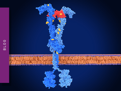
Receptor-ligand interactions are crucial for cell signalling. They are also important for drug discovery. How do microplate readers deliver benefits to both?
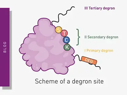
Degrons are specific sequences of amino acids or structural motifs within a protein that are important for targeted protein degradation. Find out how microplate readers can advance research into natural and engineered degrons.
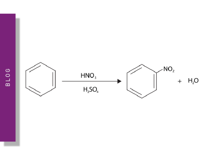
Nitration can significantly modify the structure and function of proteins in cells. But how can measurements of protein nitration reveal crucial information about biological activity and how can microplate readers help?