Introduction
Bacterial endotoxins, also known as lipopolysaccharides (LPS), are potent pyrogens found in the outer membrane of Gram-negative bacteria.1,2 Their presence in pharmaceuticals, medical devices, and biological products can trigger severe inflammatory reactions in our immune system. These inflammatory reactions may lead to serious adverse events and compromise health. Therefore, the detection and quantification of endotoxins are crucial for ensuring product safety and regulatory compliance for food, medical, and other products.
The Limulus Amebocyte Lysate (LAL) assay is a widely used method for detecting bacterial endotoxins.3-5 Two types of horseshoe crab have been used as sources of LAL: Limulus polyphemus from the North Atlantic and Tachypleus spp. from Asia. The LAL assay exploits the natural clotting response of blood cells (amebocytes) isolated from these animals to endotoxins. When exposed to endotoxins, LAL undergoes a series of enzymatic reactions that result in gel formation, which can be measured to determine the presence and concentration of endotoxins.
The PYROSTAR ES-F/Plate LAL test is sensitive, robust, suitable for small sample volumes, and is not activated by β-glucans which can lead to false positive results. It therefore ensures that products are free from harmful levels of endotoxins, safeguarding patient health and meeting stringent regulatory standards.
Assay principle
The PYROSTAR ES-F/Plate kit is based on the classic LAL assay principle. In the presence of endotoxins, the LAL responds with a clotting reaction. Clotted LAL can be detected on a microplate reader in two ways which both depend on light scattering but use different types of readouts. The LAL assay can either be performed with a light-scattering readout on a nephelometry-based microplate reader or with an turbidimetric readout on a spectrometer-based absorbance microplate reader (Figure 1). In a nephelometer, scattered light is measured due to detection taking place at an angle to the incident light. If an absorbance-based microplate reader is used, light that passes through the sample is measured - so all light that isn’t scattered. A reference measurement is used to determine the total light produced by the spectrometer, which allows an indirect statement to be made about the amount of light scattered by the sample.

Materials & methods
- 96-well plates, clear, polystyrene (Greiner, 655101)
- PYROSTAR ES-F/Plate kit (Wako, 543-10331)
- Endotoxin-free water
- NEPHELOstar Plus (BMG LABTECH)
- VANTAstar (BMG LABTECH)
Experimental Procedure
1,000 EU/mL control standard endotoxin stock was prepared in endotoxin-free water. Using the stock solution, a 10-fold dilution series was prepared with endotoxin-free water according to the kit protocol (0.001 to 100 EU/mL). A vial of PY-ROSTAR ES-F reagent containing the LAL was dissolved in 2 mL of endotoxin-free water. 50 µL of the endotoxin dilution series was pipetted into a clear 96-well microplate. The detection reaction was started by adding 50 µL of PYROSTAR ES-F reagent and the plate was immediately read on the NEPHELOstar Plus or VANTAstar for 60 min at 37°C.
Instrument Settings
|
Nephelometry, plate mode
|
||
|
Optic settings |
Laser intensity |
80 % |
|
Kinetic settings |
Number of cycles |
90 |
|
Cycle time |
40 s |
|
|
Incubation |
Target Temperature |
37 °C |
|
Absorbance, plate mode
|
||
|
Optic settings |
Spectrometer |
405 nm |
|
General settings |
Flashes |
22 |
|
Kinetic settings |
Number of cycles |
90 |
|
Cycle time |
40 s |
|
|
Incubation |
Target Temperature |
37 °C |
Here you will find the protocol files for BMG LABTECH’s Smart Control Software (CLARIOstar Plus and VANTAstar) and the Omega Software (NEPHELOstar Plus), the used plate layout and the corresponding templates for automatic evaluation of generated data in MARS. You can download these files from the overview page, import them into your BMG LABTECH software and use them to evaluate your analysis.
Results & Discussion
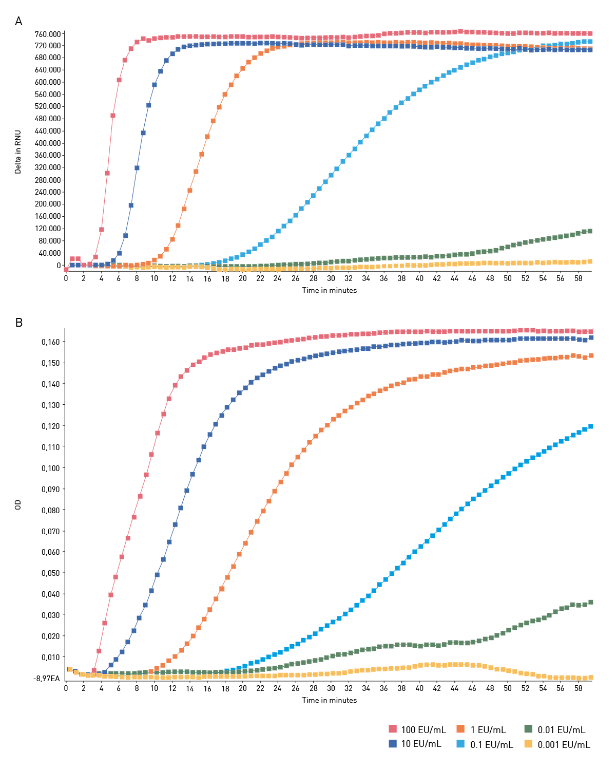
Figure 2 shows kinetic data of the PYROSTAR ES-F/Plate kit. Samples containing endotoxin displayed an increase in assay signal over time. Higher endotoxin concentrations led to a faster increase in measurement signal until the signals reached a maximum compared to samples with lower endotoxin concentrations. The nephelometric readout (figure 2A) and turbidimetric readout (figure 2B) gave comparable results regarding to the separation of endotoxin standards.
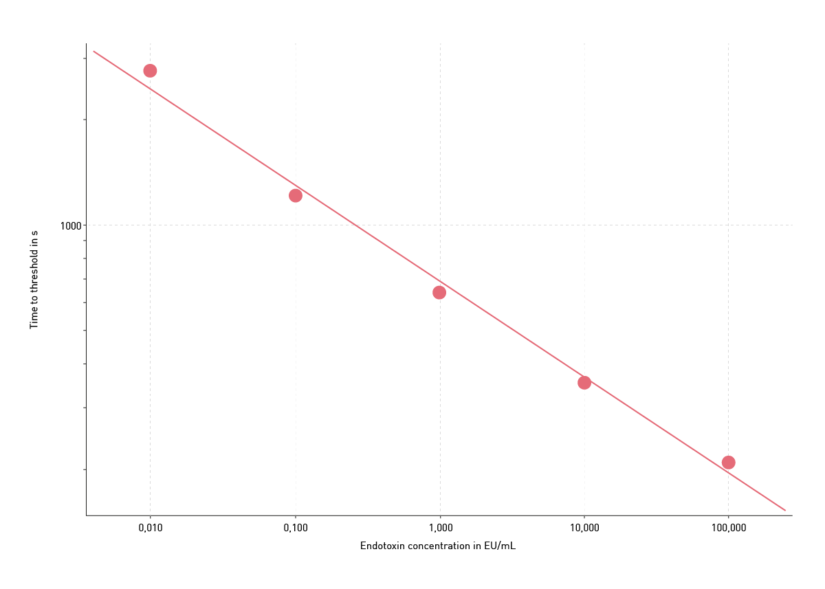
Endotoxin standard samples were used to prepare a standard curve for the LAL assay. For this purpose, first time-to-threshold calculations were performed in the MARS data analysis software. For the nephelometric readout the threshold value was set to 5% of the maximum signal, while a threshold of 0.015 OD was set for the turbidimetric LAL assay. Time-to-threshold values of the endotoxin standards were then fitted against the standard concentrations using a logarithmic linear regression fit (figure 3).
Conclusion
The PYROSTAR ES-F/Plate LAL assay could be performed with both detection modes. Both the nephelometric and the turbidimetric readout yielded comparable results with the same number of endotoxin standards, although the use of the laser-equipped NEPHELOstar Plus slightly improved overall data quality. In the end both readers, absorbance- and nephelometry-based, were well suited for measuring the PYROSTAR ES-F/Plate LAL assay.
References
- Pyrogens, Still a Danger | FDA https://www.fda.gov/inspections-compliance-enforcement-and-criminal-investigations/inspection-technical-guides/pyrogens-still-danger Accessed 03/20/2025
- Ding JL, Ho B. Endotoxin detection--from limulus amebocyte lysate to recombinant factor C. Subcell Biochem. 2010;53:187-208. doi: 10.1007/978-90-481-9078-2_9.
- Levin J and Bang FB. The role of endotoxin in the extracellular coagulation of Limulus blood. Bull. Johns Hopkins Hosp. (1964) 115:265.6
- United States Pharmacopeial Convention. Committee of Revision. “The United States Pharmacopeia.” United States Pharmacopeial Convention, Incorporated, 1985. www.usp.org
- European Pharmacopoeia Commission, and European Directorate for the Quality of Medicines & Healthcare. European pharmacopoeia. Council of Europe. www.edqm.eu/en/european-pharmacopoeia