Introduction
Bacterial endotoxins, in particular lipopolysaccharides (LPS), are structural components of the outer membrane of Gram-negative bacteria that can activate the innate immune system in a way that leads to elevated temperature and fever.1,2 Such reactions are potentially life threatening and can be triggered even by trace amounts of endotoxins which is why their detection is crucial for quality assurance in the pharmaceutical and biotechnological in-dustries. These tests are therefore an integral part of the regula-tory requirements for the safety of drugs, medical products and biological therapeutics.
Limulus Amebocyte Lysate (LAL)-based tests are an established and sensitive method for the detection of endotoxins.3,4 These tests are based on the coagulation cascade of amebocyte cells derived from horseshoe crabs (typically Limulus polyphemus but Tachypleus spp. are also used in Asia). This cascade is specifically activated by the presence of LPS triggering an enzymatic reaction that leads to clotting of LAL. Endotoxin assays, which quantify the strength of the coagulation reaction, are used to determine the amount of endotoxin present in a test sample. Over the years, several absorbance-based assays have been developed for this purpose that are still widely used today. In this application note, we highlight two absorbance-based endotoxin quantification assays that rely on distinct measurement principles to monitor a LAL clotting reaction.
Assay principle
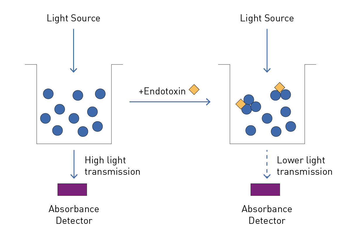
Endotoxin quantification assays with an absorbance-based read-out are typically based on two measurement principles. Both utilize the clotting reaction of LAL in the presence of endotoxin. In each case, different concentrations of endotoxin are prepared and their signals measured. These measurements are used to construct a standard curve which serves as reference for the calculation of endotoxin concentrations of unknown samples. Adding LAL starts the clotting reaction that can be quantified on a microplate reader. One way to do this is to measure the turbidity of the test sample by monitoring the light transmission through a sample. The more clotted LAL is present in the sample, the less light reaches the detector mainly due to light scattering (Figure 1). The PYROSTAR™ ES-F/Plate LAL assay kit can be used to perform a turbidimetric LAL clotting assay.
Other assays, such as the Limulus Color KY Test employ a colorimetric readout for the quantification of endotoxin in a sample. Here, a chromogenic substrate is added to the assay that is cleaved by the activated enzymes of the LAL clotting cascade in the presence of endotoxin. Higher concentrations of endotoxin lead to increased enzymatic activity and higher amounts of converted substrate. In this case, more of the yellow chromogen para-nitroaniline (pNA) is released from the substrate. The increase in light absorption due to the increase in pNA is then quantified by the microplate reader.
Materials & methods
- Clear, 96-well, polystyrene plates (Greiner, 655101)
- PYROSTAR ES-F/Plate LAL assay kit (Wako, 543-10331)
- Limulus Color KY Test (Wako, 291-53101)
- Endotoxin-free water
- SPECTROstar® Nano (BMG LABTECH)
- VANTAstar® (BMG LABTECH)
Experimental Procedure
1,000 EU/mL control standard endotoxin stock was prepared in endotoxin-free water. Using the stock solution, a 10-fold dilution series was prepared with endotoxin-free water according to the kit protocol (0.001 to 100 EU/mL). A vial of PY-ROSTAR ES-F reagent containing the LAL was dissolved in 2 mL of endotoxin-free water. 50 µL of the endotoxin dilution series was pipetted into a clear 96-well microplate. The detection reaction was started by adding 50 µL of PYROSTAR ES-F reagent and the plate was immediately read on the NEPHELOstar Plus or VANTAstar for 60 min at 37°C.
Instrument Settings
|
Absorbance, plate mode
|
||
|
Optic settings |
Spectrometer |
405 nm |
|
General settings |
Flashes |
22 |
|
Kinetic settings |
Number of cycles |
90 |
|
Cycle time |
40 s |
|
|
Incubation |
Target Temperature |
37 °C |
Here you will find the protocol files for BMG LABTECH’s Smart Control Software (CLARIOstar Plus and VANTAstar) and the Omega Software (NEPHELOstar Plus), the used plate layout and the corresponding templates for automatic evaluation of generated data in MARS. You can download these files from the overview page, import them into your BMG LABTECH software and use them to evaluate your analysis.
Results & Discussion
LAL clotting could be quantified with both the colorimetric (Figure 2A) and the turbidimetric assay kits (Figure 2B). The speed of the clotting reaction depended on the endotoxin concentration in the sample, with higher endotoxin concentrations leading to a faster increase in the absorbance units (OD) quantified by the microplate reader. In comparison, the signals derived from the colorimetric assay were significantly higher than those from the turbidimetric assay, but ultimately the lower assay threshold of 0.015 OD was achieved by the same number of standards in both assays.
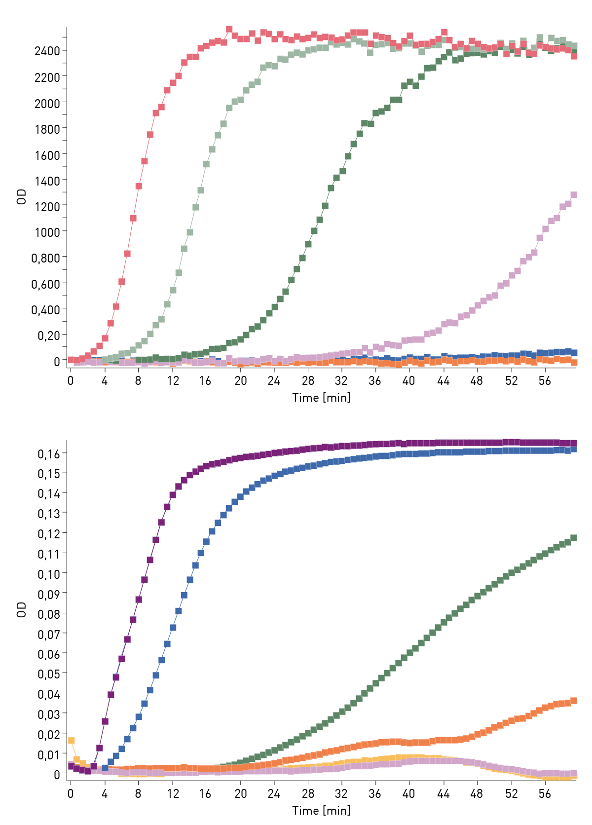
A standard curve was prepared from the standard samples with defined endotoxin concentrations using the MARS data analysis software. The standard curve was used to quantify the concentration of endotoxins in unknown samples. As a first step, the time until the lower assay threshold of 0.015 OD was reached was calculated for each sample with a kinetic calculation. Next, the time to threshold values were calculated from the kinetic test run and plotted against standard endotoxin concentrations. A logarithmic linear regression fit was used to create a standard curve (Figure 3). 
MARS automatically calculates the endotoxin concentrations of unknown samples based on the standard curve. Another advantage of MARS is that once the above calculation steps have been performed manually, they can be saved as a template and applied to future test runs or even assigned to the measurement proto-col itself to automate data evaluation. In addition, performance characteristics such as the coeffi cient of correlation R² are readily available in the standard curve fit results.
Conclusion
Both the Limulus Color KY Test and the PYROSTAR ES-F/Plate LAL assay are equally suited for the determination of endotoxin concentrations. BMG LABTECH microplate readers equipped with absorbance detection are the ideal measurement platform to perform these tests in a controlled and reliable manner. MARS data evaluation is an excellent tool for the complete and effortless evaluation of measurement results.
References
- Schwadner, R. et al. Peptidoglycan- and lipoteichoic acid- induced cell activation is mediated by Toll-like receptor 2. J. Biol. Chem. 274, 17406 17409 (1999).
- Schneier M, Razdan S, Miller AM, Briceno ME, Barua S. Current technologies to endotoxin detection and removal for biopharma- ceutical purifi cation. Biotechnol Bioeng. 2020 Aug;117(8):2588-2609. doi: 10.1002/bit.27362. Epub 2020 May 16.
- Levin J and Bang FB. The role of endotoxin in the extracellular coagulation of Limulus blood. Bull. Johns Hopkins Hosp. (1964) 115:265.6
- Levin J, Bang FB. Clottable protein in Limulus: Its localization and kinetics of its coagulation by endotoxin. Thromb. Diathes. Haemorrh. (Stuttg) (1968) 19:186.