Introduction
The quantification of viable cells in culture is a very important problem. However, the technique becomes much more valuable when the effect of growth regulatory substances and also cytotoxic agents are under study. Measurement of viable cell numbers in culture is a critical aspect in the functional in vitro bioassay of a wide range of growth factors. The nucleotide adenosine triphosphate (ATP) plays an important role in energy exchange in biological systems. It serves as the principal immediate donor of energy and is present in all metabolically active cells. ATP has been used as a tool for the functional integrity of living cells since all cells require ATP to remain alive and carry out their specialized function. Most ATP is found within living cells and links catabolic and anabolic processes. Cell injury or oxygen/substrate depletion results in a rapid decrease in cytoplasmatic ATP. Measurement of ATP is therefore fundamental to the study of living processes. A lot of methods have been used for ATP determination, but far the most successful technique is the bioluminescent method, because of its sensitivity and the wide dynamic range. ATP bioluminescence has been used for determining levels of ATP in a number of different cell types.
Assay Principle
The reaction is catalyzed by the enzyme luciferase obtained from the firefly (Photinus pyralis). The MgATP2- converts the luciferin into a form which is capable of being catalytically oxidized by the luciferase in a high quantum yield chemiluminescent reaction, according to the following equation:

Under optimum conditions, light intensity is lineary related to ATP concentration. Cellular ATP can be measured by direct lysis of the cells with suitable detergent, the released ATP is then free to react with the luciferin-luciferase and leading to light emission at 562 nm.
In this study ATP bioluminescence was used to determine whether there is a linear relationship between the number of HaCaT cells present in the culture and measured luminescence. It was attempted to use ATP-bioluminescence as a measure of proliferation and/or cell cytotoxicity depending of two different substances and avoid the use of radio-isotopes. These investigations have been carried out using the keratinocyte cell line HaCaT.
Materials & Methods
Reagents and instrumentation
HaCaT cells, propagated in 75 square cm tissue culture Hasks with weekly passage in DMEM (Seromed, FG 0435, Biochrom KG, Germany) containing 10% fetal calf serum and 1% antibiotic/antimycotic solution at 37°C and 6% CO2. Cells were seeded in microtitre plates (Greiner 96-well plates) at a density of 5000 cells.
One day after plating in the serum-free medium (Gibco- Defined Keratinocyte-SFM), in SFM supplemented hyaluronic acid, hyalogran or medium was added over a period of 24h or 48h later, cell proliferation was measured.
ATP bioluminescence
ATP releasing agent (Somalyze), Tris-acetate buffer, ATP-monitoring reagent (firefly luciferase, D-luciferin) and ATP-standard (EG+G. Wallac, Turku, Finland) are needed for the assay.
100 µL of ATP releasing agent was added to each well of a 96 well microtitre tissue culture plate (Greiner, Germany), containing 100 µL of HaCaT cells, and incubated for 5 minutes at room temperature. Then 180 µL of the cell lysate were added to wells of a 96 well white opaque microtitre plate.
This plate was loaded into a BMG LABTECH microplate reader and 20 µL of the luciferin-luciferase reagent (reconstituted with 10 mL of 0.01.M Tris-acetate buffer, 2 nM EDTA buffer ph 7.75) were added to each well by means of an injection pump, and the sample were measured.
Immediately after addition of luciferin-luciferase luminescence was monitored over a period of 10 s (Fast kinetic method). Light output was given as the integral relative light units (RLUs). ATP measurements were carried out at room temperature.
Results & Discussion
Cell numbers and the degree of cell proliferation/cytotoxicity were determinded by ATP bioluminescence and in comparison to a conventional method, the staining with dye Hoechst 33342.
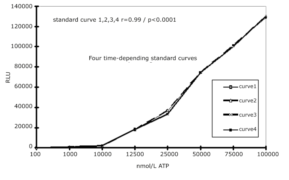
HaCaT cells were initially used to determine whether there was a correlation between cell numbers and measured ATP. The standard curve of ATP was determinded in several groups of experiments (Fig. 1) (r = 0.998; p< 0.0001). From figure 2 it can be seen that after incubation of increasing cell numbers with ATP releasing agent (100 µL) for 5 min at room temperature an increase of released ATP (r = 0.99; p< 0.0001) can be observed.
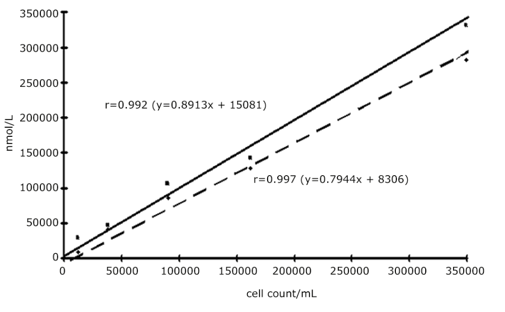 The correlation was determined from comparison of cell numbers with ATP-bioluminescence readings for each data point and Spearman’s rank correlation was determined for the group as whole. Complete medium cultured in the absence of cells served as a control and contained no detectable ATP. Incubation with 100 µL releasing agent for 5 min at room temperature was optimal for determining the amount of ATP in cultured HaCaT cells. In figure 3 and 4 the correlation between the cell numbers and the ATP concentrations of several experiments were shown after 24h and 48h, respectively.
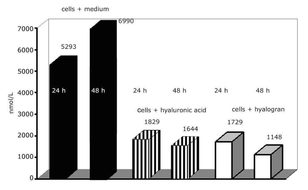
The data show that with HaCaT cell line tested there were significant correlation between increased cell number and ATP measured by the luciferin-luciferase reaction. In order to show that ATP is an indicator of proliferation and/or cell cytotoxicity the assay was carried out under addition of two different substances. These substanced are hyaluronic acid and hyalogran. Hyaluronic acid (HA) is a major component of extra-cellular matrix, distibuted ubiquitously, with the highest concentration in the soft connective tissue.
Especially the influence of hyaluronic acid is in good agreement with the fact that high molecular weight HA in high concentrations inhibits movement, adherence and phagocytosis of several cell types, while low molecular weight HA at low concentrations stimulates cellular migration, proliferation and phagocytosis.
Conclusion
This study confirmed that the concentration of ATP in the HaCaT cell line can be determined by luciferin- luciferase bioluminescence assay, and also that the monitoring of ATP bioluminescence can be used for the measurement of proliferation and/or cytotoxicity.