CLARIOstar Plus
Most flexible Plate Reader for Assay Development

Important and relevant parameters include temperature, humidity, CO2 and O2, which differ depending on the location of the tissue and its access to blood supply. Fortunately, these are all parameters that can now be measured and controlled in the laboratory for in vitro cell culture.
Historically, primary cells and transformed cell lines were cultured in 5% CO2 incubators at 37ºC, 85-90 % humidity and gassed with room air irrespective of the origin of the cells. In the human body, cells are exposed to 37ºC and notably at lower temperatures metabolism reduces rapidly, thus there is a requirement for cell-based assays to be conducted under stable, physiological temperature. CO2 tension of 5% is used to maintain the pH in cell culture growth media around pH 7.4, noting that acidification of the media at lower CO2 tension quickly leads to loss of cell viability. Primary cells and cell lines are maintained in media rich nutrients, with a high humidity 85-90 % limiting evaporation from the media and thereby obviating loss of volume that could increase the concentration of salts in the media to potentially lethal levels.
Maintaining cell viability ensures that cells can be studied over long term periods but what if the tissue from where the cells were derived are not normally exposed to atmospheric oxygen? Cells very rarely experience atmospheric oxygen levels, and thus it is questionable whether data generated from cells exposed to atmospheric oxygen levels accurately reflect reactions occurring in tissues in the body? Atmospheric O2 is 21.1% at sea level and O2 levels in the body notably vary significantly depending on the organ/tissue of interest. The table below reproduced from Carreau et al.1 provides an overview of the differences in physiological oxygen levels experienced by different tissues.
| Normal values of oxygen in various human tissues: | ||
| Tissue | mmHg | % O2 |
| Air | 160 | 21.1 |
| Trachea | 150 | 19.7 |
| Alveoli | 110 | 14.5 |
| Arterial Blood | 100 | 13.2 |
| Venous Blood | 40 | 5.3 |
| Cell | 9.9-19 | 1.3-2.5 |
| Mitochondria | <9.9 | <1.3 |
| Brain | 33.8+/-2.6 | 4.4+/-0.3 |
| Lung | 42.8 | 5.6 |
| Skin(sub-papillary plexus) | 35.2+/-8 | 4.6+/-1.1 |
| Skin(dermal-papillae) | 24+/-6.4 | 3.2+/-0.8 |
| Skin(superficial region) | 8+/-3.2 | 1.1+/-0.4 |
| Intestinal Tissue | 57.6+/-2.3 | 7.6+/-0.3 |
| Liver |
40.6+/-5.4 | 5.4+/-0.7 |
| Kidney |
72+/-20 | 9.5+/-2.6 |
| Muscle |
29.2+/-1.8 | 3.8+/-0.2 |
| Bone Marrow |
48.9+/-4.5 | 6.4+/-0.6 |
Clearly when performing assays maintaining all cells at atmospheric oxygen tension does not reflect physiological conditions, which has been recognised and addressed by a numbers of companies manufacturing incubators and workstations.
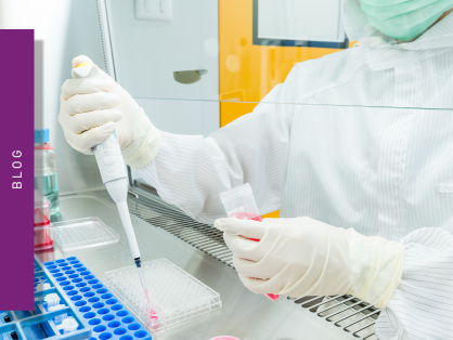
Workstations from companies like Baker-Ruskinn produce workstations providing the ability to adapt cells and perform usual laboratory bench tasks such as cell passage, cell microscopy under closely regulated O2 and CO2 conditions. While incubator manufacturers are introducing not only CO2 regulated incubators but O2 and CO2 controlled incubators to simulate physiological conditions. BMG LABTECH provides the perfect microplate reader in the CLARIOstar Plus with Atmospheric Control to perform either short end point readings or long term kinetic studies over 72 hours while also maintaining CO2 and O2 tension together with 37ºC.
In their recent Physiological Reviews article2 Keeley and Mann provide a comprehensive narrative providing a framework for researchers to follow for culture of cells in vitro under conditions relevant to an organ or tissue. This comprehensive review also highlights multiple publications describing the changes observed in all tissue types including endothelial cells, circulating cells, intestinal cells, bone marrow-derived stem cells when studied under normoxic (physiological oxygen) conditions. The review concludes with a series of guidelines that outline the process for studying tissue culture under defined physiological normoxia in vitro including:
Get further information in our topic-related scientific talk:

Thus study of cell cultures under their physiological conditions should provide more relevant phenotypic data that extrapolates to animal models and beyond. This could ultimately provide insights into disease pathology, pharmacodynamics and increase the speed and efficiency of drug development.
Most flexible Plate Reader for Assay Development
Upgradeable single and multi-mode microplate reader series
Flexible microplate reader with simplified workflows
Learn about applications for bacterial metabolism on a microplate reader.
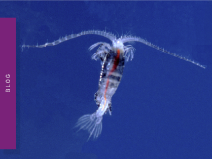
Life in the depths of the ocean operates under extreme conditions. Find out how proteins from deep-sea luminescent organisms are useful for measurements on microplate readers.
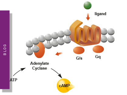
Second messengers play a pivotal role in signal transduction events in cells. But how do you measure these small, transiently lived molecules and how can microplate readers help?
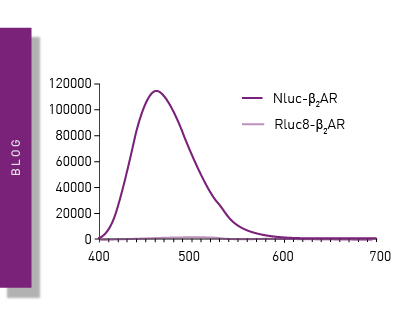
NanoBRET is used to analyse binding events, signaling pathways and receptor trafficking in live cells and has significantly expanded the range and applications of BRET assays.
Find out about the different types of cell-based assays and why they have become an indispensable tool in a such a broad variety of disciplines in this blog article.
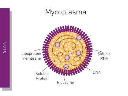
Read here about the various threats associated with mycoplasma contamination and find out about methods to prevent, detect, and eliminate mycoplasma in your cell culture.