PHERAstar FSX
Powerful and most sensitive HTS plate reader


Dr. Martin Mangold works as an Applications Specialist at BMG LABTECH headquarters in Ortenberg, Germany. He studied biology with a focus on biochemistry and cell biology at the University of Bonn before specializing in pharmaceutical sciences and drug interactions in his doctoral studies. During his time in the pharmaceutical department, Dr. Mangold gained expertise in protein sciences, binding and interaction studies, and enzyme kinetics as part of an interdisciplinary team of chemists, pharmacists and biologists. Since 2021, Dr. Mangold has been part of the BMG LABTECH team where he authors application notes, performs training courses and supports scientific customers.
The LanthaScreen™ technology is a detection method based on Time-Resolved Fluorescence (TRF), and in particular on Time-Resolved Förster Resonance Energy Transfer (TR-FRET).
The LanthaScreen technology comprises biochemical and cellular assays. It is used to study kinase and protease activity, ubiquitination, nuclear receptors, compound binding and post-translation modifications (e.g., phosphorylation and acetylation) in cell signalling.
LanthaScreen is mainly used for drug discovery and high-throughput screening (HTS) purposes. The benefit of these assays is the long fluorescence lifetime of the lanthanide donor fluorophore. This enables ratiometric measurement of acceptor emission over the donor emission after the decay of the background fluorescence. TR-FRET assays are less susceptible to compound interference than other assay formats and typically increase data quality. Moreover, it is a homogeneous approach with no washing steps required that simplifies automation.
LanthaScreen assays utilise lanthanide (terbium and europium) chelates as FRET donors. Originally based on europium as a donor fluorophore and Allophycocyanin (APC) or Alexa Fluor647 as an acceptor fluorophore, LanthaScreen was expanded to use terbium and fluorescein or GFP as donor-acceptor pair.
Because of its molecular mass of more than 100 kDaltons, allophycocyanin is typically used as a streptavidin conjugate and is coupled to a biotinylated substrate in a tri-molecular complex.
Terbium as a donor was introduced purposely to enhance flexibility. Its combination with fluorescein and GFP can be used to directly label substrates and hence overcome the steric bulk issues of the europium-APC pair. Additionally, this pair can be employed in cell-based assays, enabling the study of post-translational modifications of a target protein fused to GFP in cells. The modification is analysed after cell lysis and addition of a terbium-labelled antibody specific for the modification of interest. This approach can be for instance used to study the JAK/STAT and NF-κB pathways.
The LanthaScreen kinase activity assay uses a long-lifetime terbium or europium chelate as donor and fluorescein as acceptor. When terbium- and fluorescein-labelled molecules are brought into proximity, energy transfer takes place causing an increase in acceptor and a decrease in donor fluorescence. The TR-FRET value is determined as a ratio of the FRET-specific signal measured with a 520 nm filter to that of the signal measured with a 495 nm filter, which is specific to terbium.
In a LanthaScreen kinase activity assay, a fluorescein-labelled kinase substrate (e.g., a peptide) is typically incubated with the kinase and ATP. A terbium or europium-labelled antibody is then added and phosphorylation is detected by an increase in the TR-FRET value (fig.1). Because the substrate is directly labelled, there is no need to add streptavidin-APC. Additionally, unlike other europium-based systems, there is no requirement to add high concentrations of potassium fluoride, which can potentially disrupt antibody–product interactions.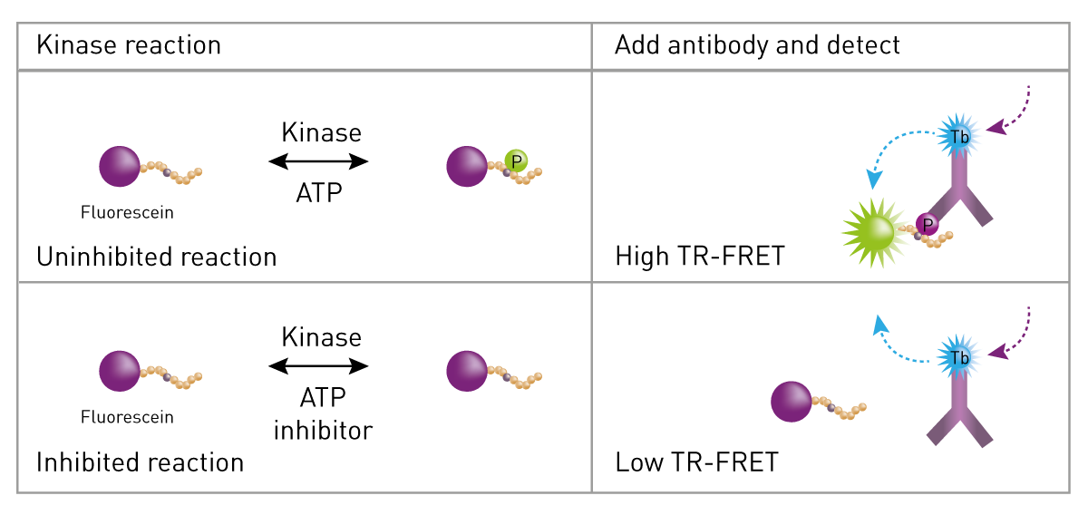
In a LanthaScreen kinase assay, kinase, fluorescein-labelled substrate, and ATP are allowed to react. Then EDTA (to stop the reaction) and terbium-labelled antibody (to detect phosphorylated product) are added. In a LanthaScreen kinase reaction, the antibody associates with the phosphorylated fluorescein labelled substrate resulting in an increased TR-FRET value. The amount of antibody that is bound to the tracer is directly proportional to the amount of phosphorylated substrate present, and in this manner, kinase activity can be detected and measured by an increase in the TR-FRET value.
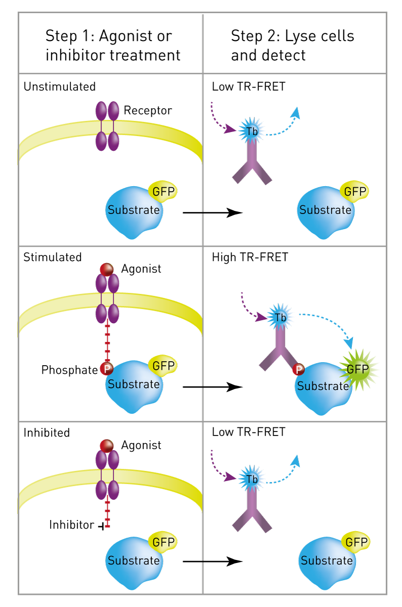
For cellular assays, the principle and methods are similar to the ones mentioned above. The substrate of the kinase to be analysed is expressed in cells as a GFP-fusion protein.
The phosphorylation of the substrate is either stimulated with an agonist to activate the pathway or treated with an inhibitor to interrupt the signalling event. No protein modification occurs in unstimulated cells.
After stimulation, cells are lysed in a buffer that contains a terbium-labelled phosphorylation site-specific antibody. The protein modification event is measured on a TR-FRET-compatible microplate reader. Little or no TR-FRET is observed with unstimulated or inhibited cells whereas stimulated cell samples display high TR-FRET (fig.2).
LanthaScreen assays are principally performed on microplate readers. The basic setup requires the TRF mode. In addition, plate readers have to be capable of measuring two emission channels in one run, either sequentially or simultaneously. Since LanthaScreen is mainly used in HTS applications, plate readers have to be compatible with 384- and 1536-well microplates.
BMG LABTECH plate readers certified for LanthaScreen detection include the PHERAstar FSX, CLARIOstar Plus and VANTAstar.
An example of a LanthaScreen kinase assay run on a BMG LABTECH plate reader can be seen in the application note "LanthaScreen TR-FRET tyrosine kinase and protein kinase C assay".
TR-FRET alternatives to LanthaScreen include LANCE, HTRF and THUNDER.
Powerful and most sensitive HTS plate reader
Most flexible Plate Reader for Assay Development
Flexible microplate reader with simplified workflows
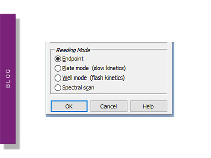
Endpoint and kinetic mode assays are used by scientists to study many processes in the life sciences. Learn about endpoint and kinetic modes on a microplate reader.
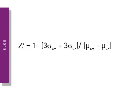
The Z prime value (Z’) is a statistical parameter that can provide practicable information on the quality of an assay. This blog looks at its usage and describes some examples of specific applications.
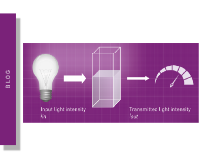
Optical density and absorbance measurements are widely used in the life sciences. This blog looks at practical applications and some of the fundamentals.
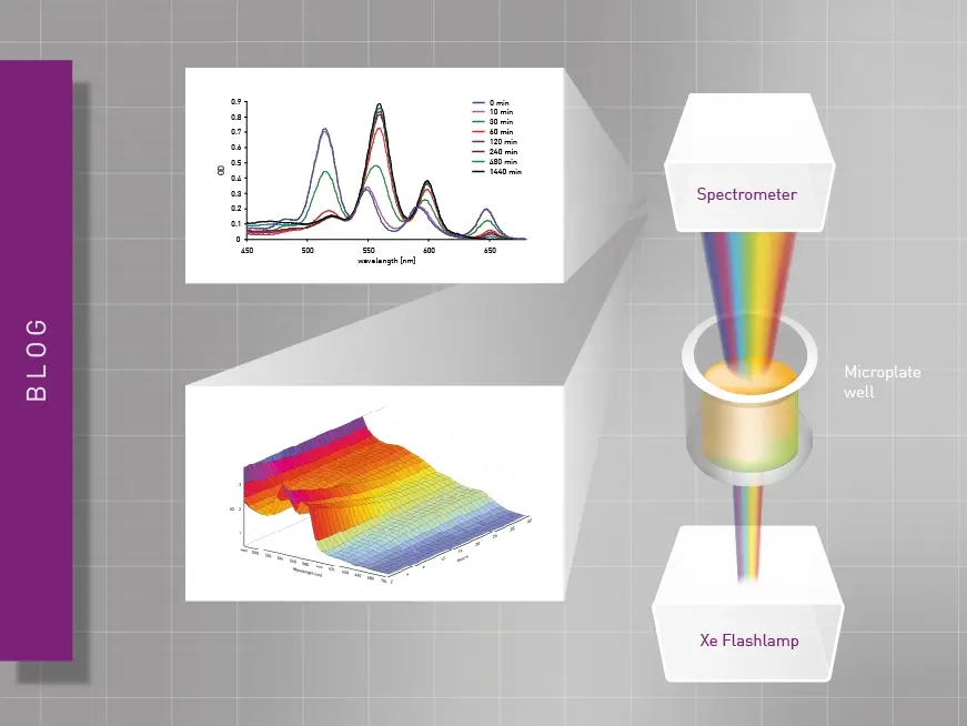
Researchers have different technology options available for absorbance measurements. This blog compares spectrometers and monochromators. What’s the difference?
ELISAs are a popular tool to detect or measure biological molecules in the life sciences. Find out how microplate readers can be used to advance research using immunoassays.