SPECTROstar Nano
Absorbance plate reader with cuvette port

The word viability denotes the ability to live. Cell viability typically refers to the ability of a cell population to live rather than to a single cell. The viability of a cell population is a relative measure expressed as percentage. An undisturbed control cell population serves to mark the 100 % cell viability. Essential cellular functions are assessed to estimate how viable a population is. Below, you will read about the need to assess cell viability and about cell viability markers that are employed to measure it in a microplate. The microplate format allows you to record 96 or more samples and controls at once. Next to cell viability assays, cell-based assays can also be employed to analyse other cellular events like cytotoxicity, ROS production or the cell’s metabolism.
Cell viability assays are one of the most abundant tests measured on microplate readers, but why is it so often necessary to know about viable cells in the microplate well?
Toxicology testing
Cell viability assays not only report on living cells, but indirectly on toxicity (decrease in cell viability). To this end, an appropriate unperturbed cell sample is measured and compared with a substance-treated cell sample. This is often used to scan novel drugs, substances, and compounds for their time-dependent and dose-dependent effects on cell viability. For instance, it is required if a cellular process is analyzed by using substances interfering with the process (e.g. inhibitors). A dose needs to be found that exhibits the desired inhibitory effect without being toxic. Another example is the use of cell viability assays to determine the dose of cancer drugs that is required to kill neoplastic cells. A measure that is often result of such basic toxicological tests is the IC50 value (inhibitory concentration 50). It describes the drug dose which is required to achieve half (50%) of the maximal inhibitory effect, in this case 50% percent cell viability (Fig. 1).

This usage of viability assays is nicely highlighted by this example of a fluorescence-based viability assay to detect the apoptotic effect of different snake venoms to cancer cell lines.
Normalization
The second purpose of cell viability measurements is to reference signals of other cellular assays to the cell viability or cell number. A viability normalization in multiplex with a functional assay helps to correct for toxicity and growth effects induced by test substances as well as for seeding differences. This is in particular important for cell death assays: it is a huge difference if 10 cells out of 20 are identified as dead or if 10 out of 1000 show signs of cell death. Hence, the acquisition of viable cells in parallel to dead cells is absolutely required in order to evaluate the toxicity of experimental conditions.
Metabolic activity
Living cells strive to convert nutrients and molecules into energy-rich compounds that they can use to fuel essential cellular activities such as the production of their building blocks, signaling, transport, and locomotion. The process of transforming molecules to “energy sources” requires many enzymes; amongst them are oxidoreductases that are frequently used to report on the metabolic activity as a measure of cell viability. A molecule that results out of these transformation processes is Adenosine triphosphate (ATP), a molecule with high energy bonds that serves as an energy supplier; and as a cell viability marker!
a) Tetrazolium-based assays
Ever heard of MTT, XTT; MTS, WST, or CCK-8? Actually, all of these cell viability assays are based on the same chemical structure: tetrazolium salts. These molecules are reduced by living cells to form colored formazans. The shift in color can be measured by absorbance microplate readers and reports on the metabolic activity of cells. Since lower cell numbers and cells with lower viability reduce less of the tetrazolium, formazan absorbance decreases with low cell viability. Tetrazolium salts differ in their water solubility and in the color of their formazan which explains different wavelengths to choose for analysis as you can read in the table below [1].
Table 1 – Summary of tetrazolium-based cell viability assays
| Tetrazolium dye | Good to know | Analysis wavelength |
Commercial kits
|
| MTT | Cell permeable/ intracellular reduction; Cell lysis and solubilization necessary before analysis | 570 nm |
CellTiter 96 Non-Radioactive Cell Proliferation Assay (MTT) (Promega); Vybrant MTT Cell Proliferation Assay Kit (ThermoFisher
|
| MTS | Combined with electron acceptor to allow tetrazolium reduction at membrane Water-soluble formazan product | 490 nm |
CellTiter 96 AQueous One Solution Cell Proliferation Assay (Promega); MTS Assay Kit (Cell Proliferation) (Colorimetric) (Abcam)
|
| XTT | Combined with electron acceptor to allow tetrazolium reduction at membrane Water-soluble formazan product | 475 nm |
CyQUANT™ XTT Cell Viability Assay (ThermoFisher); Cell Proliferation Kit II (XTT) (Sigma-Aldrich)
|
| WST | Reduction occurs primarily at the cell surface Water-soluble formazan product | 440 nm |
Cell Counting Kit - 8 (Sigma-Aldrich); Cell Proliferation Reagent WST-1 (Sigma-Aldrich)
|
b) Further cell viability assays based on the reduction potential
Another popular dye that aims at cell viability estimation by metabolic activity and cellular reduction potential is Resazurin. Upon reduction by viable cells, resazurin changes from blue to pink colored resorufin [2]. Therefore, the assay can be measured in absorbance mode by acquiring absorbance at 570 nm. However, the reduced product resorufin exhibits high fluorescence in contrast to its oxidized counterpart and is therefore preferentially measured by fluorescence with an excitation of 560/570 nm and an emission of approximately 600 nm. The assay is also known as AlamarBlue® cell viability assay, under this name ThermoFisher provides a Resazurin kit. A comparison of this kit to other popular luminescence- and absorbance-based viability kit options can be found in this application note. Promega’s resazurin-based cell viability is called CellTiter®-Blue.
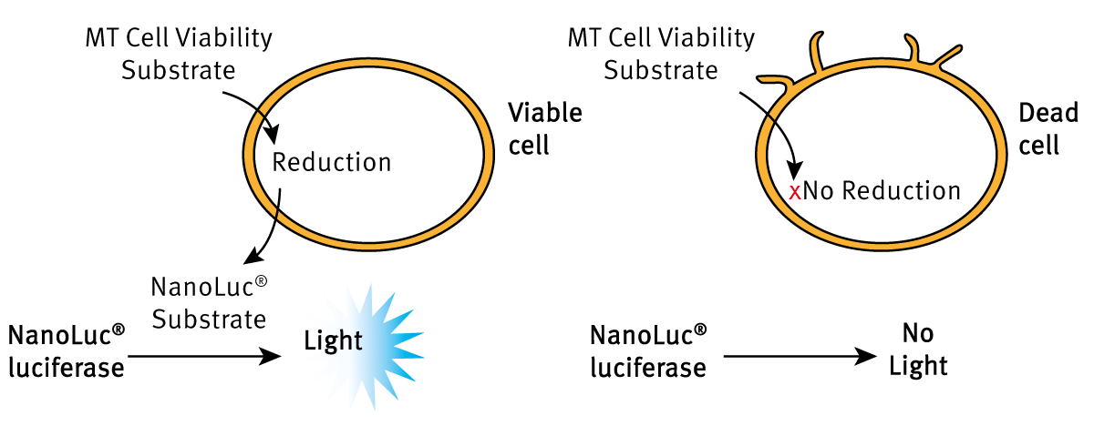
The RealTime-Glo™ MT Cell Viability assay from Promega is based on the intracellular reduction of a pre-substrate to a Luciferase substrate. The molecule is secreted out of the cells and is converted by the luciferase under the emission of light (Fig. 2). Hence, the light measured in a well directly reports on metabolic activity and cell viability and hence is specifically suited for real-time monitoring of viability.
c) ATP-based cell viability assays
A very quick and sensitive method to assess cell viability is by measuring intracellular ATP levels. ATP as the cellular energy source is proportional to the number of living cells [3]. Upon cell lysis, the released ATP is used to fuel an ATP-dependent luciferase reaction that emits light. The detected light is a direct measure of cell viability. The principle is used by kits such as CellTiter-Glo® (Promega), ATP-Glo™ (Biotium), and ATP Determination Kit (Invitrogen™, ThermoFisher). These are often employed for high-throughput cell viability measurements as they sense a few cells only and as they require the addition of a single reagent containing lysis buffer, luciferase, and substrate.
Proliferation
The increase in cell number by cell division is called proliferation. Since dividing cells are clearly viable it is used to assess cell viability. Cell division is a strictly regulated process that coordinates the replication of DNA, breakdown of nuclear envelope, division of genetic material, rearrangement of cytoplasma and membrane. Accordingly, there are many enzymes, protein modifications, and DNA marks that report on proliferation.
a) Stable fluorescent protein expression
Cell lines that constitutively express fluorescent proteins can be used to determine increases in cell number and, hence, cell viability. Nowadays, cell lines constitutively expressing fluorescent proteins (e.g. green fluorescent protein) are commercially available. They are primarily used to normalize results of other assays that are run in parallel to the cell density which is reflected by the fluorescence intensity. Furthermore, the use of two different fluorophores (e.g. GFP and RFP) in two different cell lines enables to study of influences of one cell type on another. The fluorescent proteins are typically introduced by lentiviral vectors and finally expressed under a CMV promoter. In this AppNote HeLa cells, stably expressing GFP and mcherry, were used to mimic transfection efficiency.
b) DNA replication-based proliferation assay
These cell viability assays employ nucleoside analogs that are incorporated into the newly generated DNA of proliferative cells. As this mainly occurs in dividing and living cells, nucleoside incorporation reports on cell viability. Traditionally, BrdU (5-Bromo-2´-Deoxyuridine) is added to cell culture and is incorporated into DNA instead of thymidine. The more cells divide, the more BrdU is found in the culture. Cells need to be fixed and permeabilized and their DNA requires denaturation to allow binding of an anti-BrdU antibody. Upon binding of a secondary antibody which is coupled to HRP, a substrate (TMB) can be converted to a colored molecule and detected by absorbance. The recorded absorbance reports the incorporation of BrdU and accordingly on proliferation and cell viability. A novel nucleoside analog is EdU (5-ethynyl-2´-deoxyuridine). Upon incorporation into DNA, the OregonGreen® fluorophore binds the EdU by a click reaction. In an amplification step, the signal is amplified by an anti-OregonGreen antibody labelled with HRP that catalyzes the formation of Amplex®UltroxRed. The fluorophore is very bright and provides the highest sensitivity in nucleoside incorporation cell viability assays.
Table 2 – Overview on cell viability assay.
| Cell viability assay | What does it measure? | How is it analyzed |
| Tetrazolium-based assays (MTT, MTS, XTT, INT, WST) | Metabolic activity is assessed by measuring the reduction potential and correlates with increased cell viability and cell number. Substrates are reduced to formazan, a chromophore. | Absorbance, analyzed wavelength depends on the substrate. |
| Resazurin | Metabolic activity is assessed by measuring the reduction potential and correlates with increased cell viability and cell number. Resazurin is reduced to fluorescent resorufin. | Absorbance at 570 nm or fluorescence (Excitation approx. 570 nm; Em approx. 600 nm). |
| ATP Luciferase assays | ATP is produced by living cells as an energy source. It further fuels luciferases that convert substrate under light emission which finally reports on cell viability. | Luminescence |
| RealTime Glo® | Metabolic activity is assessed by measuring the reduction potential and correlates with increased cell viability and cell number. Substrate is intracellularly reduced to a luciferase substrate. Luciferase-emitted light reports on cell viability. | Luminescence, suited for real-time monitoring |
| BrdU, EdU nucleoside incorporation assays | Report on DNA-synthesis which mainly occurs during cell division and, hence, on cell viability. | Absorbance or fluorescence |
| Constitutive fluorescent protein expression | Stable fluorescent protein expression reports on cell density and cell viability. | Fluorescence, suited for real-time monitoring |
All of the aforementioned cell viability assays can be performed in microplates. Typically a 96 well format is chosen, though it is possible to assess cell viability markers in 384 and even 1536 well plates as well. Accordingly, a microplate reader is needed to detect the optic signals coming from the assay. Of course, the assay dictates the read-out type the microplate reader needs to cover. For colorimetric cell viability determinations such as tetrazolium-based assays, single-mode absorbance readers might be sufficient. An absorbance reader such as the SPECTROstar® Nano reliably detects initial dose responses and cell viability reduction in novel experimental conditions.
However, absorbance-based cell viability assays are limited in sensitivity which necessitates the switch to fluorescent and luminescent cell viability tests. For these assays, multi-mode microplate readers are most suitable as these allow detection of absorbance, luminescence, and fluorescence. This way you have the freedom to choose the cell viability assay with the required sensitivity and analysis parameter (e.g. metabolic activity or proliferation). However, there is still a limitation: real-time monitoring of cell viability. In order to record cell viability over days, cells need a temperature and CO2-controlled environment. While BMG LABTECHs microplate readers are all capable of incubating the measurement chamber at 37 °C, only the Omega Series, the VANTAstar, and the CLARIOstar® multi-mode plate readers can be equipped with an atmospheric control unit (ACU). It regulates CO2 and, if desired, the O2 concentration inside the microplate reader to provide perfect conditions for long-term cell viability assays. The last consideration before deciding on a microplate reader is which flexibility is required: highest flexibility is achieved with a monochromator-based plate reader such as the CLARIOstar, as wavelengths for fluorescence (luminescence) measurements can be selected freely. This way, you can easily switch between assays and have many possibilities to optimize your assay. In contrast, filter-based microplate readers such as the Omega Series have built-in filters. With a standard combination of fluorescence filters, however, you can read thousands of (not only) cell viability assays without the need to exchange filters.
If you are curious to see how BMG LABTECH microplate readers measure cell viability assays, don’t miss to have a look at our cell viability and cytotoxicity application notes and peer-reviewed citations.
Absorbance plate reader with cuvette port
Powerful and most sensitive HTS plate reader
Most flexible Plate Reader for Assay Development
Upgradeable single and multi-mode microplate reader series
Flexible microplate reader with simplified workflows
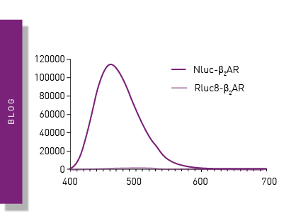
Life in the depths of the ocean operates under extreme conditions. Find out how proteins from deep-sea luminescent organisms are useful for measurements on microplate readers.
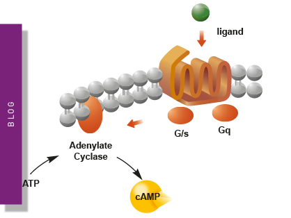
Second messengers play a pivotal role in signal transduction events in cells. But how do you measure these small, transiently lived molecules and how can microplate readers help?
NanoBRET is used to analyse binding events, signaling pathways and receptor trafficking in live cells and has significantly expanded the range and applications of BRET assays.
Find out about the different types of cell-based assays and why they have become an indispensable tool in a such a broad variety of disciplines in this blog article.
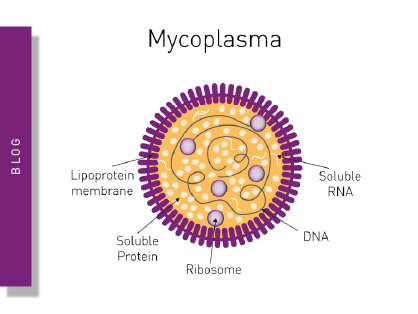
Read here about the various threats associated with mycoplasma contamination and find out about methods to prevent, detect, and eliminate mycoplasma in your cell culture.
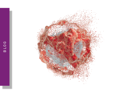
Apoptosis is a form of programmed cell death responsible for the removal of damaged or unnecessary cells. Read more about available apoptosis assays here.