Introduction
The binding sites of proteins such as antibodies are known to often contain tryptophan (Trp) residues, whose fluorescent properties may be altered upon ligand binding. Conformational changes within the binding site or simply the presence of the ligand can result in either fluorescence quenching or enhancement, which may be utilized to quantitatively investigate protein-ligand interactions. Highly stereoselective antibodies to amino acids have been used in a variety of analytical techniques for the sensitive detection of enantiomeric impurities and for enantiomer separation. The objective of this study was to test if tryptophan fluorescence can be used to determine the affinity of an anti-D-amino acid antibody toward a variety of standard and non-standard amino acids.
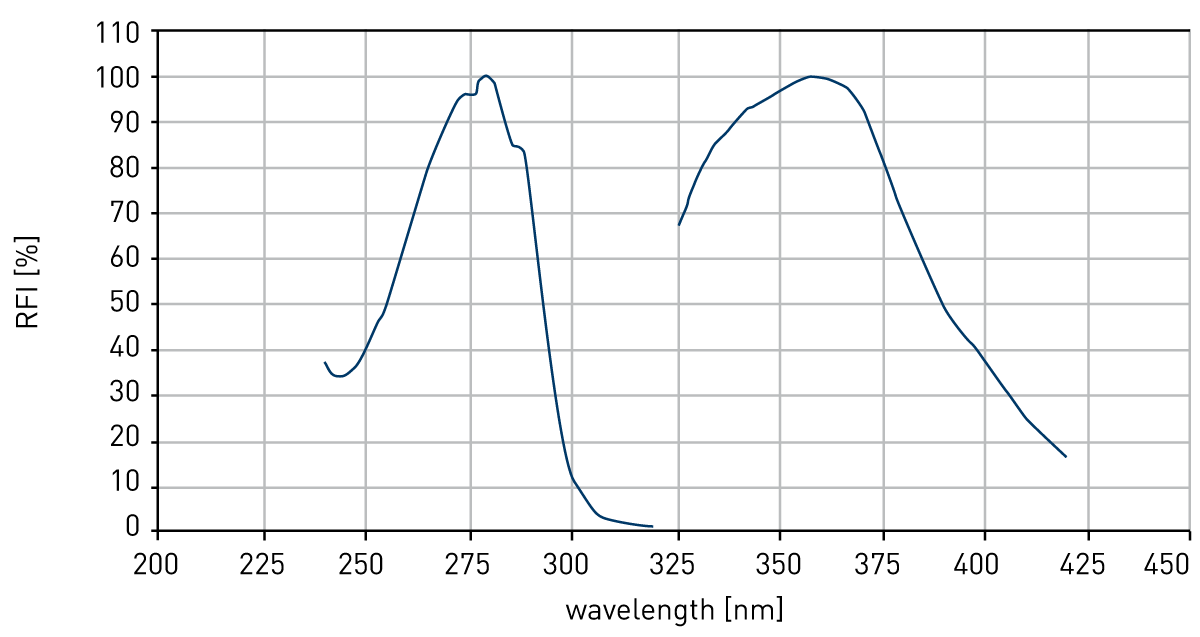
In order to examine the utility of BMG LABTECH microplate readers for measuring Trp fluorescence (Figure 1), experimental conditions were first optimized using the free amino acid as analyte.
Materials & Methods
- BMG LABTECH microplate reader
- Reacti-Bind White Opaque 96-well plates (PIERCE)
- D,L-Tryptophan (Sigma)
- D-Phenylalanine (Sigma), L -Phenylalanine (Sigma)
- Phosphate buffers (pH values between 2 and 12)
- Monoclonal anti-D-amino acid antibody
Results & Discussion
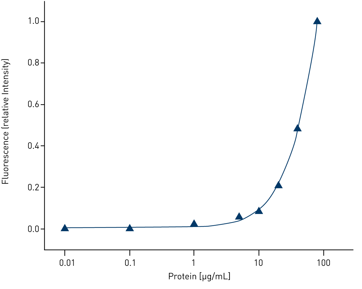
The effect of analyte concentration, temperature, and pH was investigated in order to establish optimal conditions to be used with BMG LABTECH microplate readers. As seen in Fig. 2, a clear concentration-dependent increase in Trp fluorescence was observed at concentrations ranging between about 1 mM and 1 µM. Excellent signal-to-noise ratios with minimal background fluorescence were obtained upon excitation at 280 nm and detection of fluorescence emission at 350 nm. In contrast, no fluorescence was observed using phenylalanine in the same range of concentrations (not shown).

The effect of temperature and pH was investigated using Trp at a concentration of 70 μM as analyte. As seen in Fig. 3, fluorescence intensity significantly decreases at higher temperatures.

Also changes in the pH have a considerable effect on Trp fluorescence, which is strongest at a pH around 11 (Fig. 4). Both results are in good agreement with previous reports.
 Investigating antibody stereoselectivity by measuring Trp fluorescence
As seen in Fig. 5, the BMG LABTECH plate reader can be used to determine the intrinsic Trp fluorescence of the anti-D-amino acid antibody used in this study. Increasing concentrations of the antibody in phosphate buffer, pH 7.4, resulted in increasing fluorescence emission at 350 nm upon excitation at 280 nm.
 For protein-ligand studies, a fixed concentration of the antibody was incubated with the D- or L-enantiomers of a variety of amino acids. Fig. 6 shows the results obtained with D- and L-phenylalanine, respectively.
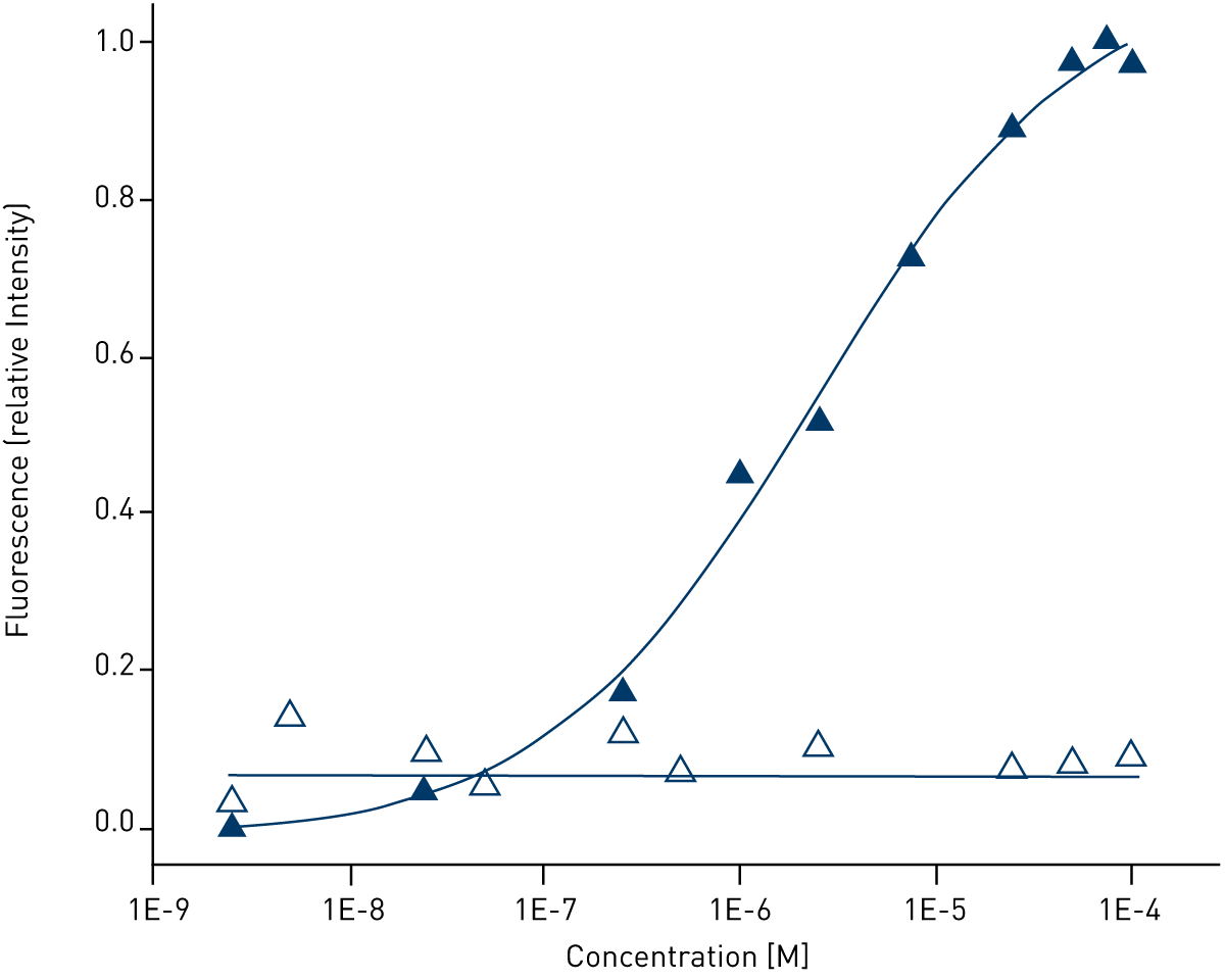 While the interaction of the antibody with D-Phe causes a concentration-dependent increase of the antibody‘s intrinsic Trp fluorescence, no such effect is observed using the L-enantiomer. Similar results were obtained with the enantiomers of cyclohexylalanine, histidine, norleucine, leucine, and norvaline (not shown). In all cases, the stereoselective interaction of the antibody with the D-enantiomers of these amino acids caused a concentration-dependant enhancement of the protein‘s intrinsic Trp fluorescence, while no change in fluorescence was caused by the L-enantiomers. As observed using other analytical techniques, the affinity of the antibody is strongest to D-amino acids having aromatic or bulky side chains, while aliphatic amino acids are bound more weakly.
Conclusion
The BMG LABTECH microplate reader allows measurement of Trp fluorescence with high sensitivity and good signal-to-noise ratios. Excitation of appropriate proteins at 280 nm and measurement of fluorescence emission at 350 nm can be employed to investigate protein-ligand interactions and to deduce, e.g., binding affinities.