SPECTROstar Nano
Absorbance plate reader with cuvette port


This author profile refers to work created by Dr Andrea Krumm during her tenure at BMG LABTECH - the Microplate Reader Company, where she served as an Applications Specialist contributing scientific content, application notes, and technical expertise. Dr Krumm is no longer employed at BMG LABTECH, but her published materials remain available here for reference and archival purposes. Dr Andrea Krumm is a biotechnology specialist and product manager known for her work in analytical instrumentation and biopharmaceutical analysis. She studied biotechnology and later earned a PhD in cancer biology, focusing on topics such as DNA repair, epigenetics, and tumor biology. After completing her doctorate, she spent several years as an Applications Specialist at BMG LABTECH, where she authored application notes, conducted workshops, and supported scientific customers. In 2020, Dr Krumm joined Tosoh Bioscience GmbH as Product Manager for Analytical Columns.
Advanced assay drug development technologies are increasingly used to evaluate cytotoxicity, enabling more efficient and accurate drug development.
This article focuses on cytotoxicity assays. However, these are often combined with viability tests for normalization purposes. You can learn more about the combination of cytotoxicity assays and cell viability assays in the following video:

The word cytotoxicity gets easier to explain (and to pronounce) if it is split into its two components: The first part “cyto” is of Greek origin and denotes the cell. The second part “toxicity” is of Latin origin and denotes the harmfulness of chemicals, drugs, organisms, or conditions to organisms or parts thereof. Hence, cytotoxicity refers to the ability of something to harm cells. Various chemical compounds can induce cytotoxic effects in cells.
There are many targets to damage cells. One such target is deoxyribonucleic acid (DNA). This biomolecule stores all the genetic information required for a functional cell, for cell division, and a copy of it is passed on to a cell’s successor. As DNA is prone to be damaged by reactive oxygen species produced by the cell itself (endogenous molecules) or erroneous replication of DNA, the cell offers effective ways to repair its DNA and escape cytotoxicity. The damage becomes a cytotoxic event if a cell notices severe or unrepairable damage. In this case, it protects itself (and the organism) by committing suicide.
Obviously, the outer barrier of a cell, the cell membrane, is crucial for cytotoxicity as well. It may be disturbed by mechanical stress or osmotic stress, which in the case of low extracellular solute concentration results in cellular water uptake and cell lysis. The membrane is another target of the immune system: Cytotoxic T cells and natural killer cells kill tumor and virus-infected cells with the help of perforin, a protein that oligomerizes in the membrane of the diseased cell to form a pore and lyse the cell.
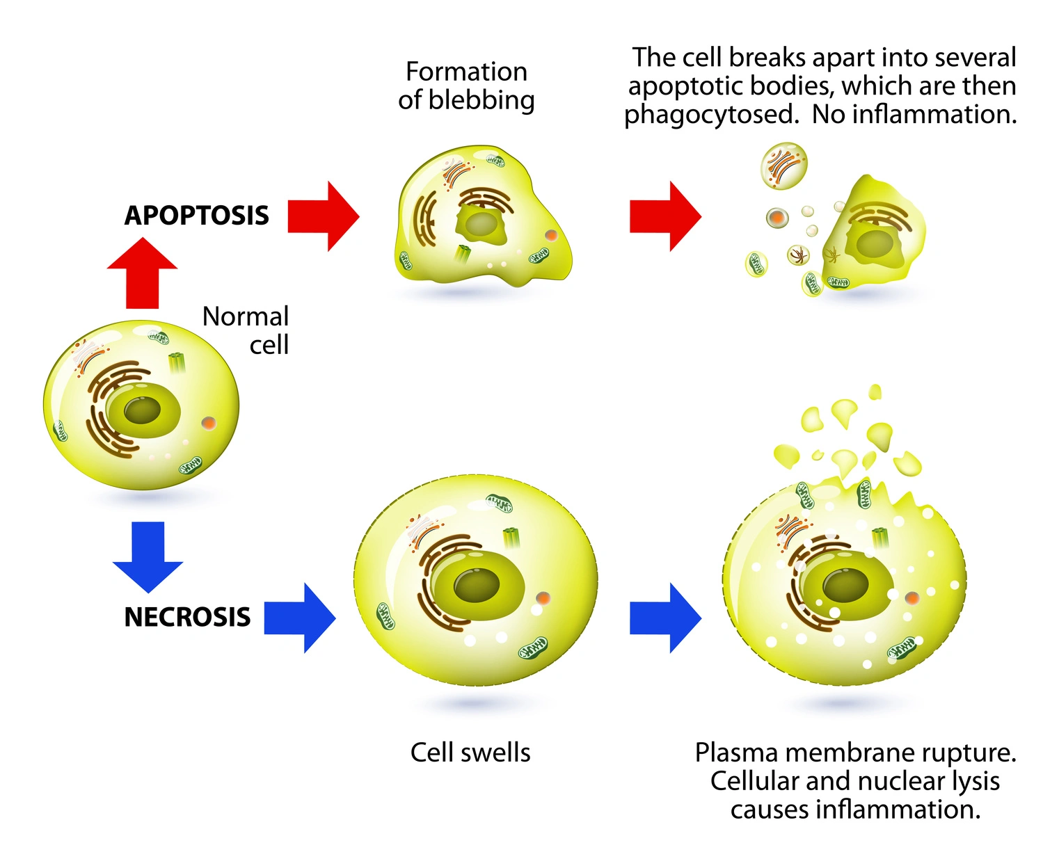
Serious cell damage results in cell death which occurs either in a programmed fashion (e.g. apoptosis) or in an unregulated fashion (necrosis). Apoptosis is a physiological process to balance cell numbers in an organism. Furthermore, it is initiated by cells noticing irreparable and cytotoxic damage to protect the organism. The cells organize the degradation and packaging of cell components and mark themselves in a way that leads to them being engulfed by macrophages. In contrast, necrosis is a traumatic and unregulated cytotoxic process in which the cell swells and loses the integrity of its membrane. In this way, cellular components are released into the extracellular space leading to local inflammation. Cytotoxic response can lead to reduced cell proliferation and cell growth, and alterations in cellular metabolism are often indicators of cytotoxicity.
There are two major reasons why cytotoxicity is studied in life science research: either you want specific cells to die and look for an adequate compound/condition or you want to exclude cytotoxicity in specific cells.
Cytotoxicity is desired in the treatment of cancer as well as in therapies for some autoimmune diseases. In cancer therapy, the selective killing of tumor cells is the main goal. Immune effector cells, such as cytotoxic T lymphocytes (CTLs) and natural killer cells, induce cell-mediated cytotoxicity by recognizing and destroying tumor target cells.
Conventional chemotherapy aims at damaging the cancer cell’s DNA and thus pushes cells into apoptosis instead of replication. The selective cytotoxicity for malignant cells is achieved by their fast and uncontrolled replication which does not leave time to repair the damaged DNA and finally leads to collapse during cell replication. Cytotoxicity assays help to find cancer-killing agents and allow for in vitro comparison of agents or conditions intended for cancer therapy. This application note for example highlights the use of a Resazurin cell viability assay to screen snake venom libraries for a potential apoptotic activity against cancer cell lines.
Cytotoxicity is desired in the treatment of cancer as well as in the therapy of some autoimmune diseases. For cancer therapy, the selective killing of tumor cells is the main goal. Conventional chemotherapy aims at damaging the cancer cell’s DNA and thus pushing it into apoptosis instead of replication. The selective cytotoxicity for malignant cells is given by their fast and uncontrolled replication which does not leave time to repair the damaged DNA and finally leads to collapse during cell replication. Cytotoxicity assays help to find cancer-killing agents and allow for in vitro comparison of agents or conditions intended for cancer therapy. This application note for example highlights the use of a Resazurin cell viability assay to screen snake venom libraries for a potential apoptotic activity against cancer cell lines.
In order to manage severe autoimmune diseases, cytotoxic drugs are used to decrease the number of immune cells that mistakenly attack the body instead of pathogens. The doses of cytotoxic drugs used for the management of autoimmune diseases such as rheumatoid arthritis are lower and are oftentimes just efficient enough to slow down the replication of immune cells but not necessarily to induce cell death. This fine line can be studied using methods that report on cytotoxicity.
Lymphocyte-mediated cytotoxicity and natural killer cell-mediated cytotoxicity are important mechanisms in immunotherapy, where effector cells such as natural killer cells and cytotoxic T lymphocytes recognize and kill specific target cells through mediated cytotoxicity.
Most drugs are not intended to be cytotoxic to cells as this might have an impact on the whole organism. For instance, novel drug candidates are tested to see whether they harm cardiomyocytes as this would result in cardiotoxicity and potentially death. In these assays, untreated cells are used as negative controls to establish baseline viability and compare the effects of test compounds. The same is true for cells of many vital organs.
The same is true for cells of each vital organ. For this reason, cytotoxicity assays may spot possible adverse effects of novel drugs at early stages of drug development that in the past required taking already approved drugs off the market.
Cytotoxicity assays make use of events that happen during the event of cell death such as loss of membrane integrity, activation of cell death-inducing enzymes called caspases, or phenotypic changes on the cell surface. Cell-based assays fundamental for these in vitro experiments, and maintaining cell health requires an appropriate cell culture medium that supplies essential nutrients and conditions.
The cell membrane loses integrity during cytotoxic events such as necrosis, but also during late stages of apoptosis. It can be detected by measuring the activity of an enzyme that leaks through the membrane: Lactate dehydrogenase (LDH). A colorimetric assay determines the abundance of LDH in cell culture supernatant by employing the LDH-dependent formation of colored formazan. The more LDH that leaks through the cell membrane, the more colored formazan is generated which indicates higher toxicity.
Fluorescent DNA dyes that do not penetrate the intact cell membrane is another way to assess membrane permeability and determine cytotoxicity.
Fluorescent DNA-binding dyes are used to detect dead cells, as these dyes only enter cells with compromised membranes, bind to DNA, and produce a fluorescent signal. The fluorescence intensity correlates with the number of dead cells present.
Examples for this kind of cytotoxicity assays are CellTox™ Green (Promega), Ethidium Homodimer-1 (as used in the Live/Dead assay, ThermoFisher Scientific), and SYBR® Green (ThermoFisher Scientific). Propidium iodide is an alternative fluorescent DNA intercalator that can be used to study cytotoxicity. For example, propidium iodide was used to study the cytotoxic effect of bacteria on a HeLa cell line in the application note: The InToxSa assay, for quantifying Intracellular Cytotoxic S. aureus Phenotypes.
Vital dyes, such as trypan blue, selectively stain dead cells but are excluded from live cells, allowing for the distinction between live and dead cells. Cell counting is important to quantify viable cells and accumulated dead cells, and flow cytometry can be used for high-throughput analysis of live and dead cell populations. Homogeneous assay formats enable simplified, one-step detection of cytotoxicity without washing steps, increasing throughput and reproducibility. The choice of assay reagent, including properties such as cell permeability and spectral characteristics, is critical for optimal detection of cell viability and cytotoxicity.
A non-permeable DNA dye is also part of a multiplexed cytotoxicity assay that discriminates between the two most abundant types of cell death: apoptosis and necrosis. A fluorescent dye indicates necrosis whereas a luminescent signal indicates apoptosis. Luminescence is generated only when phosphatidylserine is exposed to the cell surface. The exposure occurs during apoptosis and marks the cells fore digestion by macrophages. Annexin V, a phosphatidylserine-binder, is linked to incomplete parts of a luciferase. When bound to phosphatidylserine, adjacent incomplete luciferase parts form a functional enzyme capable of emitting light and indicating cytotoxicity.
Similarly, the binding of phosphatidylserine also triggers the generation of a luminescent signal in a novel approach using biocytometry. Single-cell immunophenotypes can be identified by the binding of bioparticles with their expressed antibody mimetics. The method even allows apoptotic cells specifically in subpopulations to be identified. 
Caspases are enzymes playing a major role in cytotoxicity and apoptotic cell death in particular. They constitute a group of cysteine proteases cleaving target proteins specifically after an aspartic acid side chain whereby caspases initiate and execute apoptosis. This specific cleavage is exploited for analysis of caspase activity in a cytotoxicity assay. Synthetic substrates bearing a peptide sequence that upon proteolytic cleavage by caspases release either a chromophore, a fluorophore, or a luciferase substrate. Accordingly, caspase activation can be measured in absorbance, fluorescence, and luminescence detection mode. Different protease markers, including caspases and other protease markers, are used to assess apoptosis and cytotoxicity.
A luminescent and a fluorescent caspase assay to assess cytotoxicity are described in our AN266: Promega's multiplexed cell viability and apoptosis assays.
As another target extracellular ATP can also be quantified. Cells that undergo apoptosis, driven by immunogenic cell death, release extracellular ATP. This extracellular ATP can be determined with assays such as the RealTime-GloTM Extracellular ATP Assay form Promega as described in the AppNote: Extracellular ATP measurement in real time using living cells. But not only extracellular ATP can be used to assess cell cytotoxicity. The total ATP levels of a cell culture that are directly dependent on the number of cells in a sample can also be used for this purpose as highlighted in the AppNote: The use of an ATP bioluminescence assay to quantify cell cytotoxicity
When evaluating cytotoxicity, cells exposed to test compounds are compared to untreated controls to assess cytotoxic effects and mechanisms of action.
These are just some of the ways to detect if an agent or drug is cytotoxic to cells. You can learn more about cell viability assays or cell-based assays in general in our other blog articles.
Absorbance plate reader with cuvette port
Powerful and most sensitive HTS plate reader
Most flexible Plate Reader for Assay Development
Upgradeable single and multi-mode microplate reader series
Flexible microplate reader with simplified workflows
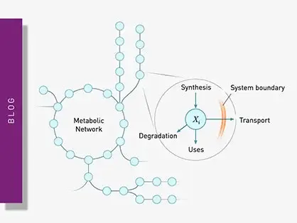
Explore metabolic flux analysis, its significance in research, and how microplate readers enhance measurements for insights.
Explore the vital role of mitochondrial metabolism in cellular function, its measurement techniques, and how microplate readers enhance research in this field.
Learn about applications for bacterial metabolism on a microplate reader.
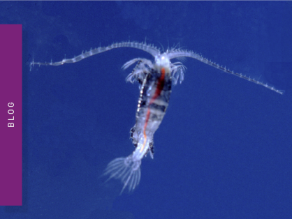
Life in the depths of the ocean operates under extreme conditions. Find out how proteins from deep-sea luminescent organisms are useful for measurements on microplate readers.
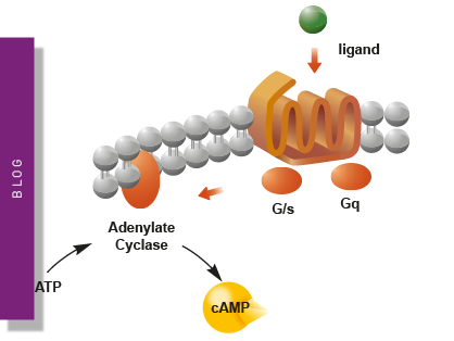
Second messengers play a pivotal role in signal transduction events in cells. But how do you measure these small, transiently lived molecules and how can microplate readers help?
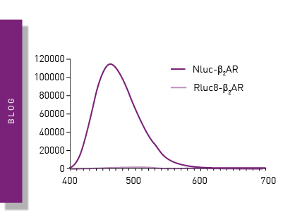
NanoBRET is used to analyse binding events, signaling pathways and receptor trafficking in live cells and has significantly expanded the range and applications of BRET assays.