Introduction
Fluorescence-based assays conquered the benches of life science labs due to many advantages: fluorophores are often non-toxic, fluorescent dyes are affordable and can be measured multiple times. The intensity of fluorescent dyes is linearly related to their concentration and significantly many assays are available. Not only did the use of fluorescence assays per se increase but in particular the use of red fluorescent dyes emitting in the red and infrared range of light has risen.
What drives the shift to red dyes?
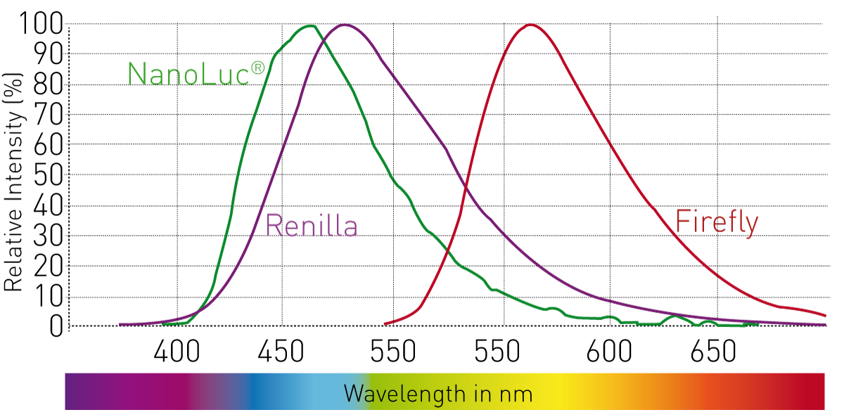
Multiplexing. Research evolves fast and requires many and exact data in a short time. Thus, multiple assays are run in parallel to detect several biological aspects in one sample. For instance, expression of a fluorescent reporter protein can be referenced to cell viability. However, luminophores and fluorophores measured in the same sample need to be spectrally separated for efficient signal separation. Many traditional assays use bright dyes emitting in the green range (AlexaFluor®488, FITC, GFP). Fluorescent dyes that are combined with such assays are often red-shifted. Similarly, red fluorescent dyes are required for multiplexing with luminescence. Luciferases emit light over broad wavelength ranges, often up to 500-600 nm (Fig. 1). Separation of luminescent and fluorescent signals is only possible when combining it with red fluorescent dyes emitting fluorescence over 650 nm.
 Autofluorescence reduction in cellular and complex samples. In complex biological samples with living cells, many unwanted fluorescent components exist that interfere with the signal of the fluorescent dye of interest (auto-fluorescence): aromatic amino acid side chains of fluorescent proteins, NAD(P)H, flavin or phenol red occupy the light spectrum from the UV to the red range. Fluorescent molecules are mainly found in cellular or complex samples such as serum and plasma.
Using fluorophores emitting further in the red range (>650 nm), like nile red or texas red prevents auto-fluorescence. Another method circumvents auto-fluorescence in two ways: long lifetime fluorophores detected when auto-fluorescence already faded and the detection of fluorescence intensity in the red. This principle is popular in homogenous assay formats that measure molecule interactions by FRET: homogenous time-resolved Förster resonance energy transfer (HTRF®, Cisbio). A long-lifetime fluorophore is used as donor and transfers energy to a red acceptor fluorophore only if this is found in proximity.
The use of red fluorescent dyes is inevitable when measuring cells or multiple assays at once. Thus, we tested the performance of the CLARIOstar Plus with dedicated detectors in measuring the red fluorescent dye AlexaFluor® 647 and compared HTRF measurements performed with standard and dedicated detectors (PMT).
Materials & Methods
AlexaFluor 647 Testing
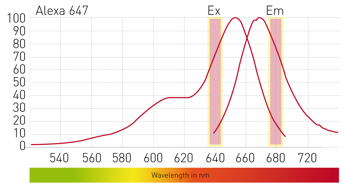
Testing of the CLARIOstar Plus was performed with the red fluorescent dye AlexaFLuor® 647, a bright fluorophore that is excited around 640 nm and emits at 680 nm (Fig. 2).
- black 384 sv microplate (Greiner #784076)
- CLARIOstar Plus (BMG LABTECH)
- AlexaFluor 647 (ThermoFisher Scientific)
- PBS (Biochrom #L1825)
The red fluorescent dye was diluted to standards of 100 nM; 20 nM; 4 nM; 0.8 nM and 0.16 nM in PBS. Eight replicates (20 µl) and 85 PBS blanks (20 µl) were pipetted into the plate for determination of the detection limit. Fluorescence of the red fluorescent dye was measured on the CLARIOstar Plus using the following instrument settings.
Instrument settings
| Optic settings | Fluorescence intensity, top optic, end point | |
| Detector | Dedicated red-shifted PMT | |
| Filters | Ex: 640-10 Dichroic: LP664 Em: 680-10 |
|
| Gain | EDR | |
| General settings | Number of flashes | 100 |
| Settling time | 0.1 s | |
Detector comparison for HTRF measurements
For comparison of standard and dedicated detector in HTRF measurements, a Cisbio kit based on Eu Cryptate donor (emission 620 nm) and XL665 (emission 665 nm) acceptor was used.
- white 96 well half area microplate (provided by Cisbio)
- CLARIOstar Plus (BMG LABTECH)
Instrument settings
| Optic settings | Time resolved fluorescence, dual chromatic | |
| Detector | Dedicated red-shifted PMT and standard PMT | |
| Filters | Ex. Ex TR Dichroic: LP TR Em 1: 665-10, Em 2: 620-10 |
|
| Integration times | 60 µs, Time: 400 µs | |
| General settings | Number of flashes | 200 |
| Settling time | 0.1 s | |
Results & Discussion
A standard curve of AlexaFluor647 was measured in fluorescence to determine its detection limit on the CLARIOstar Plus (Fig. 3). The standard curve from 100 nM down to 0.16 nM of the red fluorescent dye resulted in a highly linear correlation as indicated by an R² of > 0,9999. Calculation of the detection limit according to IUPAC standard resulted in a low limit of 0.8 pM (Table 1).
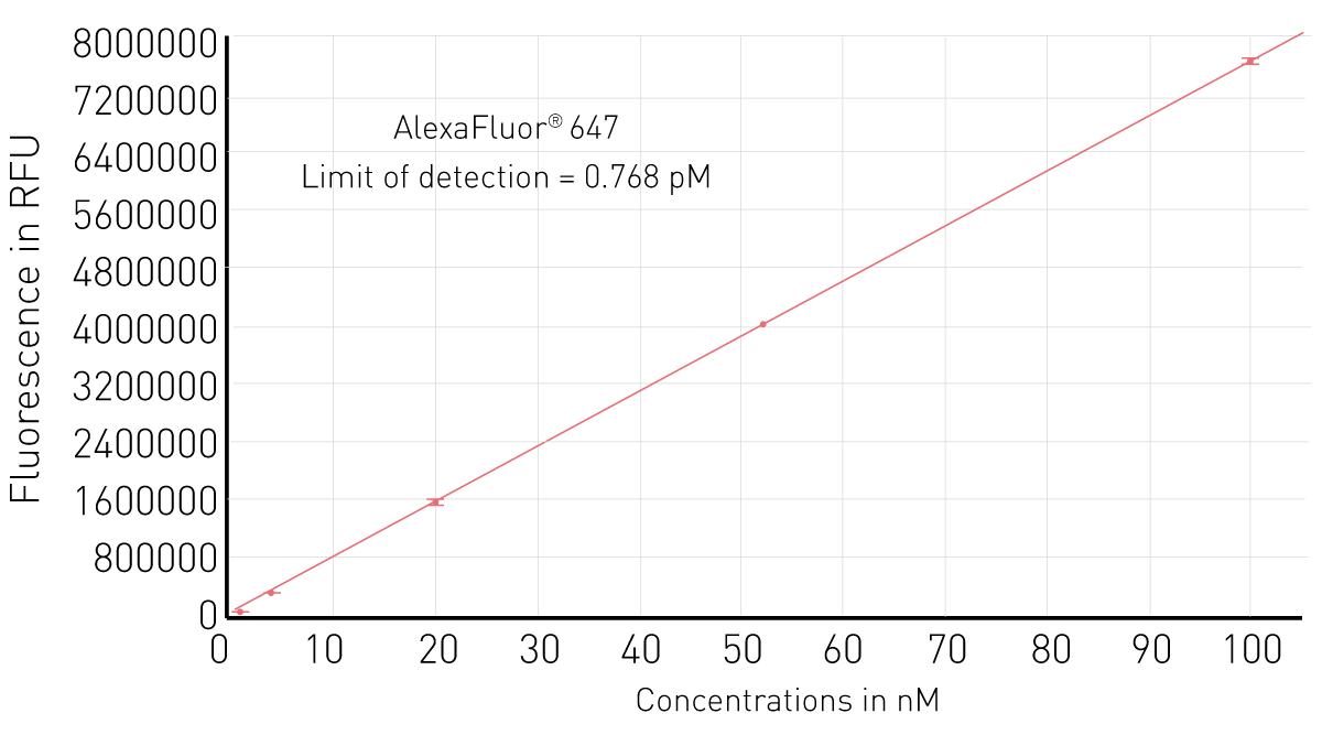
Table 1. Limit of detection calculation of the red fluorescent dye AlexaFluor647 measured on the CLARIOstar Plus
| Standard deviation of the blank (and %CV) | 19.6 (2.2%) |
| Slope of linear regression | 76676 |
| Limit of detection - LOD (3* SDBI/Slope) | 0.8 pM |
The high sensitivity is not limited to red fluorescent dyes. The LOD of 0.15 pM for FITC (data not shown) shows that the dedicated PMT is a suitable solution for multiplexed detection of red and green fluorophores.
Testing the dedicated red-shifted detection of the CLARIOstar Plus for HTRF measurements revealed improvements in assay window, measurement stability and conse-quently assay quality. The deltaF is a measure of the assay window which relates the increase in FRET ratio of positive controls to the negative control. This measure was highly improved using the dedicated detector for red fluorescent dyes as compared to the standard detector (Fig. 4) high positive control +17%, low positive control +15 %). As additionally the %CV, a measure of signal stability, of the blank was reduced using the dedicated detector the complete assay quality was improved. The Z’ value is indicative of assay quality and includes signal stability as well as distance between positive and negative controls. HTRF assays with low signals greatly benefit from the novel detector as Z’ is improved from 0.27 to 0.66 (143 %). HTRF assays with high signals display very high quality with a Z’ > 0.9, irrespective of the detector (Fig. 4).

Conclusion
Due to increased use of cell-based assays and multiplexing, fluorophores emitting in the red and far-red are on the rise. Here, we demonstrate the suitability of the CLARIOstar Plus to detect red fluorescent dyes. It displays high sensitivity with a low LOD for the dye AlexaFluor647 (0.8 pM) and significantly improved HTRF measurements. This makes the CLARIOstar Plus a reliable device for complex samples and multiplex measurements.