Introduction
Extracellular vesicles (EVs) are nanoscale particles composed of a lipid bilayer containing lipids, proteins and nucleic acids. The particles are around 50–200 nm in diameter (for small EVs) and up to 10 µm (for large EVs). EVs are secreted by all cells and play an important role in cellular communication 1. Since their discovery, the number of studies using EVs to transport biomolecules for applications in diagnostics and therapeutics has increased significantly 1.
EV isolation and purification is a significant challenge and the method used for EV isolation has a major impact on the quality of isolated EV material 2.
As a gold standard approach for EV isolation does not exist, researchers need to identify the method that suits their needs depending on the yield and specificity of EVs 2. Currently, it is not possible to predict whether EV isolation will be efficient or not due to the diversity of EV sources (for example, media, buffers, or biological fluids). Therefore, new tools are needed to assess the effectiveness of EV isolation methods.
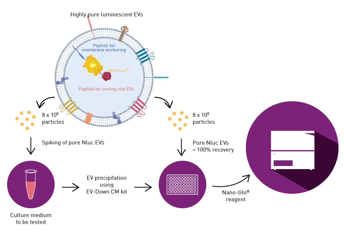
The EV-Down CM kit from the company in-EV (a subsidiary of Ciloa) is used for EV isolation from cell culture media. A fast and easy way to study the effectiveness of EV isolation is spiking culture media with pure NanoLuc® luciferase (Nluc)-labeled EVs. In this case, Nluc is used to track EVs during their isolation. The CLARIOstar® Plus is ideally suited to follow the recovery of EVs through luminescence measurements due to its sensitivity and ease of use.
Assay principle
Nluc is expressed by cells producing EVs and is incorporated into EVs between a peptide for membrane anchoring and a peptide for sorting into EVs. The incorporated Nluc luminesces upon the addition of a chemiluminescent substrate and can therefore be used to report on the amount of EVs present. This luminescent readout can be used to study the recovery of EVs after their isolation from different media.
Materials & methods
- 96-well plate, white (Thermo Scientific™, #236105)
- Nluc EVs (in-EV, #X132)
- EV-Down CM EV concentration kit (in-EV, #K214)
- Nano-Glo Luciferase Assay System (Promega, #1110)
- CLARIOstar Plus (BMG LABTECH)
Experimental Procedure
The luminescence produced by Nluc EVs was assessed using 3 x 10⁸ particles per well in 100 µL. Standard (non-labelled) EVs were used as a negative control and vehicle (EV resuspension buffer) was used as a blank.
For the recovery experiment after EV isolation, 5 x 10⁸ Nluc EVs were spiked into three different culture media before isolating EVs using the EV-Down CM kit. EV isolation was performed according to the manufacturer’s instructions and two incubation conditions were tested: overnight (as recommended) and 2 h incubation. After precipitation, the EV pellet was resuspended in phosphate-buffered saline (PBS) and the luminescence of EVs in 100 µL samples was measured in a 96-well plate to assess the recovery of EVs during the isolation step. Pure Nluc EVs were used as a reference (100% of recovery) and vehicle (EV resuspension buffer) was used as a blank. Nano-Glo Luciferase Assay Reagent was added according to the manufacturer’s instructions. Luminescence was measured on triplicate samples using the CLARIOstar Plus.
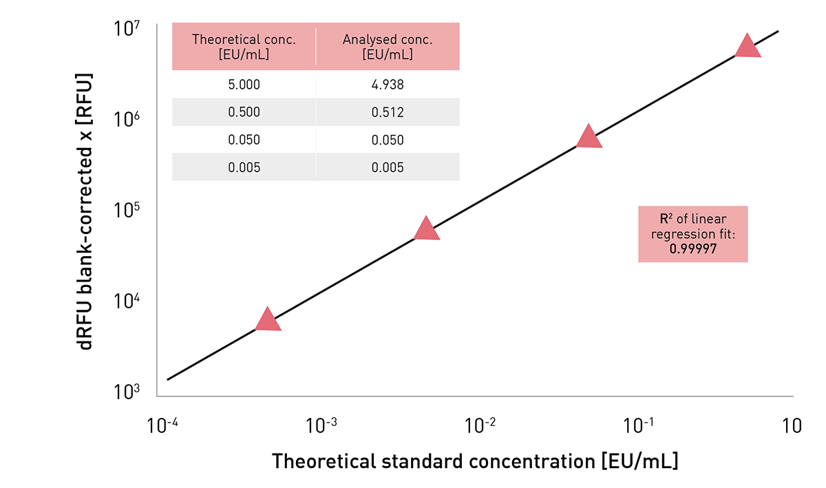
Instrument Settings – EV emission spectrum
|
Luminescence, emission scan, endpoint, top optic
|
||
|
Optic settings |
Measurement interval time |
0.8 sec* |
|
Emission wavelengths |
386-600 nm |
|
|
Emission bandwidth |
20 nm |
|
|
Scan resolution |
1 nm |
|
|
General settings |
Gain |
3600 |
|
Focal height |
Auto focus |
|
|
Spoon type |
96/384 |
|
|
Exponential smoothing with smooth intensity = 70.
|
||
Instrument Settings – EV emission spectrum
|
Luminescence, endpoint, top optic
|
||
|
Optic settings |
Filters |
470-80 nm |
|
Measurement interval time |
0.8 sec* |
|
|
General settings
|
Gain
|
EDR |
| Focal height | Auto focus | |
*To allow comparison of measurements with different measurement interval times, values are normalised to 1 sec.
Results & Discussion
The luminescence spectrum of EVs bioengineered with Nluc was compared with that of standard (non-labelled) EVs which were used here as a negative control (Fig. 2).
The results show that it is possible to detect the luminescence of EVs bioengineered with Nluc. Standard EVs, which were used as a negative control, allowed background noise to be determined and validated the specificity of the signal.
Next the recovery of EVs was determined after EV isolation. In this case, the luminescence of pure Nluc EVs was compared to the luminescence of Nluc EVs spiked in the culture media and isolated using the EV-Down CM kit (Fig. 3). 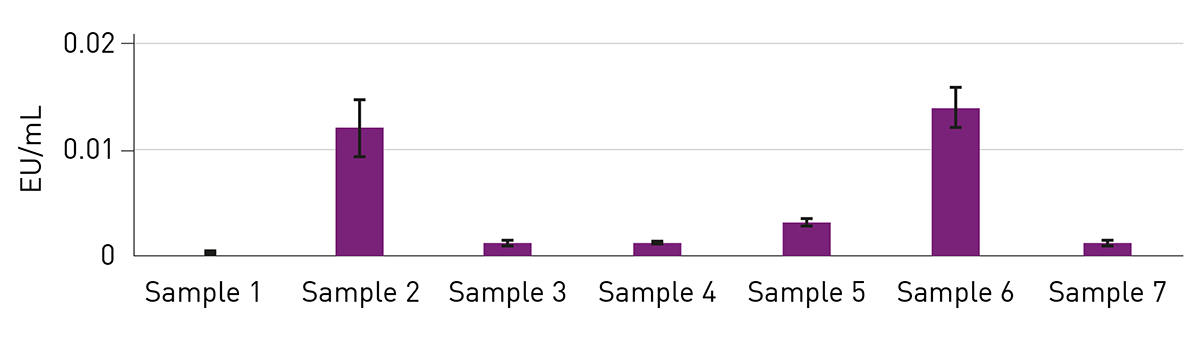 Nluc luminescence was detected after EV precipitation in the three culture media with some differences in the signal intensities. Luminescence of pure Nluc was set to 100% recovery to obtain EV recovery rates of Nluc EVs spiked in the culture media after EV isolation using the EV-Down CM kit. The recovery obtained with culture medium 1 was higher than the recovery with the other two culture media, which suggests an impact on the recovery by different media components. Moreover, the overnight incubation (recommended by the manufacturer) lead to a higher recovery during the EV isolation compared to an incubation for only 2 h, as confirmed by the higher luminescence (Fig 3).
Conclusion
Due to the diversity of EV sources and the absence of a gold standard isolation method, it is important to check recovery and quality of EVs for downstream applications. Here, the recovery of EVs using EV-Down CM kit from in-EV was assessed for EV isolation from several culture media. The use of EVs loaded with Nluc from in-EV and measurements on BMG Labtech’s CLARIOstar Plus provided a user-friendly and reliable method to evaluate EV recovery rates for the EV isolation process. This method of validation should be applicable to all EV sources (for example, culture media, biological fluids and plants) and for various EV purification methods (for example, precipitation, ultracentrifugation, tangential flow filtration (TFF) and chromatography) and thus represents a promising tool for laboratories working with EV isolation.
References
- van Niel, G., D’Angelo, G., Raposo, G. Shedding light on the cell biology of extracellular vesicles. Nat Rev Mol Cell Biol (2018) 19:213–228. doi 10.1038/nrm.2017.125.
- Welsh J.A. et al. Minimal information for studies of extracellular vesicles (MISEV2023): From basic to advanced approaches
- J. Extracell. Vesicles. (2024) 13(2):e12404. doi:10.1002/jev2.12404. [published correction appears in J. Extracell. Vesicles (2024) 13(5):e12451. doi: 10.1002/jev2.12451.