Introduction
Poly ADP-ribosylation is a post-translational modification of proteins that plays a crucial role in regulating DNA repair. Poly (ADP-ribose) polymerase (PARP) transfers ADP-ribose to itself and other nuclear proteins such as histones. The substrate for that reaction is NAD+. PARP inhibition has been demonstrated to potentiate the cytotoxicity of anti-cancer drugs and ionising radiation. Therefore much effort has been put into the development of specific PARP inhibitors.
To evaluate such inhibitors, we have first used a specific PARP activity assay, to monitor their ability to inhibit endogenous PARP activity contained within a colon cancer cell line. This assay requires protein concentration determination (BCA assay - absorbance) followed by luminescent detection.
Secondly, we have used the AlamarBlue viability assay (fluorescent read-out) to evaluate the ability of a PARP inhibitor to potentiate the effects of the chemo-therapeutic agent temozolomide to stimulate cell death in a colon cancer cell line. All measurements were performed using a FLUOstar Omega multi-detection microplate reader from BMG LABTECH.
Materials & Methods
-
Bicinchoninic Acid Protein Detection Kit from Sigma-Aldrich
-
Universal Chemiluminescent PARP assay kit including white plates from Trevigen
-
Clear and black 96 well plates from Fisher
-
Temozolomide from Sigma-Aldrich
-
AlamarBlue from Invitrogen
| Sample | Absorbance 560 nm |
| LoVo Control | 0.767 |
| LoVo 1 nM PARP Inhibitor |
0.774
|
| LoVo 3 nM PARP Inhibitor |
0.791
|
| LoVo 10 nM PARP Inhibitor |
0.778
|
| LoVo 30 nM PARP Inhibitor |
0.762
|
| LoVo 100 nM PARP Inhibitor |
0.75
|
| LoVo 300 nM PARP Inhibitor |
0.736
|
| Blank |
0.075
|
Universal Chemiluminescent PARP assay
LoVo cells were treated with PARP inhibitor at a range of concentrations (1-300 nM) for 1h before cells were harvested and lysed in PARP Buffer.
A BCA protein assay was carried out in a 96 well microplate, following manufacturer’s instructions adapted for a 96 well plate. The plate was read at 560 nm using the FLUOstar Omega.
A standard curve was generated for the BCA protein assay (Figure 1) and from this, the protein concentration of the lysates were determined, and adjusted to 40 μg per sample.
Lysates were then screened for PARP activity following manufacturer’s instructions for determining PARP activity in cell and tissue extracts.
The assay measures the incorporation of biotinylated poly (ADPribose) onto histone proteins in a 96 well strip format. Detection is carried out using a Streptavidin-Horseradish Peroxidase (HRP) conjugate. Following addition of the HRP substrate, the resulting luminescent signal was read using the FLUOstar Omega equipped with luminescence optic.
AlamarBlue Viability Assay
LoVo cells were seeded in 96 well black plates at 5000 cells per well and allowed to adhere overnight, prior to addition of compound or vehicle control.
0-300 μM temozolomide was added to cells, with and without 300 nM PARP inhibitor for 72 h. AlamarBlue 10 % (v/v) was then added to cells, and incubated for a further 6 h at 37°C. Live, metabolically active cells convert the AlamarBlue substrate to a fluorescent product, which was then detected using the FLUOstar Omega (Excitation 544 nm and Emisson 590 nm).
Results & Discussion
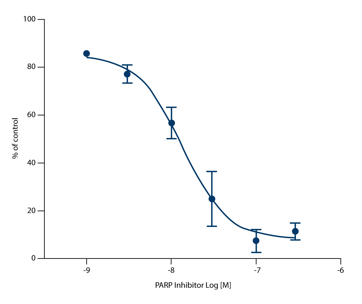
Universal Chemiluminescent PARP assay
Following luminescence measurement, data was analysed using GraphPad Prism (Figure 2). The PARP inhibitor inhibits 50 % PARP activity at a concentration of 14 nM.
AlamarBlue Proliferation Assay
Following measurement of fluorescent product, data was analysed using GraphPad Prism (Figure 3). Temo-zolomide as a single agent leads to very little cell death, with an IC50 >300 μM.
However, addition of PARP inhibitor in combination with temozolomide, leads to a significant increase in cell death, with an IC50 of 60 μM. This represents >5-fold enhancement of cell death.
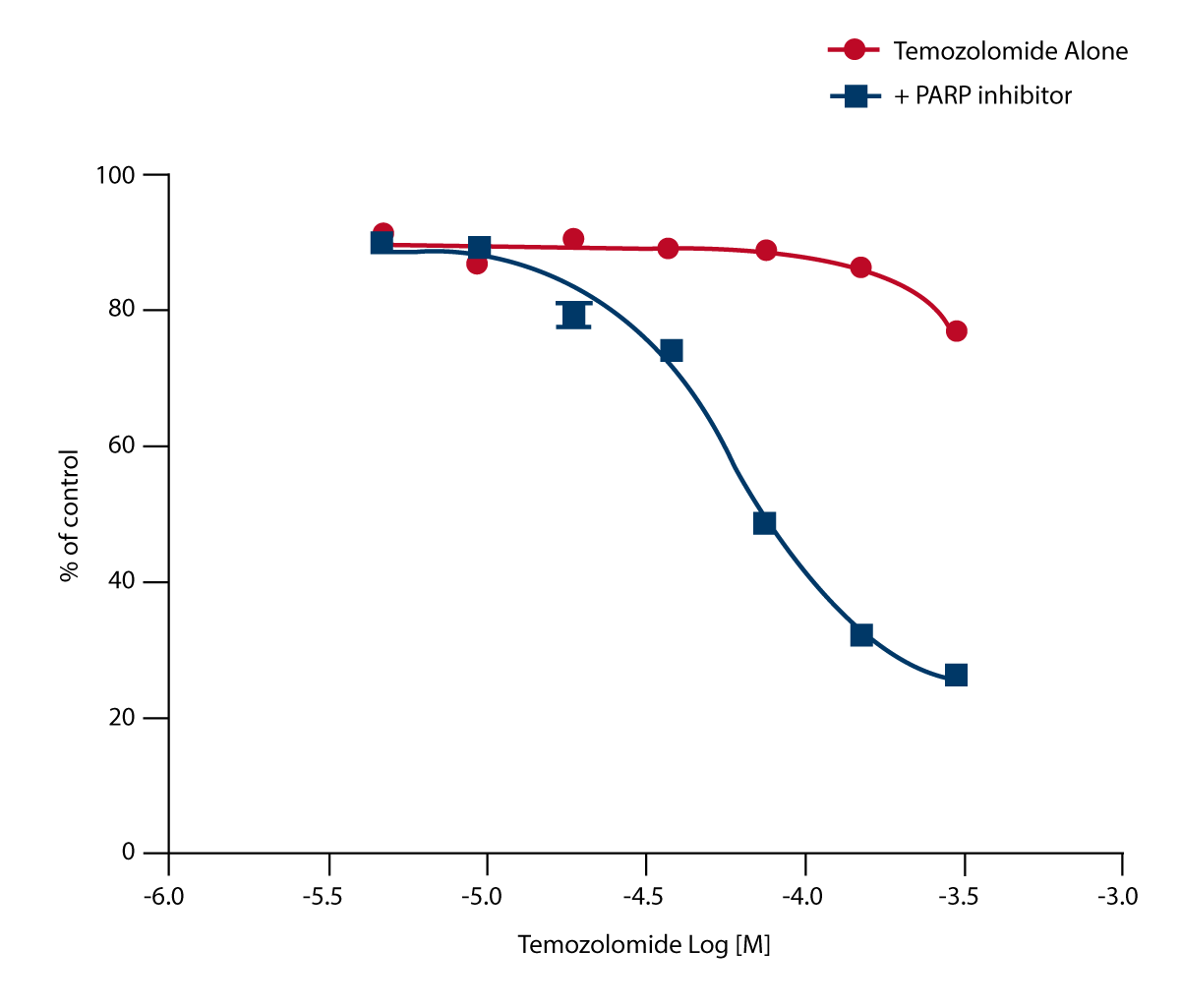 Note: PARP inhibitor alone does not stimulate cell death.
Conclusion
The ability of the FLUOstar Omega to measure absorbance, luminescence and fluorescence, facilitates simple, rapid measurement of all aspects of our evaluation, using a single machine.