SPECTROstar Nano
Absorbance plate reader with cuvette port

DNA quantification collectively refers to the different methods available to measure the amount or concentration of DNA in a sample or ways to determine its quality or purity. These types of measurements also apply to other nucleic acids like ribonucleic acids (RNAs) and are vital to the success of many of the crucial tools and approaches used in the life sciences including for example polymerase chain reaction (PCR), sequencing, and cloning.
The widespread use of next-generation sequencing and other high-throughput approaches as well as studies on transcription regulation reinforces the need for reliable and accurate sample preparation methods in molecular biology. Accurate DNA and RNA quantification (commonly referred to as nucleic acid quantification) is therefore one of the most crucial – yet overlooked - steps in sample preparation and can be accomplished through several techniques. Comprehensive solutions, including instruments and assay kits, are available to address DNA and RNA quantification needs in laboratory analysis.
DNA quantification methods in molecular biology are often based on different measurement principles and/or use completely different readouts to quantify DNA. While ultraviolet (UV) absorbance analysis measures the presence of all molecules absorbing at 260 nm, polymerase chain reaction (PCR) specifically detects the concentration of amplifiable nucleic acid molecules. Accordingly, the differences in DNA quantification techniques can have a significant impact on your results, depending on the downstream applications you want to use the analyte DNA for. It is essential to determine the concentration or purity of DNA samples to validate analytical methods and ensure suitability for downstream applications.
DNA quantification using UV absorbance has been the method of choice in molecular biology for decades. It exploits the natural absorbance of nucleic acids at 260 nm to quantify DNA1. The Lambert Beers law states that there is a logarithmic dependence between the transmission of light through samples and the product of the absorption coefficient of the substance and the pathlength² (Figure 1).
![Fig. 1: Beer’s law, with b = pathlength [cm], c = concentration of absorbing substance in solution [mol/l or M], e = substance-specific constant [cm-1 M-1] (extinction coefficient).](https://www.bmglabtech.com/hs-fs/hubfs/1_Webseite/5_Resources/Blogs/dna-quantification-fig1.webp?width=600&height=103&name=dna-quantification-fig1.webp)
For DNA and RNA, the heterocyclic rings of the nucleotides (adenine, guanine, cytosine, and thymine/uracil) are responsible for the absorption of UV light with a maximum at 260 nm. The extent of light absorption at specific wavelengths varies between dsDNA, ssDNA, and RNA samples which are reflected in different extinction coefficients:
dsDNA: 50 (μg/mL)-1cm-1
ssDNA: 33 (μg/mL)-1 cm-1
RNA: 40 (μg/mL)-1 cm-1
UV absorbance measurements performed with a spectrophotometer allow the nucleic acid concentration in samples to be calculated based on these extinction coefficients. Absorbance measurements at 260 nm combined with the measurement at 280 nm are also used to assess DNA purity, providing information about sample quality.
Since nucleic acids and their components show natural absorbance at 260 nm, no sample preparation or staining procedure is needed to quantify DNA using this spectrophotometric technique. The nucleic acid quantification based on UV absorbance is quick, easy, and robust, if you disregard the impact of pH, which may differ in water vs. buffer, on results. While UV measurements with a spectrophotometer allow quantifying DNA in highly concentrated samples up to several µg/µL, this DNA quantification method shows limited low-end sensitivity. Read more on an application example of absorbance-based DNA quantification here. Absorbance-based methods, such as UV-visible spectrophotometry, are often compared to fluorescence-based techniques, with each having distinct advantages and limitations for DNA quantification.
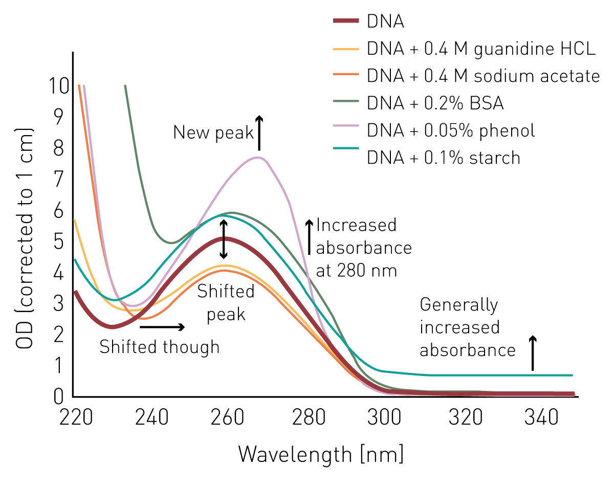
Since dsDNA, ssDNA, and RNA samples generally have comparable absorbance spectra which only differ in the extent of absorbance, it is not possible to discriminate them from each other or from free nucleotides and primers in samples by UV measurements. Moreover, neighbouring peaks in the sample derived from contaminants like proteins or phenol will distort the nucleic acid quantification reading in a sample and lead to a miscalculation of the actual nucleic acid concentration (Figure 2). Guanidine hydrochloride or sodium acetate, both salts which are frequently used for nucleic acid purification, tend to decrease the absorbance of DNA at 260 nm and consequently lead to an underestimate of the amount of DNA in samples. In contrast, cellular protein residues, or phenol extracts used to remove them, show substantial absorbance at 260 nm and thereby lead to an overestimation of the actual DNA concentration in samples.
Interestingly, what is a disadvantage for DNA quantification, can in fact be used to uncover these exact contaminants which in turn ameliorates the inaccuracies they cause in DNA quantification.
The presence of contaminants slightly changes the DNA/RNA absorbance spectrum by increasing or decreasing the absorbance at 230 nm, 280 nm, and 340 nm next to 260 nm1 (Figure 2). Phenol, proteins, and salts for example show increased absorbance at 230 nm. Other organic compounds may also maximally absorb at 230 nm, and their presence can interfere with purity assessment. Furthermore, phenol extracts, particulate substances, and several amino acids display an increased absorbance at 280 nm. Protein contamination can interfere with spectrophotometric measurements of nucleic acids, specifically affecting the A260/A280 ratio and distorting absorbance readings.
In addition to gel electrophoretic nucleic acid evaluation, contaminations may therefore be uncovered by dividing the Optical Densities (ODs) at different wavelengths. This results in the 260/230 ratio, which should be ~ 2.2 for DNA, and the 260/280 ratio, for which values of ~ 1.8 are generally targeted. Besides these purity ratios, the OD at 340 nm, which is derived from particulate substances, is often used for background correction (see application example in AN 362: DNA purity ratio – fast and easy absorbance-based evaluation of nucleic acid quality).
Tip: Full absorbance spectra can easily be used to measure OD at 260 nm for DNA quantification and at the same time calculate purity ratios which can be used to uncover contaminations.
BMG LABTECH’s single- and multi-mode microplate readers are equipped with a UV/visible spectrophotometer that can acquire absorbance spectra from 220 - 1000 nm in <1 sec/well. This enables the simultaneous acquisition of OD values at 230, 260, 280, and 340 nm in less than one second per sample, much faster than any filter-based or monochromator-based reader. Additionally, all BMG LABTECH microplate readers offer the possibility to make measurements in very small volumes, down to 2 µl of the sample using the LVis Plate.
By employing dyes that specifically bind to nucleic acids, DNA/RNA concentrations can also be quantified using fluorescence detection. Fluorometric techniques, which utilize highly sensitive fluorescent dye binding, are widely used for DNA and RNA quantification due to their ability to detect low DNA concentrations with high specificity.
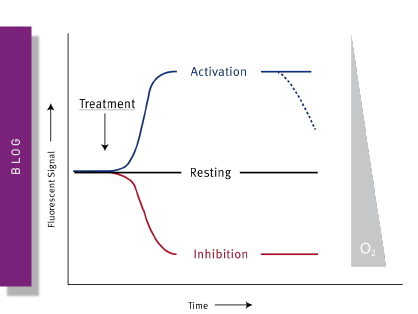
Here, dyes are excited with light of a specific wavelength and the emitted light, which correlates to the nucleic acid concentration, is captured with a fluorescence microplate reader. Different dyes show varying affinities for the different nucleic acids subspecies and can thus be used to discern between them. DNA binding of the dyes results in an increase in their fluorescent quantum yields (Figure 3). This substantial fluorescence increases upon binding and the specific affinity of the dye to DNA results in a low background signal and consequently high sensitivity and specificity using this technique. Due to the specific interaction of dye and nucleic acid, this DNA quantification method is less susceptible to the impact of potential contaminants. The sensitivity of DNA quantification is usually increased by 10-100 times compared to absorbance measurements.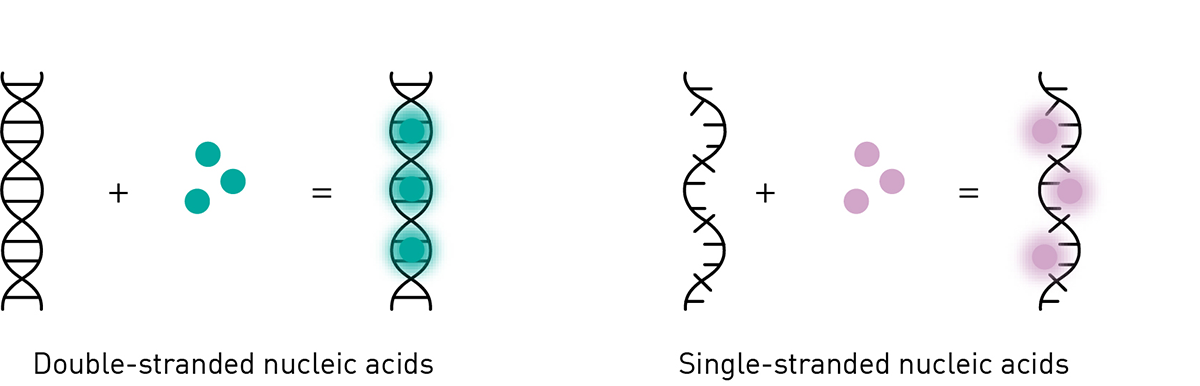
However, fluorescence-based DNA quantification requires specific fluorescent labelling dyes, a staining procedure, and a standard curve. Quantification is performed by comparing the fluorescence of the DNA sample to a standard curve generated from standards of known concentrations, allowing you to compare the sample signal to reference values. Accurate determination of DNA quantity depends on measuring within the linear range of the standard curve. Band intensity, as measured in gel-based methods, can also be compared to standards of known concentrations within the linear range for quantifying DNA fragments. Taking all this into account, the fluorescence-based DNA quantification method is significantly more expensive and time-consuming compared to other approaches in molecular biology1.
There are many different fluorescent dyes for DNA quantification on the market, like e.g. Picogreen®, Hoechst, AccueBlueTM, and QubitTM. Many of these fluorescent dyes are offered in a kit including standards of known concentration as well as suitable dilution buffers. These nucleic acid quantification kits differ in their target (e.g. dsDNA, ssDNA, RNA, and miRNA), detection range (broad range, high sensitivity kits), and the required sample volume. Picogreen® for example allows the detection of DNA down to the femtomolar range.
Tip: Check if your microplate reader is equipped with suitable filters during your assay selection. Many of the available assays contain fluorescent dyes which can be detected with standard filters. However, if you want to stay flexible and also don't want to compromise on sensitivity during nucleic acid quantification, our CLARIOstar Plus and VANTAstar® plate readers are equipped with LVF Monochromators giving you full flexibility in the wavelength-selection with filter-like sensitivity.
Fluorescence-based methods are often compared to traditional gel electrophoresis techniques, such as agarose gel electrophoresis, where DNA fragments are separated on an agarose gel and DNA quantity is estimated by comparing band intensity to standards of known concentrations. While agarose gel and gel electrophoresis methods are useful for visualizing DNA fragments and assessing DNA sample integrity, fluorometric techniques offer greater sensitivity and specificity for quantifying low DNA concentrations.
Finally, it is also possible to use amplification-based methods like polymerase chain reaction (PCR)³, real-time PCR, or loop-mediated isothermal amplification (LAMP)4 for nucleic acid quantification. These methods can be used to quantify DNA or RNA, and are suitable for measuring both DNA and RNA concentration in various samples. Approaches like PCR rely on thermal cycling and require a DNA polymerase and sequence-specific primers next to nucleotides, buffer as well as specific dyes or probes for your test samples.
Amplification-based methods like PCR have higher sensitivity compared to other fluorescence-based approaches. PCR and LAMP detect only molecules that can be amplified. The ability to amplify is an important attribute for downstream applications which is not addressed by any other method.5-7
In real-time quantitative PCR (qPCR), the absolute nucleic acid concentration is calculated based on the amplification rounds needed to reach a fluorescence threshold. qPCR techniques require costly PCR reagents and advanced instrumentation like a PCR cycler, which should be operated by trained staff. If a standard PCR is performed, the endpoint measurement of the PCR reaction can also be done using a fluorescence microplate reader and respective standards. Since LAMP assays only require a maximum temperature of 65°C in contrast to PCR, they can also be performed on BMG LABTECH readers with extended incubation function and can be read in real-time using fluorescence or absorbance-based readout. For more details on the application of an absorbance-based LAMP assay on the BMG LABTECH’s Omega series see AN356. Like LAMP the isothermal DNA detection RPA technology can also be performed at a constant temperature on a microplate reader. Recent approaches aim to significantly reduce the time requirement of PCR. The NextGenPCRTM from Molecular Biology Systems (MBS), for example, uses innovative heating technology to enable a complete PCR run in less than 2 min. By using fluorescently-labelled probes, the reaction can be evaluated on fluorescence plate readers, as in this example: AN370: NextGenPCR™ evaluated with a fluorescence microplate reader accelerates testing for SARS-CoV-2 viral RNA.
The time and human resources required for PCR are often only available in specialized laboratories, while most use standard fluorescence and absorbance methods. Absorbance and fluorescence can be considered as contrasting but complementary methods for DNA quantification. While absorbance measurements indicate contamination in a quick and easy manner, fluorescence-based measurements are highly specific and enable the detection of very low concentrations of nucleic acids, including RNA. BMG LABTECH multi-mode microplate readers can perform both kinds of DNA quantification measurements. When using BMG LABTECH readers for DNA quantification, concentration calculation is done automatically using the MARS data analysis software and can also be transferred 1:1 to subsequent evaluations using templates. Read more on a direct comparison of fluorescence- and absorbance-based DNA quantification methods in AN352.
Absorbance plate reader with cuvette port
Powerful and most sensitive HTS plate reader
Most flexible Plate Reader for Assay Development
Upgradeable single and multi-mode microplate reader series
Flexible microplate reader with simplified workflows
Gene reporter assays are sensitive and specific tools to study the regulation of gene expression. Learn about the different options available, their uses, and the benefits of running these types of assays on microplate readers.
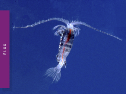
Life in the depths of the ocean operates under extreme conditions. Find out how proteins from deep-sea luminescent organisms are useful for measurements on microplate readers.
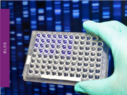
Next generation sequencing (NGS) technologies for DNA or RNA have made tremendous progress in recent years. Find out how microplate readers can advance the quality control of nucleic acids to facilitate NGS.
Measuring light scattering is an accurate and expedient way to determine drug solubility. Find out how the NEPHELOstar® Plus can be used for early drug solubility screening at high throughput.
Mitochondrial toxicity can have devastating effects on the cell and life. Find out how microplate readers can be used to assess mitochondrial health and how this impacts disease and drug discovery.
Find out how microplate readers can be used to measure histone deacetylase (HDAC) activity and assist drug discovery.