
PHERAstar FSX
Leistungsstarker und sensitivster HTS-Plate Reader auf dem Markt


AlphaScreen® (ALPHA, Englisch für Amplified Luminescent Proximity Homogeneous Assay) ist eine auf Beads basierende Chemie zur Untersuchung von Wechselwirkungen zwischen Molekülen in einer Mikroplatte. Ursprünglich als Methode für diagnostische Assays mit der Bezeichnung LOCI® (Englisch für Luminescent Oxygen Channeling Immunoassay) im Jahr 19941 entwickelt, umfasst die AlphaScreen-Technologie heute AlphaScreen, AlphaLISA® und AlphaPlexTM. Sie wird hauptsächlich für Screening-Zwecke in den Biowissenschaften eingesetzt.
Das Grundprinzip der AlphaScreen-Technologie beruht auf der Bindung zweier Ziel-Moleküle an spezifische Beads. Im Falle einer Wechselwirkung zwischen den beiden Molekülen und der daraus resultierenden räumlichen Nähe der beiden Beads findet ein Energietransfer von einem Bead zum anderen statt. Dies führt zur Erzeugung eines chemilumineszenten Signals. Die AlphaScreen-Technologie wird hauptsächlich in Hochdurchsatz-Screening-Assays eingesetzt, um biomolekulare Wechselwirkungen, die Bildung/Abspaltung eines Substrats oder Produkts, posttranslationale Modifikationen und die Quantifizierung von Analyten zu untersuchen.
Neben Interaktionsassays (einschließlich der Interaktion zwischen Ligand und Rezeptor, Proteinen sowie Protein und DNA) kann die AlphaScreen-Technologie auch für funktionelle GPCR-Assays (dem Nachweis von Second Messengern), enzymatische Assays und Immunoassays eingesetzt werden.
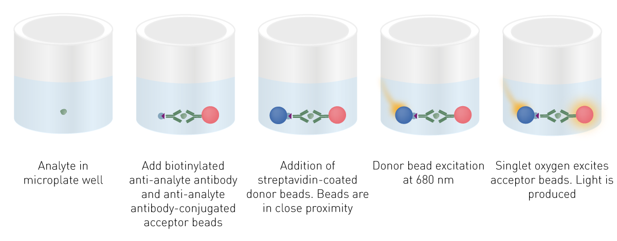
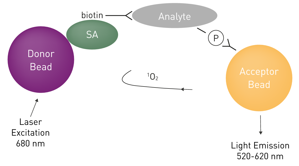
AlphaScreen-Assays basieren auf zwei Arten von hydrogelbeschichteten Beads, den sogenannten Donor- und Akzeptor-Beads. Die beiden Bead-Typen enthalten unterschiedliche Chemikalien, die für die Erzeugung des Leuchtsignals entscheidend sind. Das Donor-Bead enthält einen Photosensibilisator, der bei Anregung durch Licht bei 680 nm Sauerstoff (O2) in eine anregende Form, Singulett-Sauerstoff (1O2), umwandelt. Singulett-Sauerstoffmoleküle haben eine verkürzte Lebensdauer (4 Mikrosekunden Halbwertszeit) und können etwa 200 nm in Lösung diffundieren, bevor sie in den Grundzustand zurückfallen.
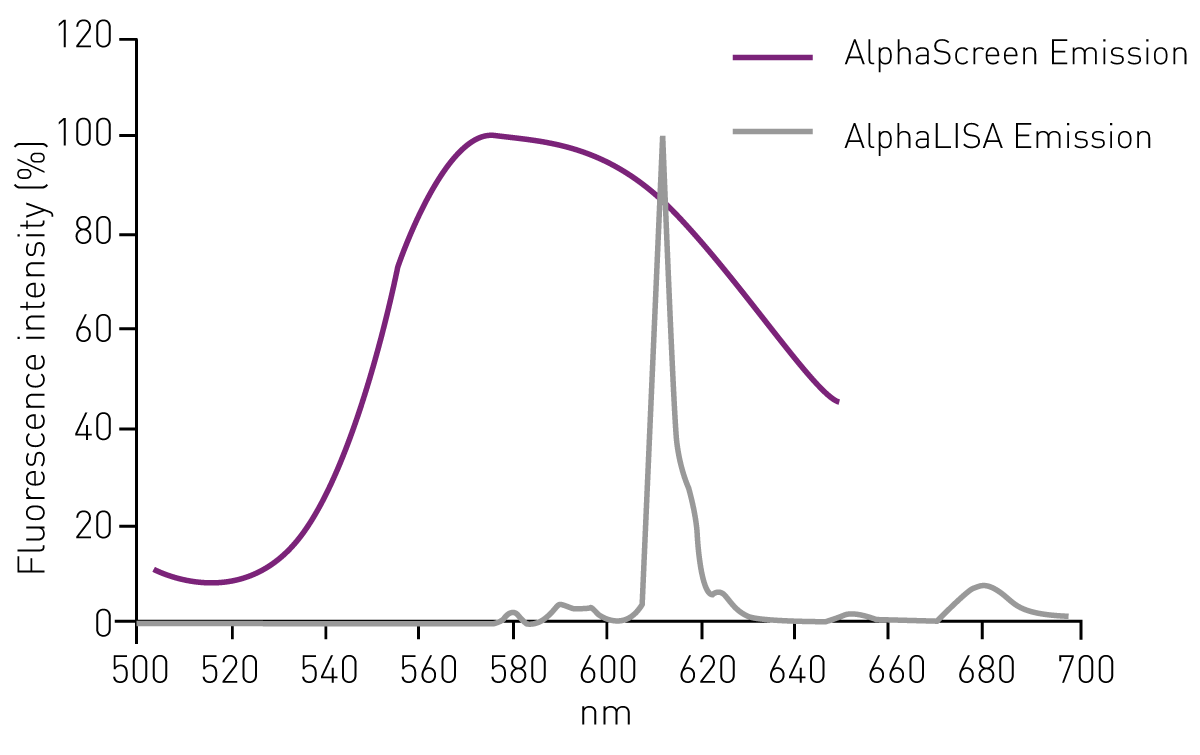
In Abwesenheit von Akzeptor-Beads fallen die Singulett-Sauerstoffmoleküle in den Grundzustand zurück, ohne einen Lichtsignal zu erzeugen. Befindet sich ein Akzeptor-Bead innerhalb von 200 nm, wird Energie von den Singulett-Sauerstoffmolekülen auf das Akzeptor-Bead übertragen, was zur Erzeugung von Licht führt. AlphaScreen-Akzeptor-Beads enthalten drei chemische Farbstoffe, Thioxen, Anthracen und Rubren. Um Licht zu erzeugen, reagieren Singulett-Sauerstoffmoleküle zunächst mit Thioxen. Das Licht wird dann auf Anthracen und Rubren übertragen und führt zu einer breiten Lichtemission von 520 nm bis 620 nm.2 Die Halbwertszeit der Signalabbaureaktion beträgt 0,3 Sekunden.
Bei Bindungsassays wird ein Bindungspartner (z. B. ein Rezeptor) mit dem Donor- und der andere (z. B. ein Ligand) mit dem Akzeptor-Bead verbunden. Wenn Rezeptor und Ligand interagieren, wird chemische Energie von einem Bead-Typ auf den anderen übertragen und ein Leuchtsignal erzeugt (Abb. 1). AlphaScreen kann auch für Verdrängungs- oder Spaltungsassays eingesetzt werden. In diesen Fällen wird eine Verringerung der Signalintensität gemessen.

Donor-Beads sind in der Regel als Streptavidin-Konjugate erhältlich, die die spezifische Bindung von Biotin an Streptavidin zur Kopplung an Biomoleküle ausnutzen. Akzeptor-Beads hingegen sind in erster Linie mit Antikörpern konjugiert. Daher erfordert der zweite Bindungspartner in der Regel die Anwesenheit eines entsprechenden Antigens, um gebunden zu werden (Abb. 2). Alternativ können beide Bead-Typen durch reduktive Aminierung direkt mit den Bindungspartnern beschichtet werden.
Da beide Arten von Beads einen Durchmesser von etwa 250 nm haben, sind sie zu klein, um in biologischen Puffern zu sedimentieren. Darüber hinaus verstopfen sie keine Spitzen oder Injektoren und können problemlos von automatisierten Geräten zur Flüssigkeitsverarbeitung gehandhabt werden. Dennoch sind sie groß genug, um gefiltert oder zentrifugiert zu werden. Die Hydrogel-Beschichtung ermöglicht die Konjugation von Biomolekülen an die Beads und verringert gleichzeitig die unspezifische Bindung und Selbstaggregation.
AlphaLISA ist eine Weiterentwicklung der AlphaScreen-Technologie, die auf denselben Donor-Beads beruht, aber eine andere Art von Akzeptor-Beads verwendet. In AlphaLISA-Beads werden Anthracen und Rubren durch Europiumchelate ersetzt. Angeregtes Europium emittiert Licht bei 615 nm mit einer viel engeren Bandbreite als AlphaScreen (Abb. 3). Daher ist die AlphaLISA-Emission weniger anfällig für Störungen durch andere Substanzen und kann gut für den Nachweis von Analyten in biologischen Proben wie Zellkulturüberständen, Zelllysaten, Serum und Plasma eingesetzt werden.
AlphaLISA ermöglicht die Quantifizierung von sezernierten, intrazellulären oder zellmembranstämmigen Proteinen. Für den Nachweis von Biomarkern wird der AlphaLISA hauptsächlich als Sandwich-Immunoassay eingesetzt. Ein biotinylierter Anti-Analyt-Antikörper bindet an das Streptavidin-Donor-Bead, während ein zweiter Anti-Analyt-Antikörper an AlphaLISA-Akzeptor-Beads konjugiert ist. In Gegenwart des Analyten kommen sich die Beads sehr nahe. Durch die Anregung der Donor-Beads werden Singulett-Sauerstoffmoleküle freigesetzt, die Energie auf die Akzeptor-Beads übertragen, die infolgedessen Licht bei 615 nm emittieren (Abb. 4). Alternativ können auch Immunoassays basierend auf dem Verdrängungsprinzip ffür diesen Zweck angepasst werden.

Im Vergleich zu klassischen ELISAs wird AlphaLISA eine bessere Sensitivität, ein größerer dynamischer Bereich und eine robustere Leistung bei reduzierten Testzeiten zu geschrieben. Da außerdem keine Waschschritte erforderlich sind (siehe unten), lässt sich die Methodik zudem leicht in automatisierte Hochdurchsatz-Screeningverfahren übertragen.

AlphaPlex ist eine Weiterentwicklung von AlphaScreen, die die Quantifizierung von bis zu drei Analyten in einem einzigen Well ermöglicht. Hierfür werden verschiedene Akzeptor-Beads verwendet, die bei unterschiedlichen Wellenlängen emittieren. In diesen Beads werden Anthracen und Rubren entweder durch Terbiumchelate (für AlphaPlex 545) oder Samariumchelate (für AlphaPlex 645) ersetzt. Biotinylierte Anti-Analyt-Antikörper binden die Streptavidin-Donor-Beads. Entsprechende Anti-Analyt-Antikörper sind zudem entweder an AlphaPlex- oder AlphaLISA-Akzeptor-Beads konjugiert. In Gegenwart der Zielanalyten löst die Anregung der Donor-Beads und die daraus resultierende Freisetzung von Singulett-Sauerstoff die chemilumineszente Lichtemission der verschiedenen Akzeptor-Beads aus.
Jede Art von Bead emittiert Licht bei einer anderen Wellenlänge. Neben 615 nm für AlphaLISA-Beads liegen die anderen Emissionsspitzen bei 545 nm für Terbium-Beads und bei 645 nm für Samarium-Beads (Abb. 5). Dementsprechend wird die Entwicklung von Triplex-Assays durch die Verwendung von AlphaLISA-, AlphaPlex 545- und AlphaPlex 645-Akzeptor-Beads in einem Assay ermöglicht.2

Die Messung der AlphaScreen-Chemie wird überwiegend mit Mikroplatten-Lesegeräten durchgeführt. Da es sich um einen unabhängigen Detektionsmodus handelt, erfordert die Assay-Detektion einen Mikroplatten-Reader mit AlphaScreen-Option. Die Technologie wird häufig im Hochdurchsatz-Wirkstoffscreening eingesetzt. Daher sollten die Mikroplatten-Reader mit 384- und 1536-Well-Platten kompatibel sein.
Der Grundaufbau des AlphaScreen-Mikroplatten-Reader besteht aus einer Lichtquelle, Anregungs- und Emissionsfiltern zur Auswahl der Wellenlänge und einem Photomultiplier-Tube-Detektor (PMT).
Da der Photosensibilisator auf den Donor-Beads spezifisch bei 680 nm angeregt wird, werden eine Xenon-Blitzlampe oder ein spezieller Anregungslaser als Lichtquelle verwendet. Ein spezieller AlphaScreen-Laser fokussiert mehr Energie bei 680 nm und übertrifft damit Xenonlampen, was zu besseren Ergebnissen mit einem breiteren Dynamikbereich und einem höheren Signal-Rausch-Verhältnis führt.
Dedizierte Laser zur Anregung von AlphaScreen sind in der Regel in High-End- und HTS-spezifischen Multimode-Mikroplatten-Readern wie dem PHERAstar FSX und dem CLARIOstar Plus erhältlich. Preiswerte Reader sind in der Regel mit einer Xenon-Blitzlampe für die AlphaScreen-Detektion ausgestattet.
AlphaPLEX umfasst die Detektion verschiedener Emissionswellenlängen. Obwohl diese sequentiell gemessen werden können, bietet die gleichzeitige Detektion von zwei Emissionen sowohl Vorteile in der Datenqualität als auch hinsichtlich der Geschwindigkeit, insbesondere bei Screening-Assays. Dementsprechend trägt die simultane Dual-Emissions-Detektion zur Steigerung des Durchsatzes bei AlphaLISA- AlphaPlex-Assays mit zwei Emissionen bei.
Unter Crosstalk versteht man das Licht, das von einem anderen als dem gemessenen Well stammt und vom Detektor somit unspezifisch gemessen wird und das Signal des gemessenen Wells negativ beeinflusst.
Da das in einem Well durch eine AlphaScreen-Reaktion erzeugte Licht diffus ist, kann es sich auf benachbarte Vertiefungen und direkt auf die Position der Detektion ausbreiten, auch wenn ein anderes Well gemessen wird. Dies führt zu verzerrten Signalen, höheren Signalschwankungen und generell zu einer geringeren Sensitivität.
Unerwünschte Lichtsignale erreichen den Detektor entweder von oberhalb der Platte und/oder durch die Wand der Wells und müssen anders verarbeitet werden (Abb. 6).
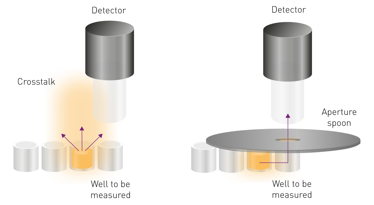
Beide Tools zur Reduzierung des Crosstalks sind auf den BMG LABTECH Mikroplatten-Readern PHERAstar FSX undCLARIOstar Plus.
Die AlphaScreen/AlphaLISA-Detektion kann mit dem PHERAstar FSX und dem CLARIOstarPlus durchgeführt werden. Da eine hochintensive Anregungsquelle für den Assay von Vorteil ist, verfügen sowohl der CLARIOstar Plus als auch der PHERAstar FSX über spezielle Festkörperlaser, die hochintensives Licht bei 680 nm erzeugen. Darüber hinaus bietet der PHERAstarFSX spezielle optische AlphaPlex-Module, die die gleichzeitige Messung von zwei Alpha-Signalen ermöglichen.
AlphaScreen bietet eine homogene, empfindliche und abwärts skalierbare Plattform, die sich besonders gut für Screening-Anwendungen mit hohem Durchsatz eignet.
Der Signalerzeugungsprozess bei AlphaScreen ist eine Kaskadenreaktion, die eine hohe Signalverstärkung bewirkt. Dies führt zu einer hohen Empfindlichkeit. Darüber hinaus ist der Hintergrund gering. Dies ist hauptsächlich darauf zurückzuführen, dass die Anregung im roten Bereich und damit bei einer höheren Wellenlänge als das Emissionssignal statt findet. Dadurch wird die Autofluoreszenz von biomolekularen Komponenten reduziert, die normalerweise im blau-grünen Bereich angeregt werden. Darüber hinaus wird durch die Zeitverzögerung zwischen Anregung und Emission das Autofluoreszenzrauschen weiter eliminiert.
Die AlphaScreen-Chemie ist homogen: Der Nachweis des gebundenen Donor-Akzeptor-Bead-Komplexes erfordert keine physische Trennung von den ungebundenen Komponenten, um den Hintergrund zu reduzieren. Folglich sind keine zwischengeschalteten Trenn- oder Waschschritte erforderlich, und der Test kann mit einem einfachen Add-and-Read-Protokoll durchgeführt werden. Dies minimiert die Handhabungsschritte und macht ihn benutzerfreundlicher und weniger zeitaufwändig als andere Methoden. Sie eignet sich daher besonders für automatisierungsgestützte Screening-Zwecke. Dies ist einer der Gründe für die große Verbreitung von AlphaScreen Anwendungen in derArzneimittelforschung und im HTS-Bereich.
Aufgrund seiner hohen Empfindlichkeit und seines geringen Hintergrunds lassen sich AlphaScreen-Assays leicht in 1536-Well-Platten überführen und können problemlos auf wenige µl reduziert werden, ohne dass die Konzentrationen der Reagenzien angepasst werden müsste oder die Zuverlässigkeit des Assays leidet: Dies wird auch in der Application Note "Miniaturisierung eines zellbasierten TNF-α AlphaLISA-Assays" bestätigt.
Weiße Mikrotiterplatten sind für AlphaScreen-Messungen am besten geeignet, da sie das Lichtsignal reflektieren. Schwarze Platten absorbieren das Licht und reduzieren dabei sowohl das Signal als den Hintergrund. Graue Platten bieten den besten Kompromiss zur Reduzierung von Crosstalk und einer hohen Signalreflexion. Ausführliche Informationen zur Plattenwahl finden Sie in unserem Artikel: "Die Mikroplatte: Nutzen in der Praxis".
Da weiße Platten eine intrinsische Phosphoreszenz haben, emittieren sie Licht, wenn sie dem Sonnen- oder Raumlicht ausgesetzt werden. Dieses unspezifische Signal wird vom Mikroplatten-Reader zusammen mit dem von der Probe emittierten Licht gemessen und führt zu einem erhöhten Blank und Hintergrund sowie zu einem reduzierten Messbereich. Es wird daher empfohlen, weiße Platten entweder im Dunkeln vorzubereiten oder sie vor der Messung etwa 15 Minuten lang im Dunkeln zu belassen.
Die AlphaScreen-Chemie ist lichtempfindlich. Idealerweise sollte sie unter gedämpften Lichtbedingungen (unter 100 Lux) vorbereitet und gemessen werden. Da dies nicht immer möglich ist, sollte ein geschlossener Raum mit grün gefilterter Beleuchtung ausgestattet werden. Leuchten in unmittelbarer Nähe des Arbeitsbereichs und/oder des Mikroplatten-Readers sollten mit Grünfiltern ausgestattet werden. Es hat sich gezeigt, dass die Verwendung von Grünfiltern fast genauso effektiv ist wie die Durchführung des Tests im Dunkeln (Abb. 7).
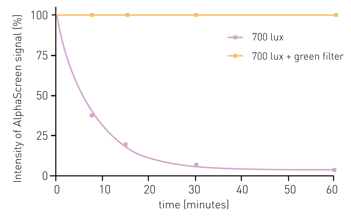
Abb. 7: Der Signalverlauf über verschiedene Belichtungszeiten bestätigt die Wirksamkeit von Grünfiltern zum Schutz der Signalstabilität von AlphaScreen.3
Platten, die über einen längeren Zeitraum inkubiert werden, sollten vor Licht geschützt werden - z.B. abgedeckt mit einem dunklen Tuch, Aluminiumfolie oder einem Karton. Vermeiden Sie es, die Mikroplatte oder den Mikroplatten-Reader direkter Sonneneinstrahlung oder intensivem Licht auszusetzen.
Die AlphaScreen-Chemie ist temperaturempfindlich. Daher sollten starke Schwankungen der Raumtemperatur vermieden werden, da diese sich negativ auf die Signalerzeugung, -intensität und -stabilität auswirken. Normalerweise verändert sich das AlphaScreen-Signal etwa um 8 % pro °C.3
Damit während der Messung keine unerwünschten Gradienten in der Signalintensität über die gesamte Platte entstehen, sollten Reagenzien und Mikrotiterplatten an die Raumtemperatur angepasst werden. Bei Platten kann dies leicht durch Inkubation in einem THERMOstar-Inkubator erreicht werden. Der PHERAstar FSX mit AAS-System ermöglicht es sogar, das Lesegerät aktiv auf eine beliebige Temperatur zwischen 18° und 45°C einzustellen und so die Inkubationskammer des Geräts auf Raumtemperatur oder darunter abzukühlen. In dieser Application Note wird demonstriert wie das AAS-System ermöglicht, eine stabile Temperatur aufrechtzuerhalten, die Verdunstung zu reduzieren und die Abkühlzeit der Gerätes von hohen zu niedrigen Temperaturen zu verringern.
Zellkulturmedien wie RPMI 1640, MEM und DMEM wirken sich negativ auf die Signalintensität aus. Bereits 10 % Kulturmedium im Assay führt zu einer Signalreduktion von etwa 30 %. In ähnlicher Weise reduziert 10 % fötales Kälberserum das Signal um etwa 25 %.3 Es wird daher empfohlen, die Zellproben vor der Untersuchung mit der AlphaScreen-Chemie mit einem geeigneten Puffer wie PBS zu spülen.
Die AlphaScreen-Technologie kann zur Analyse von Bindungen oder biochemischen Vorgängen in 96-, 384- und 1536-Well-Plattenformaten für verschiedene biologische Fragestellungen eingesetzt werden. Die Technologie ist vor allem im Bereich des Arzneimittelscreenings weit verbreitet.
AlphaScreen wird in verschiedenen Assays eingesetzt, darunter z.B. GPCR-Funktionsassays (cAMP, IP3, und phospho-ERK1/2), enzymatischen Assays (Tyrosinkinase, Helikase, Protease, Phosphatase), Interaktionstests (Zytokin-Bindungstests, funktionelle Nuklearrezeptor-Assays, Liganden-Rezeptor-Bindungstests, Protein/Protein, Protein/DNA,Protein/Peptid) oder ELISA-ähnlichen Immunoassays zur Quantifizierung von Analyten (TNF-α,IgG).
In der Application Note PHERAstar measures AlphaScreen assay to develop selective inhibitors for the human YEATS domains zeigen wir ein Beispiel für einen AlphaScreen-basierten Interaktionsassay.
YEATS-Domänen sind epigenetische Regulatoren. Das YEATS-enthaltende ENL wurde als ein Hauptauslöser für verschiedene Arten von akuter Leukämie identifiziert. ENL stellt daher ein Ziel der Arzneimittelforschung dar. Um nach selektiven Inhibitoren zu suchen, wurde ein Histon 3 (H3) - YEATS-Domänen-Bindungstest mit dem AlphaScreen Histidin-Detektionskit durchgeführt. Das Donor-Bead ist über Streptavidin an acyliertes H3 und das Akzeptor-Bead an die YEATS-Domäne gekoppelt. Wenn Protein und Peptid interagieren, werden die Beads zusammengebracht und ein AlphaScreen-Signal wird erzeugt (Abb. 8). Inhibitoren verringern das Emissionssignal entsprechend, indem sie die H3-YEATS-Interaktion unterbrechen.

Neben Interaktionuntersuchungen können auch Immunoassays durchgeführt werden. Die Reaktionen vieler GPCRs lassen sich mit den derzeitigen Assay-Formaten nicht leicht messen. Die ERK-Phosphorylierungskaskade führt zur Phosphorylierung sowohl von zytoplasmatischen als auch von nukleären Proteinen, die die Gentranskription regulieren. Daher kann sie zum Screening auf zelluläre Veränderungen, ausgelöst durch GPCR-Aktivität ausgelöst, verwendet werden.
Zu diesem Zweck wurde der AlphaScreen SureFire® ERK1/2-Phosphorylierungsassay für Zelllysate verwendet. Dabei handelt es sich um einen Sandwich-Immunoassay. Ein phosphoryliertes zelluläres Protein befindet sich als Bindeglied zwischen einem Anti-Analyten-Antikörper, der mit einem Streptavidin-beschichteten Donor-Bead assoziiert ist, und einem Anti-Phospho-Antikörper, der mit einem Protein A-konjugierten Akzeptor-Bead assoziiert ist (Abb. 9). Die Phosphorylierung des Analyten führt zu einer Verstärkung des Lichtsignals. Dies wird in der Application Note An AlphaScreen SureFire Phospho-ERK1/2 assay gezeigt.
AlphaScreen, AlphaLISA, AlphaPlex und SureFire® sind eingetragene Marken von revvity.
Leistungsstarker und sensitivster HTS-Plate Reader auf dem Markt
Flexibelster Plate Reader für die Assayentwicklung
