SPECTROstar Nano
Absorbance plate reader with cuvette port

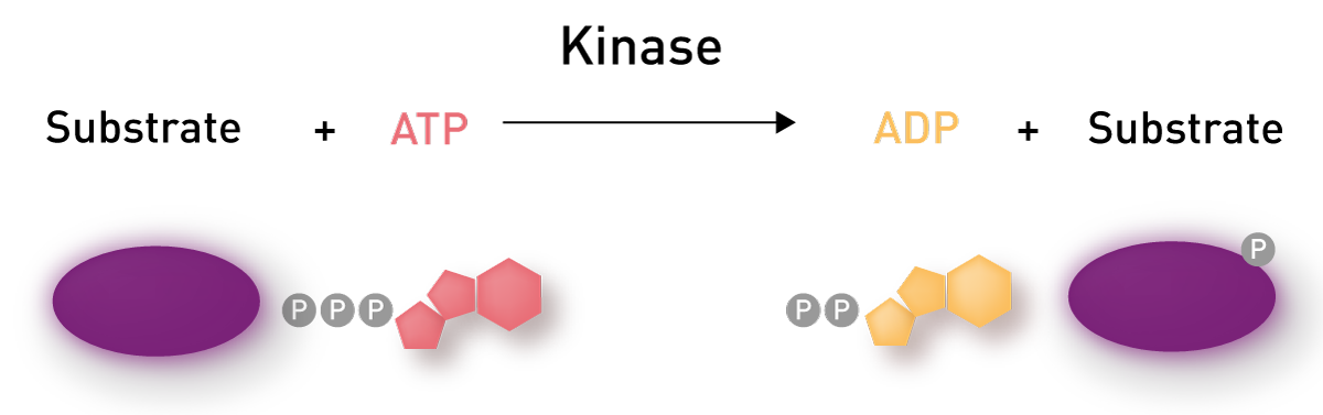
In the most basic of terms, a kinase is an enzyme that can transfer a phosphate group from ATP to a biomolecule (fig. 1). Although this biomolecule could be a protein, lipid, or carbohydrate most research is performed on protein kinases so that will be our focus. The transfer of the phosphate group is called phosphorylation. In protein phosphorylation, the transfer can only occur onto 3 amino acid residues, serine, threonine, and tyrosine. Kinases, for the most part, are divided into those that can phosphorylate serine/threonine and those that can phosphorylate tyrosine. In addition, kinases are not able to phosphorylate every possible residue but are restrained by a variety of sequence motifs around the phosphorylatable residue. These sequences determine if a kinase, specifically the kinases active site, can interact with the protein, coordinate ATP-hydrolysis and phosphate transfer that characterize the phosphorylation event.
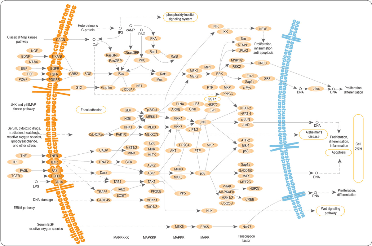
Addition of phosphate causes changes to the local environment in the protein due to charge on the phosphate group as well as simply addition of a ‘bulky’ extension. These local changes can result in conformational changes in the protein that alter its function. This altered function includes changes in the ability of the protein to interact with binding partners. The classic example is transcription factors whose ability to bind specific regulatory elements in DNA are changed by phosphorylation events. Changes in protein function can also be seen in changes to enzyme activity, including changes to kinase activity. From the changes in protein function, extensive webs of interaction and interplay between various kinase signaling pathways have been drawn and are enough to make your head spin (fig. 2).
The final cellular effect of these pathways is the phosphorylation of proteins that are important in the regulation of vital processes such as differentiation, proliferation, and apoptosis. As evidenced by the complex signaling webs associated with kinases, their activity is a tightly regulated process. This regulation is important due to the essential nature of the cellular processes in which they are involved. The involvement of kinases in essential processes also explains the interest in them as targets of therapeutics. Especially to remedy diseases where dysregulation of kinases has been identified as a key component. In particular, cancers are frequently associated with kinase malfunction so the discovery of drugs that target kinases is desirable, and to achieve this assays that can accurately represent kinase activity are required. Using these assays, the ability of test compounds to inhibit the kinase activity can be determined.
In general, the search for kinase inhibitors requires a very targeted approach to decrease the possibility of confounding effects. The targeted approach usually requires the isolation of the kinase from the complex signaling milieu found within cells. Therefore, the typical kinase assay is referred to as an in vitro kinase assay which has been optimized for enzyme/substrate concentrations and buffers conditions, including optimized ATP and co-factor concentrations.
In vitro kinase assays
As we alluded to in the previous section there are several steps involved in the process of a protein being phosphorylated. Protein kinase assays have taken advantage of this fact to develop assays that, monitor interactions with the kinase active site, evaluate conversion of ATP to ADP, and attachment of a phosphate group onto the substrate. Choice of which approach you should use depends on the question you wish to answer, the type of sample to which you have access, and the detection capability you prefer. Fortunately, there are options that employ all the detection capabilities available on BMG LABTECH multimode microplate readers: absorbance, fluorescence, luminescence, fluorescence polarization (FP), time-resolved fluorescence (TRF), and AlphaScreen.
Kinase binding assays
There is a huge selection of binding assays available. To give you an idea, here two popular and commonly used kinase binding assays are being highlighted.
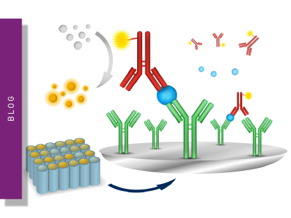
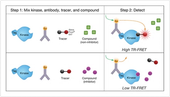
The LanthaScreenTM Eu Kinase Binding assay platform (ThermoFisher Scientific) was designed as an HTS platform that can identify any compound that alters binding to the active site (aka ATP-site) on a kinase. The platform employs a fluorescently labeled tracer of which there are now six. The tracers are based on known inhibitors that bind to kinases. Specificity for a kinase is achieved using a tagged version of the kinase. This tag can bind a Europium-labeled antibody such that when antibody and tracer are both bound to the kinase a high TR-FRET signal is observed. Potential inhibitors are discovered based on their ability to displace the tracer resulting in a decreased TR-FRET (fig. 3). Currently over 300 validated assays are listed. These include several important mutant versions of some of the kinases. Furthermore, it has been shown that this assay platform can be used to study both active and inactive kinases and to assess the role of phosphorylation on the interaction with the tracer1. And as we saw in figure 2 the regulation of kinases by phosphorylation is essential.
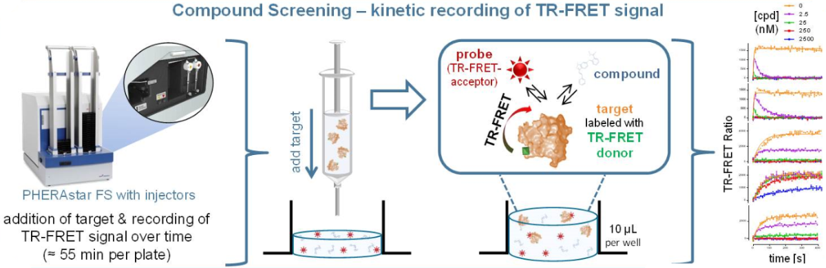
The TR-FRET-based method to measure kinases is further used to analyze the kinetic binding parameters of kinase interactions. To this end, the binding parameters association rate and dissociation rate are initially determined for a combination of Europium-labeled kinase and a fluorescently labeled tracer. Then an unknown potential kinase inhibitor is added to the measurement to investigate whether it competes with the known tracer. If this is the case, the association curve of the tracer changes, and the association and dissociation rate of the unknown inhibitor can be calculated (fig. 4). Details on the kinetic kinase assay are found in our application note “Binding kinetics: high throughput assay for kinase inhibitors”.
Kinase activity assays based on ATP-ADP conversion
A few different assays seek to assess kinase activity based on the amount of ATP that has been consumed. Unlike the kinase binding assay, these are truly kinase activity assays as they measure the conversion of ATP to ADP as a part of the phosphorylation event. These types of assays can be applied to any kinase, or really any enzyme that converts ATP to ADP as a part of their activity.
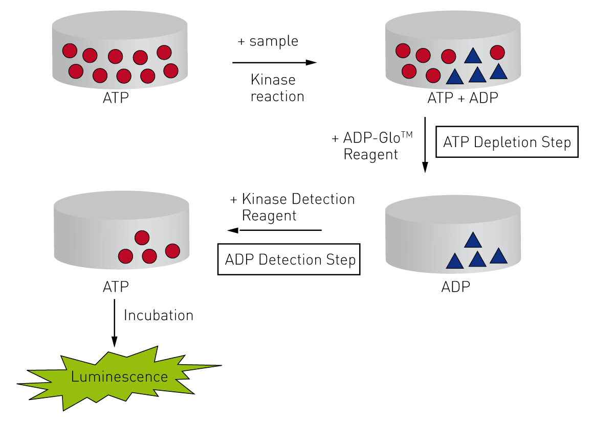
Kinase-Glo® and ADP-Glo™ both use the fact that firefly luciferase/D-luciferin requires ATP to generate light. Thus, light production can be linked to the amount of ATP that was consumed by a kinase under a particular condition. This is most straightforward in Kinase-Glo which measures residual ATP after a kinase reaction was carried out. So, a highly active kinase will deplete ATP and have little light production. In a kinase inhibition assay, the presence of inhibition would be observed as a higher light signal due to more ATP left because of less kinase activity2. ADP-Glo measures the amount of ADP generated by first adding a reagent that stops kinase activity and depletes residual ATP. Then a second reagent is added that stops ATP degradation, converts ADP to ATP, and has the luciferase/luciferin reagents to generate light (fig. 5). The result is a signal that tracks in the opposite direction of Kinase-Glo. High light production for active kinases is reduced by inhibitors.
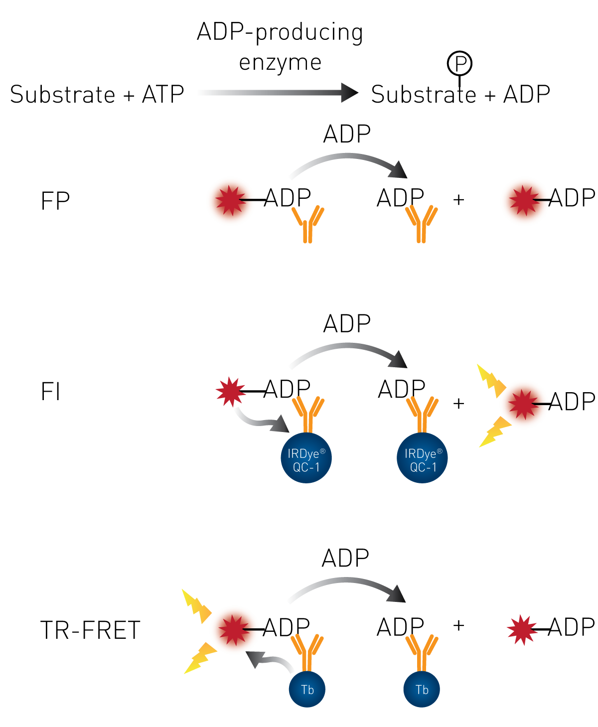
BellBrook Labs provides users with the ability to detect the levels of ADP in their Transcreener® assays using your choice of 3 different detection modes, fluorescence, FP, and TR-FRET. Regardless of detection mode the assay principle for each center on a fluorescently labeled ADP and an antibody to ADP (Fig. 6). The increase in ADP production in the presence of active kinase displaces the fluorescently labeled ADP from the antibody resulting in a quantifiable change in signal intensity4-7. An example of how the assay is performed on a high-throughput reader is found in our application note “PHERAstar FSX certified for Transcreener assays”.
Both ADP-Glo and Kinase-Glo benefit from a very straightforward detection of the luminescent signals. However, the enzymes used to produce light, and in the case of ADP-Glo additional enzymes can all be affected by the chemical being tested so additional confirmation is required. Still, for the highest throughput in kinase activity assays and kinase inhibition assays, there is a place for these Glo assays. Transcreener assays benefit from having fewer variables that you need to consider when performing the assays.
Furthermore, they can be used to monitor the kinetic reactions, which can be useful in determining not only whether inhibition is occurring but also the mechanism of action of the inhibitor.
Kinase activity assays based on substrate conversion
An alternative approach to measure kinase activity utilizes the monitoring of substrate conversion. An example is the pyruvate kinase activity assay. This coupled enzyme assay comprises two different enzymatic reactions. The first one is the transfer of a phosphate group from phosphoenolpyruvate to ADP by pyruvate kinase, yielding pyruvate and ATP. The second enzymatic reaction then uses pyruvate and a fluorescent peroxidase substrate to generate a fluorescence signal. Since it is dependent on the generation of pyruvate, this fluorescence signal can be used as a readout for the kinetic measurement of pyruvate kinase activity. Alternatively, the product can be detected with an absorbance measurement since the product also allows a colorimetric readout.
Substrate phosphorylation assays
Most of this last type of kinase activity share some common features. A kinase reaction step is performed first, and the presence/quantity of phosphorylation is then determined with a phospho-specific antibody. There are many options that are essentially modified ELISA’s where a peptide substrate is immobilized in the wells of a microplate. The kinase assay is performed directly in the wells and then a phospho-specific antibody is used as a part of a procedure that quantifies the amount of phosphorylation. As with other ELISA’s these tend to be labor-intensive and not well-suited high throughput. They do provide an affordable option to perform kinase assays such as PKA activity assays8.
Options that employ more advanced detection capabilities are also available like LanthaScreen Kinase Activity assay and HTRF® KinEASE assay that both use peptide substrates that are modified to enable FRET when a lanthanide labeled antibody binds to the phosphorylated substrate in these homogeneous TR-FRET assays. For the LanthaScreen platform, a variety of different substrate peptides are available to provide some kinase specificity including PKC and tyrosine kinases9. The KinEASE platform offers 3 general substrates which can be used somewhat universally for most kinases10. THUNDER™ cellular kinase assays, on the other hand, utilize a TR-FRET antibody pair. While one antibody binds to a generic epitope on the target of interest, the TR-FRET is only achieved, if the second antibody, which specifically recognizes a phosphorylated epitope, also binds to the target.
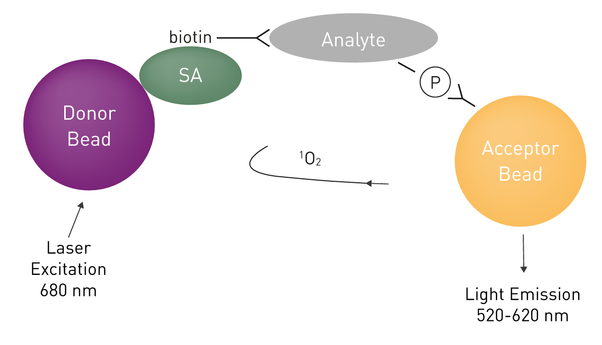
Two other platforms share the ability to use an antibody to total protein as a part of the assay. This enables them to be used to detect cellular phosphorylation events in addition to in vitro kinase assays. DELFIA® kinase assays have the potential to use tagged peptide or protein substrates for in vitro kinase assays or using cellular lysates as the source of the kinase. Plates are coated appropriately to bind the peptide and phosphorylation is recognized by an Eu-labeled anti-phospho antibody. If available, an appropriate antibody to total substrate protein can be used to coat the plates, thus, opening the door to assess cellular protein phosphorylation events11. The SureFire® Kinase Assays are based on Alpha technology and are consequently homogeneous, unlike DELFIA. Using SureFire Kinase Assays is further simplified by the wide number of verified antibody combinations, including those for phospho-ERK detection12. (fig. 7).
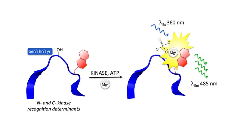
 The final type of platform is quite unique, using peptide sensors that are optimized for detection of kinases (and phosphatases) by using chelation-enhanced fluorescence that has been integrated with the Sox chromophore. The assay principle for the AssayQuants PhosphoSens® platform is depicted in figure 8.
 The platform can be used in homogeneous kinetic assay useful for characterizing inhibitors, especially mechanism of action and residence time. AssayQuant also offers slightly modified assays that can employ crude cell lysates or be used in an endpoint fashion for inhibitor screening13. Our scientific talk “Kinetic or endpoint monitoring of protein kinase and phosphatase activity” explains how the method can be targeted to a specific kinase and used in drug development.
Table 1: Kinase assays suited to microplate readers
| Assay Name | Principle | Detection Mode (s) | Homogeneous | Universal or kinase-specific | Endpoint or kinetic |
| LanthaScreen™ Kinase binding | Kinase binding | TR-FRET | Yes | Kinase specific | Endpoint or kinetic |
| Kinase-Glo® | ATP conversion | Luminescence | No | Universal | Endpoint |
| ADP-Glo™ | ATP conversion | Luminescence | No | Universal | Endpoint |
| Transcreener® | ATP conversion | Fluorescence, FP, TR-FRET | Yes | Universal | Kinetic or endpoint |
| ELISA-like | Substrate phosphorylation | Absorbance, fluorescence | No | Kinase specific | Endpoint |
| LanthaScreen™ Kinase Activity | Substrate phosphorylation | TR-FRET | Yes | Kinase specific | Endpoint |
| HTRF® KinEASE | Substrate phosphorylation | TR-FRET | Yes | Universal | Endpoint |
| DELFIA® kinase | Substrate phosphorylation | TRF | No | Kinase specific | Endpoint |
| SureFire® | Substrate phosphorylation | AlphaScreen AlphaLISA | Yes | Kinase specific | Endpoint |
| PhosphoSens® | Substrate phosphorylation | Fluorescence, TRF | Yes | Kinase specific | Kinetic or endpoint |
| Pyruvate Kinase Activity | Substrate conversion | Absorbance, fluorescence | Yes | Kinase specific | Kinetic or endpoint |
As we have shown throughout and summarized in Table 1 there are many possible ways to measure commercially available kinase assays. Having a multimode microplate reader like the Omega series, VANTAstar, CLARIOstar® Plus or PHERAstar® FSX will allow you to make choices based on what is best for the questions that you are trying to answer, rather than having limitations and trying to make an assay work that is not optimal for your purposes. If you are considering a high throughput screen for kinase inhibitors you should really look no further than the PHERAstar. It measures two emission wavelengths at the same time. This is required for the detection modes such as fluorescence polarization (FP) and TR-FRET, which are important in measuring kinases. The “simultaneous dual emission” capability reduces the read time by half and makes the PHERAstar FSX readers’ sensitivity and speed unparalleled.
Clearly, there are a lot of options for your kinase research. We hope to make your choice of a microplate reader the easiest part of your project.
https://journals.sagepub.com/doi/abs/10.1177/1087057109339207
https://journals.sagepub.com/doi/full/10.1016/j.jala.2006.07.001
Shultz et. al. (2009): Promega's ADP-Glo kinase assay, BMG LABTECH App Note 202.
Maurer et. al. (2016): PHERAstar FSX certified for Transcreener assays, BMG LABTECH App Note 291.
Ganske et. al. (2014): Transcreener ADP2 FI assay performed on BMG LABTECH microplate readers, BMG LABTECH App Note 274.
Kumar et. al. (2014): Transcreener ADP2 FP assay certification for BMG LABTECH instrumentation, BMG LABTECH App Note 269.
Kumar et. al. (2012): Methyltransferase, acetyltransferase, kinases, and GTPases can all be measured with Transcreener assays, BMG LABTECH App Note 225.
Hoffmann et. al. (2014): LanthaScreen TR-FRET tyrosine kinase and protein kinase C assay, BMG LABTECH App Note 268.
Absorbance plate reader with cuvette port
Powerful and most sensitive HTS plate reader
Most flexible Plate Reader for Assay Development
Upgradeable single and multi-mode microplate reader series
Flexible microplate reader with simplified workflows
ELISAs are a popular tool to detect or measure biological molecules in the life sciences. Find out how microplate readers can be used to advance research using immunoassays.
Light scattering offers distinct advantages for scientists interested in immunology. Find out how the NEPHELOstar Plus is used for high-throughput immunological tests.
Reactive oxygen species (ROS) may have physiological as well as pathological effects. Here we explain what ROS are and how they can be measured on microplate readers.