Introduction
G-protein coupled receptors (GPCRs) are a large family of receptors which have a broad involvement in cellular responses affecting many important body functions both in health and disease. On activation, GPCRs signal through different G-proteins (e.g. Gs, Gi or Gq) to elicit a cellular response which can be mediated through the activation of a number of different signaling cascades which include intracellular Ca2+ mobilization and the formation or inhibition of adenylate cyclase: Although these technologies have been successful in many cases they have had their limitation. Responses for many GPCRs such as Gi coupled GPCRs are not easily measured in current assay formats and other methods have been investigated as alternative readout. One such pathway is the ERK phosphorylation cascade, which results in the phosphorylation of both cytoplasmic and nuclear proteins regulating gene transcription. Hence this type of assay has the ability to measure cellular changes activated by Gs, Gi and Gq coupled receptors giving potential to use a single assay format for screening.
Here a microplate reader equipped with a flash lamp is used to detect ERK phosphorylation using an AlphaScreen® SureFire® proximity-based assay. ERK phosphorylation is demonstrated at receptors which signal via Gi and Gq G-proteins.
Assay Principle
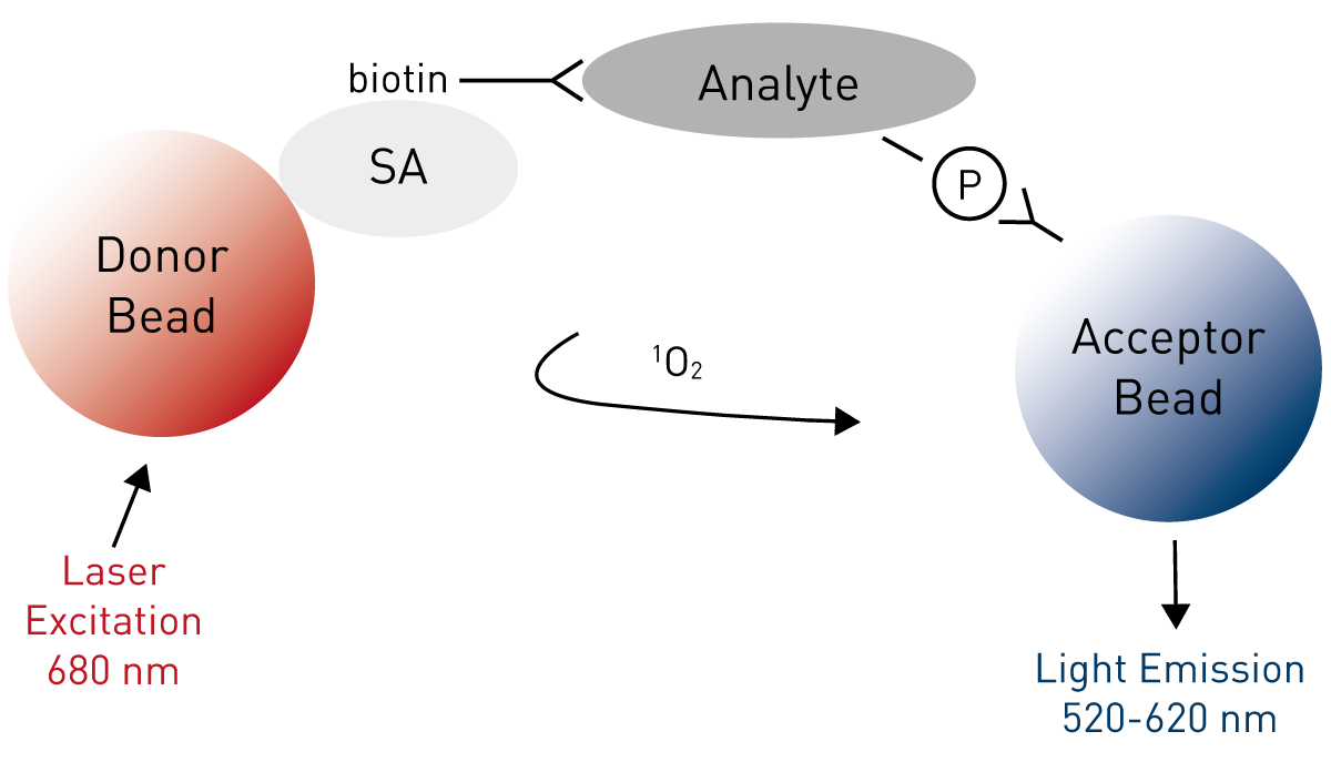
The AlphaScreen® assay uses the diffusion of singlet state oxygen from Donor to Acceptor beads. Upon excitation, at 680 nm of Donor beads, ambient oxygen is converted into singlet oxygen released at a rate of up to 60,000 molecules per second. Singlet oxygen molecules have a short lifetime (4 μs in aqueous solutions) and diffuse of no more than 200 nm. When a biomolecular interaction brings the Donor and Acceptor beads in proximity, the singlet oxygen reaches the Acceptor bead and a cascade of chemical reactions is initiated producing a greatly amplified luminescence signal in the range of 520-620 nm. The AlphaScreen® SureFire® ERK1/2 phosphorylation assay is based on an immuno-sandwich assay principle (Figure 1).
A phosphorylated cellular protein (analyte) is sandwiched between a streptavidin (SA)-coated Donor bead associated with an analyte antibody and a Protein A-conjugated Acceptor bead associated with an antiphospho specific antibody. Phosphorylation of the analyte results in an increase in the luminescence signal.
Materials & Methods
- Microplate reader from BMG LABTECH equipped with the AlphaScreen® option
- AlphaScreen® SureFire® ERK1/2 phosphorylation assay kit, PerkinElmer
- AlphaScreen® Protein A general IgG detection kit, PerkinElmer
- Proxiplate™-384 plus, white, 384-shallow well plate, PerkinElmer
- 96-well half area tissue treated cell culture plate, Corning
CHO cells stably expressing either a Gq or Gi coupled GPCR were plated at a cell density of 25K per well in 96 well half area tissue culture treated plates and cells were allowed to attach for 4h at 37°C. After 4h media was removed and replaced with 45 μL of DMEM/F12 w/o FBS to allow quiescence of the cells. Cells were incubated for 18h and then the assay performed. 5 μL of test compound was added and incubation time varied for time course experiments while kept constant, 5 min, for concentration response curve experiments. Following the incubation period, culture medium was removed and cells lysed with lysis buffer. Lysates were then transferred to a white 384-shallow well Proxiplate™ and AlphaScreen® beads were added in a single pipetting step in accordance with the kit protocol. The plate was incubated for 2h and read on the microplate reader. Data were expressed as % maximal response obtained with 0.3% FBS.
Instrument Settings
| Measurement method: | AlphaScreen |
| Reading Mode: | Endpoint |
| Filters: | Ex AS/Em AS |
General Settings
| Excitation time: | 0.30 s |
| Integration start: | 0.34 s |
| Integration time: | 0.60 s |
Results & Discussion
Initial experiments were designed to look at the optimal time of stimulation for both receptors.
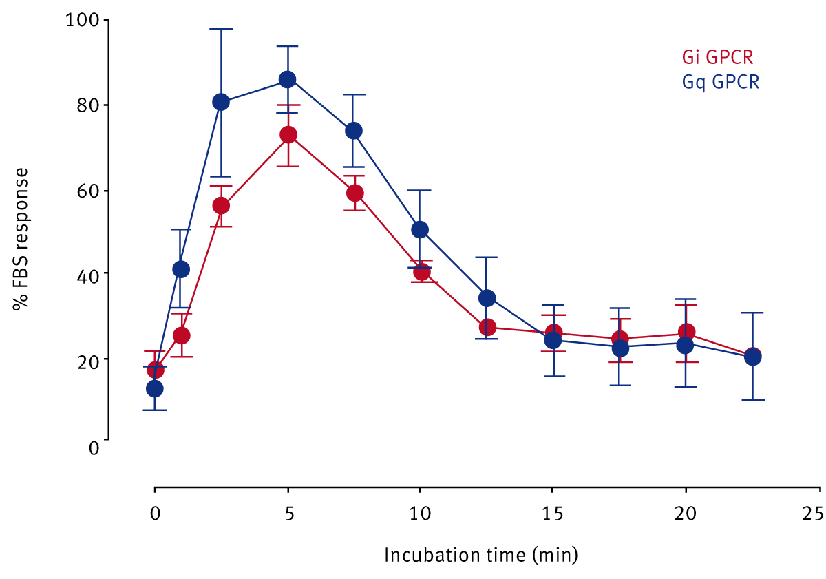
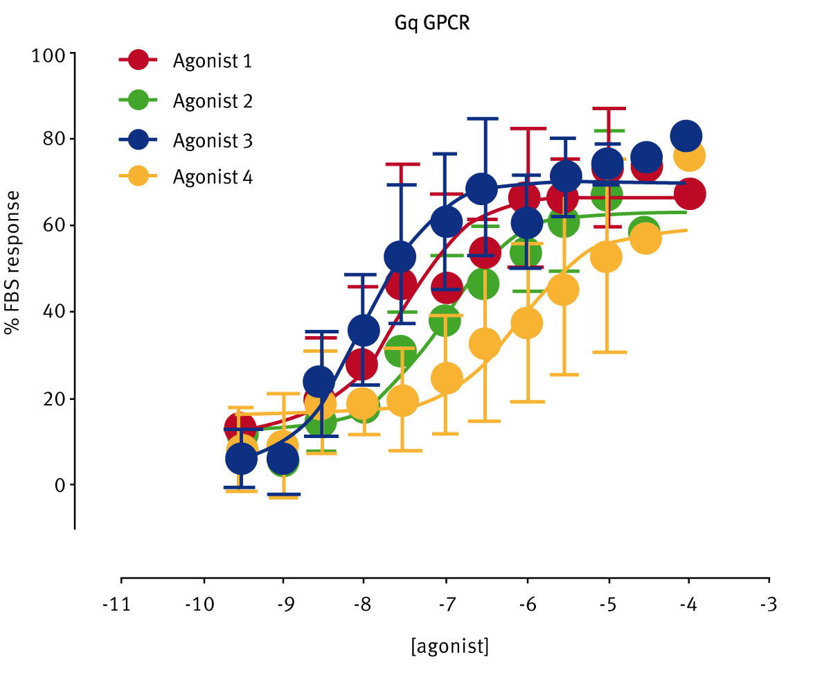
Agonist stimulation gave a transient increase in pERK1/2, peaking at 5 min for both Gi and Gq receptor activation. Using the 5 min stimulation time point standard agonists were profiled in the concentration response curve format (Fig. 3A und B).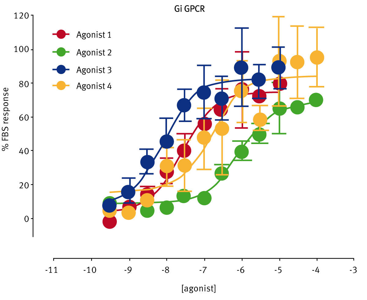
The AlphaScreen® SureFire® ERK1/2 phosphorylation assay system is able to detect G-Protein coupled receptor signaling and can be robustly measured using the BMG LABTECH microplate reader.
Conclusion
The AlphaScreen® SureFire® ERK1/2 phosphorylation assay was successfully performed in 384-well format.